Modulation of intestinal tight junctions by
Zonula occludens toxin permits enteral
administration of insulin and other
macromolecules in an animal model.
A Fasano, S Uzzau
J Clin Invest.
1997;
99(6)
:1158-1164.
https://doi.org/10.1172/JCI119271
.
The intestinal epithelium represents the major barrier to absorption of orally administered
drugs and peptides into the systemic circulation. Entry of molecules through the paracellular
pathway is restricted by tight junctions. We have previously reported that these structures
can be modulated by Zonula occludens toxin (Zot). In the present report, we show that Zot
reversibly increases rabbit intestinal permeability to insulin by 72% (P = 0.034) and
immunoglobulins by 52% (P = 0.04) in vitro. When tested in vivo, Zot induced a 10-fold
increase of insulin absorption in both the rabbit jejunum and ileum, whereas no substantial
changes were detected in the colon. Similar results were obtained with immunoglobulins,
whereby Zot induced twofold and sixfold increases of IgG absorption in the jejunum and
ileum, respectively. In diabetic rats, bioavailability of oral insulin coadministered with Zot
was sufficient to lower serum glucose concentrations to levels comparable to those
obtained after parenteral injection of the hormone. The survival time of diabetic animals
chronically treated with oral insulin + Zot was comparable to that observed in parenterally
treated rats. These studies offer an innovative strategy for the oral delivery of drugs and
proteins normally not absorbed through the intestine.
Research Article
Find the latest version:
J. Clin. Invest.
© The American Society for Clinical Investigation, Inc. 0021-9738/97/03/1158/07 $2.00
Volume 99, Number 6, March 1997, 1158–1164
Rapid Publication
Modulation of Intestinal Tight Junctions by Zonula Occludens Toxin Permits
Enteral Administration of Insulin and Other Macromolecules in an Animal Model
Alessio Fasano*‡ and Sergio Uzzau*
*Division of Pediatric Gastroenterology and Nutrition, and ‡Gastrointestinal Pathophysiology Section, Center for Vaccine Development, University of Maryland, School of Medicine, Baltimore, Maryland 21201
Abstract
The intestinal epithelium represents the major barrier to ab-sorption of orally administered drugs and peptides into the systemic circulation. Entry of molecules through the para-cellular pathway is restricted by tight junctions. We have previously reported that these structures can be modulated by Zonula occludens toxin (Zot). In the present report, we show that Zot reversibly increases rabbit intestinal perme-ability to insulin by 72% (P5 0.034) and immunoglobulins by 52% (P5 0.04) in vitro. When tested in vivo, Zot induced a 10-fold increase of insulin absorption in both the rabbit je-junum and ileum, whereas no substantial changes were de-tected in the colon. Similar results were obtained with im-munoglobulins, whereby Zot induced twofold and sixfold increases of IgG absorption in the jejunum and ileum, re-spectively. In diabetic rats, bioavailability of oral insulin coadministered with Zot was sufficient to lower serum glu-cose concentrations to levels comparable to those obtained after parenteral injection of the hormone. The survival time of diabetic animals chronically treated with oral insulin 1
Zot was comparable to that observed in parenterally treated rats. These studies offer an innovative strategy for the oral delivery of drugs and proteins normally not absorbed through the intestine. (J. Clin. Invest. 1997. 99:1158–1164.) Key
words: permeability • oral delivery • drugs • diabetes •
im-munodeficiency
Introduction
Intestinal absorption of numerous compounds routinely used for the treatment of common diseases is profoundly limited by their physicochemical characteristics. With the exception of those molecules which are transported by active or facilitated transcellular mechanisms, absorption of large and more hydro-philic macromolecules is almost exclusively limited to the
paracellular pathway (1). Under normal conditions, however, this pathway is restricted to molecules with molecular radii , 11 Å (2). The utility of the paracellular route for oral drug delivery has remained unexplored due to a limited under-standing of tight junction (tj)1 physiology and the lack of
sub-stances capable of increasing the tj permeability without irre-versibly compromising intestinal integrity and function (3–6). We have recently demonstrated that Zonula occludens toxin (Zot), a protein elaborated by Vibrio cholerae (7, 8), induced modifications of cytoskeletal organization that lead to the opening of tj related to the PKCa-dependent polymerization of actin monomers into actin filaments strategically localized to regulate the paracellular pathway (9). We have also shown that this modulation is reversible, time- and dose-dependent, and is confined to the small intestine, since Zot does not affect the colon permeability (7, 10). The selective effect of the toxin on the small intestine seems related to the regional distribution of the Zot receptors that are present in the jejunum and ileum, but not in the colon (10). The aforementioned properties make this moiety a potential tool for modulating the tj permeability. In the present report, we have demonstrated that Zot may be used to enhance the intestinal absorption of orally adminis-tered macromolecules through the paracellular pathway.
Methods
Zot purification. Purified Zot was obtained as previously described (10). Briefly, zot gene was fused in frame with the maltose binding protein (MBP) malE gene, using vector pMal-c2 (10) to create a MBP-Zot fusion protein. The fusion product was expressed in Es-cherichia coli, and obtained by disrupting the cells using a french press. The MBP-Zot fusion protein was then purified by affinity chro-matography using an amylose column (MBP-fusion purification sys-tem, New England Biolabs, Beverly, MA). Purified Zot was finally obtained by cleaving the fusion protein with factor Xa (10). The amount of toxin obtained was assessed by the Bradford method (11).
In vitro Ussing chambers experiments. Experiments were carried out as previously described (12) following an experimental protocol approved by our institutional animal welfare committee. Briefly, adult male New Zealand white rabbits (2–3 kg) were killed by cervical dis-location. Segments of rabbit intestine (including jejunum, ileum, and colon) were removed, rinsed free of the intestinal content, opened along the mesenteric border, and stripped of muscular and serosal layers. Eight sheets of mucosa so prepared were then mounted in Lu-cite Ussing chambers (1.12 cm2 opening), connected to a voltage
clamp apparatus (EVC 4000 WPI, Sarasota, FL), and bathed with freshly prepared buffer containing (mM): NaCl, 53; KCl, 5; Na2SO4,
30.5; mannitol, 30.5; Na2HPO4, 1.69; NaH2PO4, 0.3; CaCl2 1.25; MgCl2
Address correspondence to Alessio Fasano, M.D., Division of Pediat-ric Gastroenterology and Nutrition, University of Maryland, School of Medicine, 23 S. Greene Street, Box 140, Baltimore, MD 21201. Phone: 410-328-0812; FAX: 410-328-1072; E-mail: afasano@umabnet. ab.umd.edu
Received for publication 27 September 1996 and accepted in re-vised form 9 January 1997.
1.1; NaHCO3, 25. The bathing solution was maintained at 378C with
water-jacketed reservoirs connected to a constant-temperature circu-lating pump and gassed with 95% O2/5% CO2. Potential difference
(PD) was measured, and short-circuit current (Isc) and tissue resis-tance (Rt) were calculated as previously described (7). Once the tis-sues reached a steady state condition, paired tistis-sues, matched on the basis of their resistance, were exposed luminally to either 125I-insulin
10211M (2 mCi 5 10212 M) (Amersham Corp., Arlington Heights, IL)
or 156.25 ng 125I-IgG (1 mCi 5 83.3 ng), alone or in the presence of
1.1 3 10210 M Zot. 1-ml aliquot from the serosal side and 50 ml from
the mucosal side were immediately obtained to establish baseline val-ues. Samples from the serosal side were then collected at 20-min in-tervals for the following 80 min. The reservoirs were then emptied, washed twice with Ringer’s solution, and refilled with fresh Ringer’s solution containing only 10211 M 125I-insulin or 156.25 ng 125I-IgG
pre-viously added to the chambers. Two further 1 ml samples from the se-rosal side of each chamber were finally collected at 20-min intervals to establish whether the effect of Zot on the permeability of the drugs tested was reversible.
In vivo perfusion experiments. Intestinal perfusion was carried out as previously described, with minor modifications (10, 13). More specifically, after a 24-h fast, 2.5–3 kg adult male New Zealand white rabbits were anesthetized with 50 mg ketamine per kg body weight, followed by injection of 7.0 mg xylazine per kg body weight i.m. Body temperature was kept at 378C by a lamp. The abdominal cavity was opened by a midline incision, and three distinct segments of the intes-tine: (a) the proximal jejunum below the ligament of Treitz, (b) the distal ileum, and (c) the proximal colon, were cannulated in the same animal. A second cannula was placed 10–15 cm below each proximal cannula. The segments were rinsed free of intestinal contents with 0.9% (wt/vol) NaCl warmed to 378C. The proximal cannulae were connected by a polyvinyl tube to a peristaltic pump (model WPI SP220 I; World Precision Instruments, Inc.), and the three segments were si-multaneously perfused at a rate of 0.4 ml/min with the perfusion solu-tion (10) containing either 143 pmol/ml of 125I-insulin, or 8.33 ng/ml 125I-IgG. All the solutions were made isotonic by adjustment with
NaCl, and the pH was fixed at 7.4 by gassing with 95% O2/5% CO2.
Eluates were collected in 20-min aliquots from the distal cannu-lae. An initial equilibrium period of 30 min was allowed, followed by three consecutive 20-min collection periods for baseline measure-ment of the net transport of water and either insulin or IgG, in each segment studied. Subsequently, 1.1 3 10210 M Zot in PBS was added
to the perfusion solution, and a second perfusion period (30 min equilibration 1 33 20 min collection) was carried out. To establish whether the effect of Zot on intestinal permeability was reversible, a third perfusion period was performed with the same solution used in the first period. At the end of each perfusion period, the mesenteric vein draining each segment perfused was cannulated, and a blood sample was obtained to measure the amount of 14C-PEG-4000 and
the macromolecule tested (either 125I-insulin or 125I-IgG) that reached
the bloodstream. At the end of the experiment, the animal was killed, and the segments perfused were isolated, measured, dried and weighed. Water absorption was calculated as previously described (10).
In vivo experiments in BB/Wor diabetic rats. Acute, type 1 dia-betes mellitus male BB/Wor rats (12, 13) were obtained from the De-partment of Pathology of the University of Massachusetts. The ani-mals were anesthetized with a mixture of ketamine (113.2 mg/kg body wt [bw]) and acepromazine (0.68 mg/kg bw). The jugular vein was cannulated as previously described (14) to allow frequent blood drawing. The rats were kept fasting overnight. The following day, the animals were sedated by methoxyflurane inhalation, and the esopha-gus was cannulated to place the tip of the rigid cannula in the gastric antrum. The rats then received, via the cannulated esophagus, one of the following treatments: (a) oral ultralente® human insulin
(Humu-lin® U; range 5-30 U, Eli Lilly and Co., Indianapolis, IN) in 400 ml of
NaHCO3 solution (1.5 g/100 cm3, pH 8.2) so as to neutralize the
gas-tric acidity, (b) oral Humulin® U (range 5–30 U) plus purified Zot
(range 1.1–4.4 3 10210 mol) in 400 ml NaHCO
3 solution, (c) oral Zot
1.1 3 10210 mol in 400 ml of NaHCO
3 solution, (d) parenteral
Humu-lin® U (range 1.2–2.4 IU) (the dose was established on the basis of the
animal weight and glucose level, following the animal supplier recom-mendations) plus oral NaHCO3 solution (400 ml), or (e) oral NaHCO3,
400 ml. The glucose level of each animal was assessed before treat-ment, and at 30 min intervals thereafter, using a micromethod assay (One Touch® II blood glucose meter; Lifescan Inc., Milpitas, CA).
30 min after the beginning of the study, the rats were allowed food. To avoid possible biases, each animal was scheduled to receive all five treatments listed above on different days. Since some animals died before the completion of the entire protocol, only some rats received all five treatments listed above. The casualties were not significantly related to any of the treatments received, but were mainly because of the mechanical occlusion of the jugular cannulation. The different groups of treatment were comparable in terms of animal body weight and baseline glycemic levels.
In another set of experiments, the diabetic animals were divided in three groups of treatment: (a) parenteral Humulin® U (dose range
0.8–1.6 IU), (b) oral Humulin® U 10 IU1 Zot 1.1 3 10210 mol, and (c)
oral Humulin® U 10 IU. The body weight, temperature, and blood
glu-cose level (before treatment and 60 min thereafter) were daily moni-tored. Once the rats died, segments of the small and large intestine were isolated, fixed in paraformaldehyde 4%, and processed for light microscopy examination.
Statistical analysis. All values are expressed as means6standard error (SE). The analysis of differences was performed by Student’s
t-test for either paired or unpaired variates when applicable. For the experiments with BB/W or diabetic rats, the level of glucose among animals who received oral insulin alone was compared with those that received oral insulin 1 Zot by repeated measures analysis of vari-ance. In addition, individual t-tests were applied to each post-admin-istration timepoint. A P value , 0.05 was considered to be statisti-cally significant.
Results
In vitro animal model
Insulin and immunoglobulin G (IgG) were selected as proto-type molecules to be tested for oral delivery, based on the rela-tive size and structure, biological activities, and therapeutic relevance of these proteins. Rabbit ileum was mounted in Ussing chambers, and in vitro transepithelial transport of
125I-insulin and 125I-IgG from the mucosal (i.e., intestinal
lu-men) to the serosal (i.e., bloodstream) boundaries in the pres-ence and abspres-ence of purified Zot was measured. Zot (1.1 3 10210 M) reversibly increased the intestinal absorption of both
insulin (3.8460.41 fmol/cm2 per min vs. 6.6061.4 fmol/cm2 per
min, untreated vs. Zot-treated tissues, respectively, P5 0.034,
n5 4) (Fig. 1 C) and IgG (7.9761.17 pg/cm2 per min vs.
12.161.8 pg/cm2 per min, P5 0.04, n5 4) (Fig. 1 D) in a
time-dependent manner. Zot permeabilizing effect peaked at 80 min, and was completely reversible within 20 min from the withdrawal of the toxin from the Ussing chambers (see Fig. 1). This Zot-induced increase in absorption coincided with a re-duction in tissue resistance (Fig. 1, A and B). No significant changes of Rt were observed in control tissues after 80 min in-cubation, however, the tissues exposed to insulin alone showed an increase of Rt above the baseline when the solutions were changed (see Fig. 1 A).
In vivo animal model
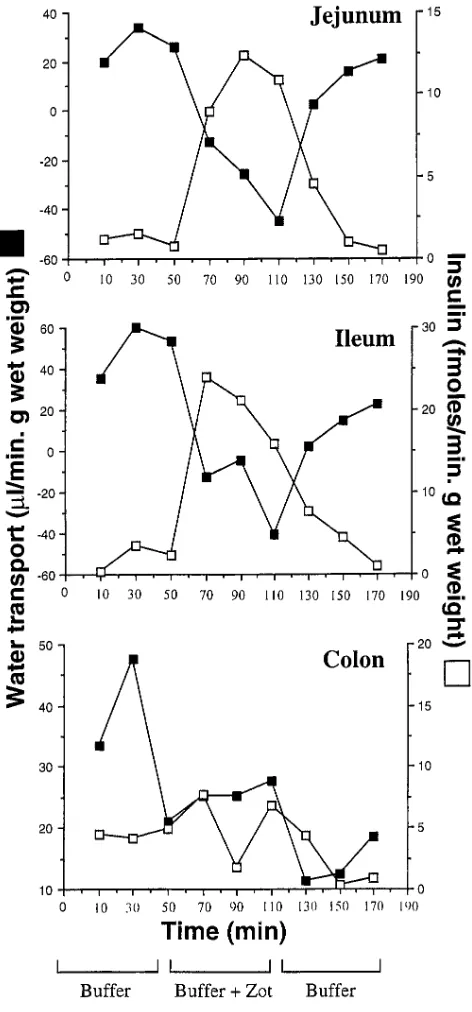
coin-cides with the regional distribution of Zot receptor(s) within the intestine (10). Therefore, three distinct segments of rabbit intestine—the jejunum, the distal ileum, and the colon—were simultaneously perfused in the same animal. When added to the perfusion solution, Zot (1.1 3 10210 M) increased the
pas-sage of 125I-insulin across both the jejunum and distal ileum
10-fold, whereas no substantial changes were observed in the co-lon (Fig. 2). The increased absorption of insulin was reciprocal with a shift of water absorption toward secretion (Fig. 2), a change that has been related to the permeabilizing effect of Zot on the paracellular pathway in vivo (10). This effect was detectable as soon as 20 min after Zot perfusion in the small intestine, and was completely reversible within 60 min of its withdrawal (Fig. 2). Zot also reversibly increased the serum concentration of both 125I-insulin and the non-absorbable marker 14C-polyethylene glycol (PEG)-4000 from the jejunum and the
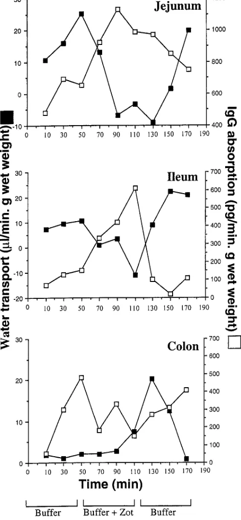
ileum, but not from the colon (Fig. 3). Similar results were ob-tained with IgG, whereby Zot (1.1 3 10210 M) induced twofold
and sixfold increases of 125I-IgG absorption in the jejunum and
ileum, respectively (Fig. 4). Again, no increases in absorption were detected in the colon (Fig. 4). Zot also increased the con-centration of both 125I-IgG and 14C-PEG-4000 in the efferent
mesenteric vein of the perfused small intestinal segments (data not shown).
Oral delivery of insulin in BB/Wor diabetic rats
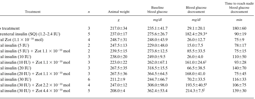
Acute treatment. We subsequently evaluated the bioactivity of insulin after enteral co-administration with Zot to acute type 1 diabetic male BB/Wor rats (15, 16). Insulin was orally administered to animals with or without Zot, and the blood glucose levels of the rats were serially measured. After oral ad-ministration of insulin alone (given at doses between 5 and 30 IU), blood glucose levels of treated animals were not apprecia-bly lowered (Table I). In contrast, when insulin at doses as low as 10 IU was orally coadministered with Zot 1.1 3 10210 mol
(5 mg), a significant reduction in blood glucose concentration was observed (Table I and Fig. 5). This decrement was compa-rable to that seen with a conventional dose of SQ insulin
(Ta-Figure 1. Reversible effect of purified Zot on tissue resistance (A and B) and transepithelial transport of insulin (C) and IgG (D) in rabbit ileum in vitro. Paired tissues, matched on the basis of their resistance, were exposed luminally to either 10211 M 125I-insulin (Amersham) (left) or 156.25
ng 125I-IgG (right), alone (s) or in the presence of 1.1 3 10210 M Zot (h). After 80 min of incubation, the Ringer’s solutions were replaced with
[image:4.612.58.539.59.434.2]ble I), and returned to baseline by 6 h post-administration (Fig. 5). There was a significant difference in mean glucose level between the oral insulin treatment and the oral insulin 1 Zot treatment (P5 0.002), as well as a significant time x group interaction (P5 0.03), reflecting the drop in glucose level in the presence of Zot. By t tests, the difference in glucose level reached statistical significance between 60 and 120 min post administration (Fig. 5). Administration of Zot alone did not
al-ter the blood glucose levels (Table I and Fig. 5). Mean (6SE) time to reach the blood glucose nadir after oral treatment with insulin 1 Zot (97612 min) was not different from that ob-served in the rats receiving parenteral insulin (90619 min). Furthermore, increased amounts of either Zot (up to 4.4 3 10210 mol) or insulin (up to 30 IU) each induced a
dose-depen-dent decrement of blood glucose (Table I). No toxicity was ob-served in the treated animals.
Prolonged treatment. To establish whether repeated Zot administration had any adverse effect on the survival of instru-mented animals, survival times were compared in diabetic rats daily treated with oral insulin 10 IU, with or without 1.1 3 10210 mol Zot. After the administration of insulin 1 Zot,
post-operative survival time (average 84 h) and decrease in blood glucose levels 60 min after treatment (average 31%) were com-parable to those observed in parenterally treated animals (96 h survival and 44% blood glucose decrement, respectively). In contrast, rats treated with oral insulin alone (10 IU) survived for only 60 h after jugular vein cannulation, and their blood glucose levels increased by 19% 60 min after treatment. None of the animals treated with insulin 1 Zot experienced diar-rhea, fever, or other systemic symptoms, and no structural changes could be demonstrated in the small intestine on histo-logical examination (data not shown).
Discussion
The intestinal epithelium represents the largest interface (more then 2,000,000 cm2) between the external environment and the
internal host milieu, and constitutes the major barrier through which molecules either can be absorbed or secreted. The para-cellular route is the dominant pathway for passive transepithe-lial solute flow in the small intestine, and its permeability
[image:5.612.316.555.57.242.2]de-Figure 2. Effect of purified Zot on water (j) and insulin (h) trans-port, as determined by the in vivo perfusion assay, in rabbit jejunum, distal ileum, and colon. Note the reversible increment of insulin ab-sorption that Zot induced in the small, but not in the large, intestine. This effect coincided with the decreased absorption of water evoked by the toxin. A representative experiment is shown.
Figure 3. Serum concentrations of 125I-insulin (j) and 14C-PEG-4000
(h) in the presence (period II) or absence (periods I and III) of Zot in the perfusion solution. At the end of each period (see methods sec-tion), the efferent mesenteric vein of the perfused segment was can-nulated to measure the amount of 125I-insulin and 14C-PEG-4000 that
[image:5.612.60.296.59.565.2]pends on the regulation of intercellular tj (17). A century ago, these structures were thought to be a secreted extracellular ce-ment forming an absolute and unregulated barrier within the paracellular space (18). Physiological studies of the past sev-eral decades have shown that the tj is a dynamic structure whose physiological regulation remains largely undefined (17). In the past few years, we have witnessed an explosion in re-search aimed at creating new drug delivery systems (19).
Un-fortunately, attempts so far to find ways to increase paracellu-lar transport by loosening intestinal tj have been hampered by unacceptable side effects induced by the potential absorption enhancing agents. (3–6). For the most part, these agents fall within two classes: (a) calcium chelators, and (b) surfactants (5). Both types have properties which limit their general utility as a means to promote absorption of various molecules. In the case of calcium chelators, Ca21 depletion induces global changes
in the cells, including disruption of actin filaments, disruption of adherent junctions, and diminished cell adhesion (6). In the case of surfactants, the potential lytic nature of these agents may cause exfoliation of the intestinal epithelium, irreversibly compromising its barrier functions (5).
Considering these limitations, it was worth examining whether our previous findings on Zot modulation of tj perme-ability could be applied to develop alternative approaches to enhancing drug absorption through the paracellular route. We have recently demonstrated that Zot activates a complex intra-cellular cascade of events that regulate intestinal permeability (9). Zot induces a dose- and time-dependent PKCa-related polymerization of actin filaments strategically localized to reg-ulate the paracellular pathway (9). These changes are a pre-requisite to opening of tj, and are evident at a toxin concentra-tion as low as 1.1 3 10213 M (10). The toxin exerts its effect by
[image:6.612.320.554.56.289.2]interacting with a specific surface receptor that is present in the small intestine, but not in the colon (10). Our previous data suggest that the regional distribution of Zot receptors coin-cides with the different permeabilizating effect of the toxin on the various tracts of intestine tested (10). We have also demon-strated, both in vivo (10 and present paper) and in vitro (7, 10,
[image:6.612.59.295.67.576.2]Figure 4. Effect of purified Zot on water (j) and IgG (h) transport as determined by the in vivo perfusion assay in rabbit jejunum, distal ileum, and colon. The experiment was carried out as described in the legend to Fig. 2. In this set of experiments the insulin in the perfusion solution was substituted with 125I-IgG 8.33 ng/ml.
and present paper) that the effect of Zot on tissue permeability occurs within 20 min of the addition of the protein to the intes-tinal mucosa, and is readily reversible once the toxin is re-moved. The in vivo experiments in BB/wor diabetic rats pre-sented in this study also showed a transient permeabilization effect of the toxin; the blood glucose levels of the animals orally treated with insulin 1 Zot decreased within 30 min of co-administration of the compounds, reached its nadir after 90 min, and returned to baseline values after 6 h (see Fig. 5).
Zot displays multiple properties that make it the most promising tool currently available to enhance drug and pep-tide transport through the intestinal mucosa. Zot (a) is not cy-totoxic and does not affect the viability of the intestinal epithe-lium ex vivo (7, 9); (b) fails to completely abolish the intestinal transepithelial resistance (7, 9, and present paper), (c) inter-acts with a specific intestinal receptor whose regional distribu-tion within the intestine varies (10), (d) is not effective in the large intestine where the presence of the colonic microflora could be potentially harmful if the mucosal barrier was com-promised (10 and present paper), (e) does not induce acute systemic side-effects (for at least 80–90 h) when orally adminis-tered (present paper), and (f) induces a reversible increase of tissue permeability (7, 9, and present paper). Our results dem-onstrate that coadministration of Zot with biologically active ingredients enhances intestinal absorption of the active mole-cule, and that this enhancement is effective for both relatively small (insulin, 5733 D) and large molecules (IgG, 140–160 kD). Furthermore, the experiments in BB/Wor diabetic rats demon-strate that orally delivered insulin can retain its biological ac-tivity without provoking severe hypoglycemia within the range of the insulin administered (i.e., up to 15 times more than the effective parenteral insulin dose). These findings have impor-tant practical implications, since the insulin therapeutic index (i.e., the ratio between the median toxic dose and the median therapeutic dose) is relatively low.
Our data suggest that the modulation of intestinal tj may be used for the oral administration of molecules normally not ab-sorbed through the intestine. Further studies are needed,
how-ever, to establish the possible clinical application of this system for the treatment of diseases that currently require frequent and long-life parenteral drug administration.
Acknowledgments
We thank Dr. Stuart A. Chalew, Dr. Simeon E. Goldblum, and Dr. Stephen S. Savarino for their comments on the manuscript. We are also indebted to Mrs. Klara Margaretten, Dr. Michael Salmonoff, and Ms. Carol Lauderbaugh for their invaluable technical assistance. Fi-nally, we thank Dr. Steven S. Wasserman for assistance with the sta-tistical analysis.
This work was funded in part by National Institute of Health grants DK48373 and AI35740 to A. Fasano.
References
1. Madara, J.L., and J.S. Trier. 1986. Functional morphology of the mucosa
of the small intestine. In Physiology of the Gastrointestinal Tract. L.R.
Johnson, editor. Raven Press, New York. 1209–1250.
2. Bakker, R., and J.A. Groot. 1989. Further evidence for the regulation of
the tight junction ion selectivity by cAMP in goldfish intestinal mucosa J.
Membr. Biol. 111:25–35.
3. Lee, V.H.L., A. Yamamoto, and V.B. Kompella. 1991. Mucosal
penetra-tion enhancers for facilitapenetra-tion of peptide and protein drug absorppenetra-tion. Crit. Rev.
Ther. Drug Carrier Syst. 8:91–192.
4. Muranishi, S. 1990. Absorption enhancers. Crit. Rev. Ther. Drug Carrier
Syst. 7:1–33.
5. Hochman, J., and P. Artursson. 1994. Mechanisms of absorption
en-hancement and tight junction regulation. J. Controlled Release. 29:253–267.
6. Citi, S. 1992. Protein kinase inhibitors prevent junction dissociation
in-duced by low extracellular calcium in MDCK epithelial cells. J. Cell Biol. 117:
169–178.
7. Fasano, A., B. Baudry, D.W. Pumplin, S.S. Wasserman, B.D. Tall, J.M.
Ketley, and J.B. Kaper. 1991. Vibrio cholerae produces a second enterotoxin,
which affects intestinal tight junctions. Proc. Natl. Acad. Sci. USA. 88:5242–
5246.
8. Baudry, B., A. Fasano, J.M. Ketley, and J.B. Kaper. 1992. Cloning of a
gene (zot) encoding a new toxin produced by Vibrio cholerae. Infect. Immun.
60:428–434.
9. Fasano, A., C. Fiorentini, G. Donelli, S. Uzzau, J.B. Kaper, K. Marga-retten, X. Ding, S. Guandalini, L. Comstock, and S.E. Goldblum. 1995. Zonula Occludens Toxin modulates tight junctions through protein kinase
C-depen-dent actin reorganization, in vitro. J. Clin. Invest. 96:710–720.
10. Uzzau, S., C.R. Fiore, K.T. Margaretten, and A. Fasano. 1996. Modula-Table I. Effect of Oral Insulin, Alone or Coadministered with Purified Zot on the Blood Glucose Levels of BB/Wor Diabetic Rats
Treatment n Animal weight
Baseline blood glucose
Blood glucose decreament
Time to reach nadir blood glucose
decreament
g mg/dl mg/dl min
No treatment 3 217.0634 235.1641.7 29.1620.1 180660 Parenteral insulin (SQ) (1.2–2.4 IU) 5 237.0617 275.6626.7 182.4629.3* 90619 Oral Zot (1.1 3 10210 mol) 4 248.7631 248.0643.9 26.0612.7 7569
Oral insulin (5 IU) 2 247.5613 229.0648.0 15.067.5 78617 Oral insulin (5 IU) 1 Zot 1.1 3 10210 mol 2 239.5615 273.8612.5 85.5633.5 75615
Oral insulin (10 IU) 3 238.0620 249.069.5 26.064.0 110650 Oral insulin (10 IU) 1 Zot 1.1 3 10210 mol 3 223.0622 262.0667.1 161.0624.6‡ 93628
Oral insulin (20 IU) 3 267.5635 318.5615.5 66.5638.5 140670 Oral insulin (20 IU) 1 Zot 1.1 3 10210 mol 3 267.5636 364.5664.5 168.0641.0 75645
Oral insulin (30 IU) 6 211.269 244.7666.7 70.2633.5 116633 Oral insulin (30 IU) 1 Zot 2.2 3 10210 mol 4 247.0612 308.0698.0 193.5640.5§ 106675
Oral insulin (30 IU) 1 Zot 4.4 3 10210 mol 5 208.064 362.4653.4 214.367.5i 139630
Oral insulin, when orally coadministered with Zot, decreases serum glucose concentration of diabetic rats to levels comparable to those obtained with
the parenteral administration of the hormone. *P5 0.01 vs. no treatment, P5 0.007 vs. oral insulin (10 IU), P5 0.003 vs. oral Zot (1.1 3 1021 mol); ‡P5
[image:7.612.66.557.76.268.2]tion of intestinal tight junctions: a novel mechanism of intestinal secretion. Gas-troenterology. 110:370a. (Abstr.)
11. Bradford, M.M. 1976. A rapid and sensitive method for the quantitation of microgram quantities of protein utilizing the principle of protein-dye
bind-ing. Anal. Biochem. 72:248–254.
12. Fasano, A., F.R. Noriega, D.R. Maneval, Jr., S. Chanasongcram, R. Rus-sell, S. Guandalini, and M.M. Levine. 1995. Shigella enterotoxin 1: an entero-toxin of Shigella flexneri 2a active in rabbit small intestine in vivo and in vitro.
J. Clin. Invest. 95:2853–2861.
13. Guandalini, S., A. Fasano, F. Albini, G. Marchesano, A. Nocerino, M. De Curtis, F.F. Rubaltelli, A. Pettenazzo, and A. Rubino. 1988. Unconjugated bilirubin and the bile from light exposed Gunn rats inhibit intestinal water and
electrolyte absorption. Gut. 29:366–371.
14. Harms, P.G., and S.R. Ojeda. 1974. A rapid and simple procedure for
chronic cannulation of the rat jugular vein. J. Appl. Physiol. 36:391–392.
15. Bellgrau, D., and A.C. Lagarde. 1990. Cytotoxic T-cell precursors with low-level CD8 in the diabetes-prone Biobreeding rat: implications for
genera-tion of an autoimmune T-cell repertoire. Proc. Natl. Acad. Sci. USA. 87:313–317.
16. Haber, B.A., S. Chin, E. Chuang, W. Buikhuisen, A. Naji, R. Taub. 1995. High levels of glucose-6-phosphatase gene and protein expression reflect
an adaptive response in proliferating liver and diabetes. J. Clin. Invest. 95:832–841.
17. Anderson, J.M., and C.M. Van Itallie. 1995. Tight junctions and the
mo-lecular basis for regulation of paracellular permeability. Am. J. Physiol. 269:
467–475.
18. Cereijido, M. 1992. Evolution of ideas on the tight junction. In Tight
Junctions. CRC Press, Inc., Boca Raton, FL. 1–13.
19. Langer, R. 1990. New methods of drug delivery. Science (Wash. DC).