R E S E A R C H A R T I C L E
Open Access
Cervical spine reposition errors after
cervical flexion and extension
Xu Wang
1,3, René Lindstroem
1, Niels Peter Bak Carstens
2and Thomas Graven-Nielsen
4*Abstract
Background:Upright head and neck position has been frequently applied as baseline for diagnosis of neck problems. However, the variance of the position after cervical motions has never been demonstrated. Thus, it is unclear if the baseline position varies evenly across the cervical joints. The purpose was to assess reposition errors of upright cervical spine.
Methods:Cervical reposition errors were measured in twenty healthy subjects (6 females) using video-fluoroscopy. Two flexion movements were performed with a 20 s interval, the same was repeated for extension, with an interval of 5 min between flexion and extension movements. Cervical joint positions were assessed with anatomical landmarks and external markers in a Matlab program. Reposition errors were extracted in degrees (initial position minus reposition) as constant errors (CEs) and absolute errors (AEs).
Results:Twelve of twenty-eight CEs (7 joints times 4 repositions) exceeded the minimal detectable change (MDC), while all AEs exceeded the MDC. Averaged AEs across the cervical joints were larger after 5 min’intervals compared to 20 s intervals (p< 0.05).
Conclusions:This is the first study to demonstrate single joint reposition errors of the cervical spine. The cervical spine returns to the upright positions with a 2° average absolute difference after cervical flexion and extension movements in healthy adults.
Keywords:Spine, Neck, Fluoroscopy, Cervical vertebrae, Position sense, Motion, Flexion and extension, Repositioning
Background
The upright head and neck position is the most frequent human posture of daily life. This position is baseline for scientific studies and diagnosis [1–3]. The natural head position can be compared within or between subjects [4], and the initial head position may influence move-ments of the cervical spine [5, 6]. The variation of the upright posture after neck movements between cervical joints or cervical regions are unknown, and it is unclear if cervical spine motion should be regarded in single joint units, as multi-joint units or as regional units with respect to joint reposition.
Cervical x-rays include the upright posture, and change in flexion and extension x-rays are assessed from
the upright neck posture. Such as in pre- and post-surgical evaluation of cervical spine motions [1–3, 7–9]. Knowledge of the variance in cervical upright joint repo-sitioning, is a prerequisite for assessment of dynamic cervical joint motion; however, the variance has never been investigated. Thus, it is unclear how much the up-right neck varies in scientific investigation and diagnosis. Impaired proprioception has been demonstrated in pa-tients with cervical disorders and forward head postures [10–14]. Cervical radiculopathy patients showed im-paired head reposition acuity compared with healthy controls [2]. Head or neck repositioning were also im-paired in older adults [15], and patients with cervical spondylosis [16], cervicogenic dizziness [17], whiplash [18, 19], muscle fatigue [20], and non-traumatic neck pain [21–23]. In general, reposition acuity has been used to evaluate proprioception [24]. Subjects with pain in the upper cervical region demonstrated additional * Correspondence:[email protected]
4Center for Neuroplasticity and Pain (CNAP), SMI, Department of Health and
Science Technology, Faculty of Medicine, Aalborg University, Fredrik Bajers Vej, 7D-39220 Aalborg, Denmark
Full list of author information is available at the end of the article
impairment in sensorimotor control compared to sub-jects with pain in the lower cervical region [21].
Head and neck positions can be resolved with respect to the horizontal plane or other anatomical structures [2, 8]. For both assessments it is unknown if head and neck reposition errors mainly occur in the suboccipital region or in the cervical regions below.
The sub-occipital anatomy, muscle density and func-tion are different compared with that of the lower cer-vical spine [25]. The distinct osseous shapes of occiput, atlas, and axis underlie functional differences. The upper cervical spine contributes with almost 60% of free and unrestricted cervical spine axial rotation [26].
The acuity of cervical joints’ position sense in healthy subjects is important. This is because reposition errors of the upright position are reflected in dynamic motion of the cervical spine and in clinical studies, where the upright cervical spine serves as baseline [6].
Memory of position or time delay effects influence the cervical joint position sense [27, 28]. Time delay is hy-pothesized to affect the reposition of cervical joints. However, the effect of time delay on neck reposition is unknown.
The aim of this study was to assess healthy cervical spine reposition errors in the upright position. It was hy-pothesized that 1) all single cervical joints demonstrate reposition errors between end-range flexion and exten-sion movements and 2) the reposition error is increased with longer time delays.
Methods Participants
Six healthy females (age: 24.3 ± 3.8 years; height: 163.5 ± 6.0 cm; weight: 56.8 ± 7.5 kg; body mass index: 21.2 ± 2.4 kg/m2; mean ± standard deviation) and 14 healthy males (27.6 ± 5.4 years; 179.1 ± 6.6 cm; 74.1 ± 6.6 kg; 23.0 ± 1.5 kg/m2) without neck symptoms within the last 3 months were included. Exclusion criteria were possible pregnancy and any neck disorder. All participants were recruited from university students and staff through bul-letins and a website.
Ethics, consent and permissions
The study was conducted in accordance with the Declar-ation of Helsinki and approved by the North Denmark Region ethics committee (N20140004). Participants signed a written informed consent form.
Experimental procedure
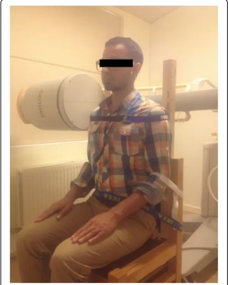
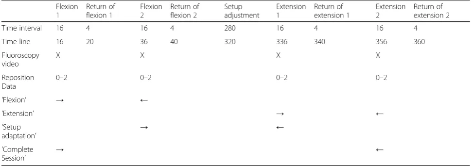
Static baseline fluoroscopy images from two repetitive flexion and two repetitive extension motions were ex-tracted from a larger study of cervical dynamic motion. For data acquisition subjects were seated in a chair with
hips, knees and ankles at 90°. Shoulders, elbows and waists were fixed by straps (Fig. 1).
Subjects were instructed to sit in their normal upright head and neck position and return to this initial upright head and neck position as precisely as possible after cer-vical motion and stay in that position for approximately 4 s until beginning of the second repeated motion. The return motion to the upright position was not recorded in order to reduce radiation exposure (Table 1). Timing and execution of experimental tasks were practiced sev-eral times before recording, subjects were instructed to move their head and neck through their entire range of flexion or extension and return to the initial upright position. To reduce out of plane motion subjects were provided a line in the sagittal plane on the wall, ceiling and floor. Subjects were instructed visually to follow this line from a mark at their eye height, and return to this mark.
Two flexion movements followed by two extension motions were recorded from upright to end range. The static position at the beginning of each motion was re-corded for 2 s, and the baseline image was extracted from these 2 s. The outcome image was recorded with 4 s delay at the beginning of the second repeated motion (Table 1). Thus the recordings included the baseline
[image:2.595.304.539.401.695.2]positions from consecutive motions. The static images were screened for motion wobble, and the extracted video images represent the static positions found in ac-cordance with the experimental procedures.
The diameter of the fluoroscopic screen was too small to accommodate acquisition of both flexion and exten-sion motions without a 300-s change in the experimental setup. The experimental chair was moved with the ject fixed and without active participation from the sub-ject. Subjects were at the end of change in set up reminded to return to the previously memorized baseline position before data acquisition (Table 1). Subjects were instructed to return to and remain in the memorized upright baseline position through the experimental session. Flexion and extension move-ments were free and unrestricted. The flexion and ex-tension time was approximately 16 s including 2 s of static imaging at upright and end-range, and after data acquisition the subjects returned to upright at their own pace.
Fluoroscopic recordings
Fluoroscopy videos (see Additional file 1) were re-corded from the upright position to the end range of either the flexion or extension movement at 25 frames per second with an average of 45 KV, 208 mA, 6.0 ms X-ray pulses and average source-to-subject (C7) distance of 92.4 cm. The video sequences were digitalized and stored on a computer. The aver-age radiation dose for the variability study of cervical motion patterns was calculated to be 0.48 mSv by PCXMC, the radiation dose for the static images in-cluded in this study is calculated to 0.06 mSv [29].
Image analysis
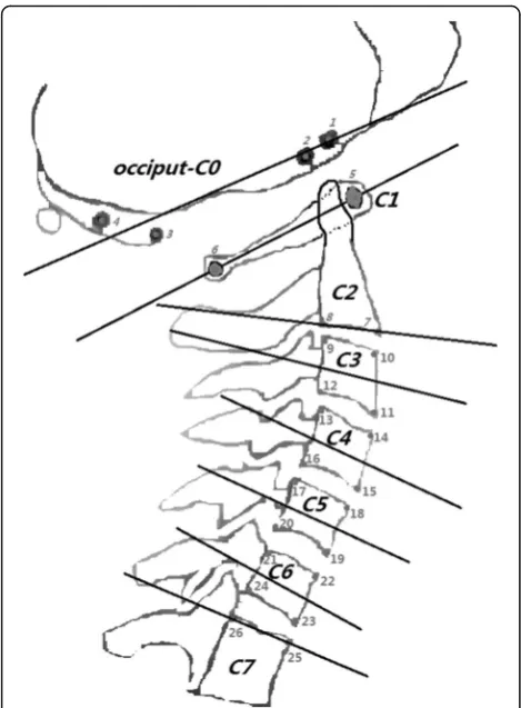
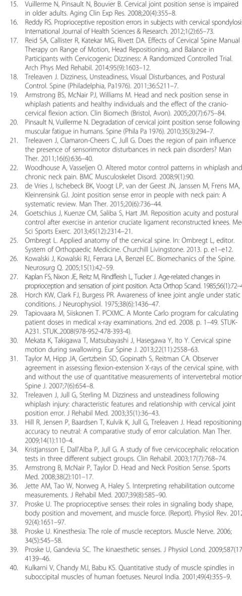
Each image was manually marked and analyzed in a custom-designed Matlab-based program. Calculations of joint rotation were according to a previous validated method [7, 9, 30, 31]. The cervical joint motion was de-termined from the vertebral midplanes [3, 7]. Change in cervical joint motions were recorded as reposition errors in degrees. The analysis returned individual joint angles in degrees. The midplanes were resolved with respect to the horizontal plane, and change in the midplanes of C0 and C7 demonstratedachange in head position and the thoracic spine below C7, respectively. Anatomical struc-tures were marked on all vertebrae except for C0, which were marked with 4 external markers (Fig. 2).
Reposition error
[image:3.595.55.543.99.270.2]Data collection yielded four upright baseline positions (flexion1, flexion2, extension1, extension2) for C0/C1, C1/C2, C2/C3, C3/C4, C4/C5, C5/C6, and C6/C7 (Table 1). The upright positions were used to calculate four reposition errors (‘flexion’,‘extension’,‘setup adapta-tion’and ‘complete session’) as illustrated in Table 1. Re-position errors were analyzed as CEs and AEs. The CEs were the change in joint disc midplanes, and the AEs were the absolute values of the CEs. Previously, constant errors (CEs) and absolute errors (AEs) have been applied in studies of reposition errors [32–35]. CE represents the average magnitude of reposition errors demonstrat-ing multidirectional under and over estimations of target position [33]. The AE represents the average AE and is calculated as the absolute value of CE [33]. The joints moved in extension with positive change and in flexion with a negative change, thus the sign indicates the direc-tion of the errors.
Table 1Experimental procedures
Flexion 1
Return of flexion 1
Flexion 2
Return of flexion 2
Setup adjustment
Extension 1
Return of extension 1
Extension 2
Return of extension 2
Time interval 16 4 16 4 280 16 4 16 4
Time line 16 20 36 40 320 336 340 356 360
Fluoroscopy video
X X X X
Reposition Data
0–2 0–2 0–2 0–2
‘Flexion’ → ←
‘Extension’ → ←
‘Setup adaptation’
→ ←
‘Complete
Session’ → ←
Statistical analysis
Reposition errors are presented as mean and standard error of measurement (SEM), and measurement errors are presented with SEM. Reposition errors and measure-ment errors were tested for normality with the Shapiro-Wilk test demonstrating skewness of reposition errors but normal distribution of measurement errors.
For each joint CEs or AEs in the four comparisons (‘flexion’,‘extension’,‘setup adaptation’and ‘complete ses-sion’) were compared using the Friedman test and if sig-nificant followed by post-hoc assessments by the Wilcoxon test with Bonferroni corrections. Kruskal-Wallis test was performed for difference of CEs or AEs between joints in each task (‘flexion’,‘extension’, ‘setup adaptation’ and ‘complete session’) and post-hoc Mann Whitney U-test with Bonferroni corrections were
applied to assess specific differences. The significance level was set atP< 0.05.
One investigator (XW) analyzed an upright image three times for intra-rater reliability. Intra-class correla-tions coefficient (ICC 3,1) assessed reliability of image marking. The minimal detectable change (MDC) for the measurement error was further calculated by 1.96 ×√2 × SEM [36]. Statistical analysis was performed in SPSS (IBM Statistics 22).
Results
In total, 140 cervical spine joints were analyzed among the 20 participants. However, the shoulder shadow in two subjects obscured C5/C6 and C6/C7 yielding 136 joints for analysis. The intra-rater analysis results across joints are presented in Table 2. The intra-rater reliability test demonstrated the average SEM marking error across the three images to be between 0.13° and 0.42°. The ICC of the intra-rater image marking for the three images was 0.998. The reported reposition errors occurred mainly within the C0 to C7 range, as the average changes of C0 and C7 with respect to the horizontal plane across the four upright positions were mean (SEM) 0.53° (1.64°) and 1.04° (1.07°), respectively.
[image:4.595.56.291.86.405.2]Constant errors
Table 3 presented CEs from the four tasks. Twelve of 28 joints in Table 3 exceeded the joint MDC in Table 2.
The average CEs across all joints were small for all tasks (Table 3). Head and neck showed small average CEs the Mean (SEM) of ‘flexion’ and ‘extension’ were 0.21° (0.28°) and 0.01° (0.30°), respectively. The range of all the four taskswas−21.1° to 14.3°. The range showed that cervical joints displayed a large variation of reposi-tioning errors at a single joint.
Analysis of differences in CEs within joints was signifi-cant for the ‘setup adaptation’ task (Kruskal Wallis, P< 0.05), and post-hoc test showed that the C0/C1 repos-ition error was larger than C2/C3 (P< 0.05) (Table 3). Friedman test showed no significant effect of time delay on constant reposition errors.
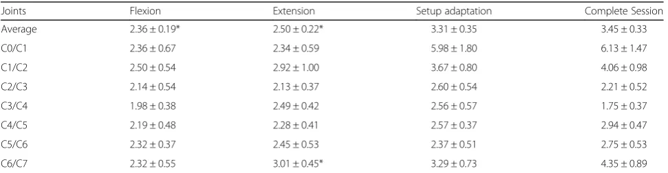
Absolute errors
All AEs in Table 4 exceeded the MDC. Average AEs were larger compared to CEs. Absolute ‘flexion’ and Fig. 2shows the analysis marking points. Four metal balls on pliable
[image:4.595.306.539.664.706.2]wires attached to a pair of glasses served as external markers for occiput (C0), the central areas of the medullary cavities of the anterior and posterior arch were marked on atlas (C1). Two points in proximity to the inferior vertebral plate were marked on axis (C2). The third to the sixth cervical vertebrae (C3-C6) were marked with 4 points in proximity to the vertebral plates. The seventh cervical vertebra (C7) was marked with two points in proximity to the superior vertebral plate. The mid-planes were calculated from the marking points, and joint angles were further calculated between mid-planes. The author XW created the skeletal illustrations
Table 2Minimal detectable change
Joints C0/C1 C1/C2 C2/C3 C3/C4 C4/C5 C5/C6 C6/C7 Overall
MDC 0.35° 0.70° 0.70° 0.73° 1.17° 1.17° 0.58° 0.73°
SEM 0.13° 0.25° 0.25° 0.26° 0.42° 0.42° 0.21° 0.26°
‘extension’ were 2.36° (0.19°) and 2.50° (0.22°), respect-ively (Table 4).
The average AEs found for ‘flexion’,‘extension’,‘setup adaptation’ and ‘complete session’ were different from each other (Table 4; Friedman, P< 0.05). Post-hoc test showed that the average AEs were smaller for ‘flexion’ (P< 0.05) and ‘extension’ (P< 0.05) compared to ‘complete session’. The result showed the increased AEs for 340 s time delay (‘complete session); however, a simi-lar increase was not found for 300 s time delay (‘setup adaptation’).
The CEs and AEs of the upper cervical region (C0– C2) showed larger reposition errors compared to the middle and lower cervical regions (Table 3 and Table 4).
Discussion
The study demonstrates that the position of head and cervical joints varies when repositioned in the memo-rized upright postures. Twelve out of twenty-eight CEs exceeded the MDC, and all AEs exceeded the MDC. The first study hypothesis was confirmed with respect to AEs, as all single cervical joints demonstrated reposition errors which exceeded the MDC. In contrast the MDC was only exceeded by 12 out of 28 CEs.
The study gave conflicting evidence on the second hy-pothesis, which tested if reposition errors are increased
with longer time delays, as one (340 s AE) of the two long time delays showed significantly different reposition errors, while the other (300 s AE) showed no difference. Likewise, no increased reposition errors were demon-strated with time for CEs.
Constant errors & absolute errors
The average AE of the flexion movements in this study was 2.36°, similar results were documented by Artz et al. after flexion movement with reposition errors from 1.61° to 2.25° [8]. The CE was also calculated as the average CE across all cervical joints and in ‘flexion’it was 0.21° ± 0.28° which is in contrast to the larger average AE for all joints (2.36° ± 0.19°) in‘flexion’.
This study showed large reposition errors for individ-ual joints; however, these large reposition errors were frequently counterbalanced by large reposition errors in other cervical joints in order to acquire a suitable head position. The average CEs were close to zero, this is in line with a previous study of head and neck reposi-tioning, this study found the reposition error suitable for group comparisons within or between patients [4].
[image:5.595.56.540.99.224.2]Proprioception from muscle spindles is a factor in motor control [37–39] and muscle spindles are more frequent in the upper cervical region compared to the lower cervical region [40]. However, the larger numbers
Table 3Constant reposition errors of cervical joint
Joints Flexion Extension Setup adaptation Complete Session
Average 0.21 ± 0.28 0.01 ± 0.30 0.12 ± 0.45 0.34 ± 0.44
C0/C1 1.74 ± 0.88 1.74 ± 0.68 −4.82 ± 1.98* −1.35 ± 2.01
C1/C2 0.05 ± 0.79 −0.66 ± 1.20 1.66 ± 1.09 1.06 ± 1.33
C2/C3 −0.70 ± 0.71 −0.52 ± 0.60 1.96 ± 0.67 0.74 ± 0.71
C3/C4 −0.63 ± 0.58 −0.65 ± 0.69 0.25 ± 0.81 0.27 ± 0.54
C4/C5 1.49 ± 0.61 −0.37 ± 0.66 −0.32 ± 0.69 0.80 ± 0.80
C5/C6 −0.31 ± 0.67 0.27 ± 0.79 0.42 ± 0.76 0.38 ± 0.85
C6/C7 −0.29 ± 0.78 −1.14 ± 0.81 1.89 ± 0.98 0.46 ± 1.37
Mean (± SEM) of reposition errors in degrees from tasks defined in Table1(‘flexion’,‘extension’,‘setup adaptation’and‘complete session’). Significantly different from C2/C3 (*,P< 0.05)
Table 4Absolute reposition errors of cervical joint
Joints Flexion Extension Setup adaptation Complete Session
Average 2.36 ± 0.19* 2.50 ± 0.22* 3.31 ± 0.35 3.45 ± 0.33
C0/C1 2.36 ± 0.67 2.34 ± 0.59 5.98 ± 1.80 6.13 ± 1.47
C1/C2 2.50 ± 0.54 2.92 ± 1.00 3.67 ± 0.80 4.06 ± 0.98
C2/C3 2.14 ± 0.54 2.13 ± 0.37 2.60 ± 0.54 2.21 ± 0.52
C3/C4 1.98 ± 0.38 2.49 ± 0.42 2.56 ± 0.57 1.75 ± 0.37
C4/C5 2.19 ± 0.48 2.28 ± 0.41 2.57 ± 0.37 2.94 ± 0.47
C5/C6 2.32 ± 0.37 2.45 ± 0.53 2.37 ± 0.51 2.75 ± 0.53
C6/C7 2.32 ± 0.55 3.01 ± 0.45* 3.29 ± 0.73 4.35 ± 0.89
Mean (± SEM) of reposition errors in degrees across‘flexion’,‘extension’,‘setup adaptation’and‘complete session’.‘Extension’was different compared with
[image:5.595.56.542.588.714.2]of muscle spindles in the upper cervical region are not reflected in the reposition errors found in the upper cer-vical region. Larger reposition errors were demonstrated in the upper cervical region compared to the lower cer-vical region. The results are in agreement with the re-sults found by Treleaven et al. in neck pain disorders [21]. Thus, larger upper cervical region reposition errors have been demonstrated both in healthy subjects and pa-tients with neck pain. Treleaven et al. suggested that upper and lower regions should not be grouped in re-position error studies of whiplash as the grouping may decrease homogeneity. This study supports this sugges-tion for healthy subjects. This study also suggests that the cervical spine should not be regarded as a single unit of motion, the cervical spine should be regarded as a complex structure with multiple units of motion.
The reposition errors documented in this study appear to reflect the normal variance of cervical motions, and the reposition errors may furthermore be reflected in re-peated dynamic neck movements to and from the up-right position. The average absolute upup-right head and neck reposition error (2.36°) may profoundly influence the cervical joint motion pattern, as 2.36° may be a large proportion of the total joint motion.
The results show a variance in reposition of head and neck, the variance may be attributed to normal variance of motor control; however, the variance may also be in-fluenced by other factors such as posture. Forward head posture was associated with larger reposition errors after flexion and extension movements, when compared to a control group [12]. However, the sitting posture did not vary significantly for postural repositioning errors of cervico-thoracic angle compared to control groups [13]. The joint reposition errors demonstrated in this study may improve the understanding of cervical joint position sense. The study suggests the need to control for normal variance of sensor positions in studies of cervical repos-ition errors, and the study further suggests that results across multiple joints may have varying contributions from those joints.
Time delay and reposition error
No significant difference of the head and neck was found for CEs of 20 s, 300 s or 340 s time delays. For AEs avaged across joints, twenty seconds delay gave smaller er-rors than 340 s delay although no difference was found between 20 s delay and 300 s delay.
Study limitations
The largest confounder was the measurement error and this was reflected in the MDC. The orientation of C0 and C7 with respect to horizontal changed; however, the average change was small. Other confounders, such as image distortion, out of plane motions and natural
variance were not reflected in the MDC; however, only upright images were used, so errors due to image distor-tion and out of plane modistor-tion were minimal, and the ob-jective of the study was to assess natural variations. The inter-rater examiner was not blinded, as the marking procedure required the shape of the initial marking of a specific vertebra to be reference for subsequent mark-ings of that vertebra in a marking session. The investiga-tor was not blinded to his own ratings, as the marking procedures required the shape of the initial marking of a vertebra as reference for subsequent markings of that vertebra in a marking session. The study investigated re-positioning of single cervical joints after free and unre-stricted motion with visual tracking of a line on the floor wall and ceiling. Most reposition studies of the head and neck are conducted blindfolded. The nature of neck pain is recurrent. Thus, no neck pain within the last 3 months may not ensure that all included necks were healthy.
Conclusion
This is the first study to investigate reposition errors of single cervical joints after flexion and extension move-ments. The average CEs after flexion and extension movements were 0.21° and 0.01°, respectively. The aver-age AEs after flexion and extension movements were 2.36° and 2.50°, respectively. This results indicated that in healthy subjects the cervical spine returns to the neu-tral upright position with an error of approximately 2.5° after flexion and extension movements.
Additional file
Additional file 1:Fluoroscopic video of cervical motion. A flexion video of cervical flexion movement recorded by fluoroscopic video technology. Find the file at http: (MP4 4148 kb)
Abbreviations
BMI:Body mass index; SEM: Standard error of measurement; ICC: Intra-class coefficient correlation; MDC: Minimal detectable change; CE: Constant error; AE: Absolute error
Acknowledgement Not applicable
Funding
Danish Chiropractors’Foundation supported this study. XW has been awarded a scholarship provided by the China Scholarship Council (File NO. 201306170027) to pursue his PhD study at Aalborg University. Center for Neuroplasticity and Pain (CNAP) is supported by the Danish National Research Foundation (DNRF121).
Availability of data and materials
Data will be shared when formal request is approved by Professor Thomas Graven-Nielsen ([email protected]).
Authors’contributions
contributed to funding, study design. RL, NPBC and TGN made critical revisions and suggestions for interpretation of manuscript. All authors read and approved the final version of the report.
Competing interests
The authors declare that they have no competing interests.
Consent for publication
[image:7.595.295.535.89.679.2]Written consent form was signed by the participant for publication of the figure in this manuscript.
Ethics approval and consent to participate
The study protocol was conducted in accordance with the Declaration of Helsinki and approved by the North Denmark Region ethics committee (N20140004). Every participant provided a written informed consent form.
Author details
1SMI, Department of Health and Science Technology, Faculty of Medicine,
Aalborg University, Fredrik Bajers Vej 7D-3, 9220 Aalborg E, Denmark.
2Vejgaard Kiropraktisk Klinik, Aalborg, Denmark.3Department of Orthopedics,
The Second Hospital of Jilin University, Ziqiang 218, Changchun 130041, Jilin, People’s Republic of China.4Center for Neuroplasticity and Pain (CNAP), SMI,
Department of Health and Science Technology, Faculty of Medicine, Aalborg University, Fredrik Bajers Vej, 7D-39220 Aalborg, Denmark.
Received: 26 October 2016 Accepted: 15 February 2017
References
1. Auerbach JD, Anakwenze OA, Milby AH, Lonner BS, Balderston RA. Segmental Contribution Toward Total Cervical Range of Motion. Spine (Philadelphia, Pa1976). 2011;36(25):E1593–9.
2. Wibault J, Vaillant J, Vuillerme N, Dedering A, Peolsson A. Using the cervical range of motion (CROM) device to assess head repositioning accuracy in individuals with cervical radiculopathy in comparison to neck- healthy individuals. Man Ther. 2013;18(5):403–9.
3. Wu SK, Kuo LC, Lan HC, Tsai SW, Su FC. Segmental percentage contributions of cervical spine during different motion ranges of flexion and extension. J Spinal Disord Tech. 2010;23(4):278–84.
4. Cuccia AM, Caradonna C. The natural head position. Different techniques of head positioning in the study of craniocervical posture. Minerva Stomatol. 2009;58(11–12):601–12.
5. Sato T, Masui K. Effect of Initial Head Position on Range of Active Cervical Flexion. J Phys Ther Sci. 2012;24(11):1141–4.
6. Walmsley RP, Kimber P, Culham E. The effect of initial head position on active cervical axial rotation range of motion in two age populations. Spine (Philadelphia, Pa1976). 1996;21:2435–42.
7. Frobin W, Leivseth G, Biggemann M, Brinckmann P. Sagittal plane segmental motion of the cervical spine. A new precision measurement protocol and normal motion data of healthy adults. Clin Biomech (Bristol, Avon). 2002;17(1):21–31.
8. Artz NJ, Adams MA, Dolan P. Sensorimotor function of the cervical spine in healthy volunteers. Clin Biomech (Bristol, Avon). 2015;30(3):260–8. 9. Anderst WJ, Donaldson WF, Lee JY, Kang JD. Cervical spine intervertebral
kinematics with respect to the head are different during flexion and extension motions. J Biomech. 2013;46(8):1471–5.
10. Stanton TR, Leake HB, Chalmers KJ, Moseley GL. Evidence of Impaired Proprioception in Chronic, Idiopathic Neck Pain: Systematic Review and Meta-Analysis. Phys Ther. 2016;96(6):876–87.
11. Tjell C, Tenenbaum A, Rosenhall U. Auditory function in whiplash-associated disorders. Scand Audiol. 1999;28(4):203–9.
12. Lee M, Lee H, Yong M. Characteristics of Cervical Position Sense in Subjects with Forward Head Posture. J Phys Ther Sci. 2014;26(11):1741–3. 13. Edmondston SJ, Chan HY, Ngai GCW, Warren MLR, Williams JM, Glennon S,
Netto K. Postural neck pain: An investigation of habitual sitting posture, perception of‘good’posture and cervicothoracic kinaesthesia. Man Ther. 2007;12(4):363–71.
14. Bexander CSM, Hodges PW. Cervico-ocular coordination during neck rotation is distorted in people with whiplash-associated disorders. Exp Brain Res. 2012;217(1):67–77.
15. Vuillerme N, Pinsault N, Bouvier B. Cervical joint position sense is impaired in older adults. Aging Clin Exp Res. 2008;20(4):355–8.
16. Reddy RS. Proprioceptive reposition errors in subjects with cervical spondylosis. International Journal of Health Sciences & Research. 2012;1(2):65–73. 17. Reid SA, Callister R, Katekar MG, Rivett DA. Effects of Cervical Spine Manual
Therapy on Range of Motion, Head Repositioning, and Balance in Participants with Cervicogenic Dizziness: A Randomized Controlled Trial. Arch Phys Med Rehabil. 2014;95(9):1603–12.
18. Treleaven J. Dizziness, Unsteadiness, Visual Disturbances, and Postural Control. Spine (Philadelphia, Pa1976). 2011;36:S211–7.
19. Armstrong BS, McNair PJ, Williams M. Head and neck position sense in whiplash patients and healthy individuals and the effect of the cranio-cervical flexion action. Clin Biomech (Bristol, Avon). 2005;20(7):675–84. 20. Pinsault N, Vuillerme N. Degradation of cervical joint position sense following
muscular fatigue in humans. Spine (Phila Pa 1976). 2010;35(3):294–7. 21. Treleaven J, Clamaron-Cheers C, Jull G. Does the region of pain influence
the presence of sensorimotor disturbances in neck pain disorders? Man Ther. 2011;16(6):636–40.
22. Woodhouse A, Vasseljen O. Altered motor control patterns in whiplash and chronic neck pain. BMC Musculoskelet Disord. 2008;9(1):90.
23. de Vries J, Ischebeck BK, Voogt LP, van der Geest JN, Janssen M, Frens MA, Kleinrensink GJ. Joint position sense error in people with neck pain: A systematic review. Man Ther. 2015;20(6):736–44.
24. Goetschius J, Kuenze CM, Saliba S, Hart JM. Reposition acuity and postural control after exercise in anterior cruciate ligament reconstructed knees. Med Sci Sports Exerc. 2013;45(12):2314–21.
25. Ombregt L. Applied anatomy of the cervical spine. In: Ombregt L, editor. System of Orthopaedic Medicine. Churchill Livingstone. 2013. p. e1–e12. 26. Kowalski J, Kowalski RJ, Ferrara LA, Benzel EC. Biomechanics of the Spine.
Neurosurg Q. 2005;15(1):42–59.
27. Kaplan FS, Nixon JE, Reitz M, Rindfleish L, Tucker J. Age-related changes in proprioception and sensation of joint position. Acta Orthop Scand. 1985;56(1):72–4. 28. Horch KW, Clark FJ, Burgess PR. Awareness of knee joint angle under static
conditions. J Neurophysiol. 1975;38(6):1436–47.
29. Tapiovaara M, Siiskonen T. PCXMC. A Monte Carlo program for calculating patient doses in medical x-ray examinations. 2nd ed. 2008. p. 1–49. STUK-A231. STUK..2008(978-952-478-393-4).
30. Mekata K, Takigawa T, Matsubayashi J, Hasegawa Y, Ito Y. Cervical spine motion during swallowing. Eur Spine J. 2013;22(11):2558–63. 31. Taylor M, Hipp JA, Gertzbein SD, Gopinath S, Reitman CA. Observer
agreement in assessing flexion-extension X-rays of the cervical spine, with and without the use of quantitative measurements of intervertebral motion. Spine J. 2007;7(6):654–8.
32. Treleaven J, Jull G, Sterling M. Dizziness and unsteadiness following whiplash injury: characteristic features and relationship with cervical joint position error. J Rehabil Med. 2003;35(1):36–43.
33. Hill R, Jensen P, Baardsen T, Kulvik K, Jull G, Treleaven J. Head repositioning accuracy to neutral: A comparative study of error calculation. Man Ther. 2009;14(1):110–4.
34. Kristjansson E, Dall'Alba P, Jull G. A study of five cervicocephalic relocation tests in three different subject groups. Clin Rehabil. 2003;17(7):768–74. 35. Armstrong B, McNair P, Taylor D. Head and Neck Position Sense. Sports
Med. 2008;38(2):101–17.
36. Jette AM, Tao W, Norweg A, Haley S. Interpreting rehabilitation outcome measurements. J Rehabil Med. 2007;39(8):585–90.
37. Proske U. The proprioceptive senses: their roles in signaling body shape, body position and movement, and muscle force. (Report). Physiol Rev. 2012; 92(4):1651–97.
38. Proske U. Kinesthesia: The role of muscle receptors. Muscle Nerve. 2006; 34(5):545–58.
39. Proske U, Gandevia SC. The kinaesthetic senses. J Physiol Lond. 2009;587(17): 4139–46.