of Respiratory Viruses
Slinporn Prachayangprecha,aClaudia M. E. Schapendonk,bMarion P. Koopmans,b,cAlbert D. M. E. Osterhaus,b,dAnita C. Schürch,b Suzan D. Pas,bAnnemiek A. van der Eijk,bYong Poovorawan,aBart L. Haagmans,bSaskia L. Smitsb,d
Center of Excellence in Clinical Virology, Department of Pediatrics, Chulalongkorn University and Hospital, Bangkok, Thailanda
; Department of Viroscience, Erasmus Medical Center, Rotterdam, the Netherlandsb
; Virology Division, Centre for Infectious Diseases Research, Diagnostics and Screening, National Institute for Public Health and the Environment, Bilthoven, the Netherlandsc
; Viroclinics Biosciences, Rotterdam, the Netherlandsd
Efficient detection of human respiratory viral pathogens is crucial in the management of patients with acute respiratory tract
infection. Sequence-independent amplification of nucleic acids combined with next-generation sequencing technology and
bioinformatics analyses is a promising strategy for identifying pathogens in clinical and public health settings. It allows the
char-acterization of hundreds of different known pathogens simultaneously and of novel pathogens that elude conventional testing.
However, major hurdles for its routine use exist, including cost, turnaround time, and especially sensitivity of the assay, as the
detection limit is dependent on viral load, host genetic material, and sequencing depth. To obtain insights into these aspects, we
analyzed nasopharyngeal aspirates from a cohort of 81 Thai children with respiratory disease for the presence of respiratory
vi-ruses using a sequence-independent next-generation sequencing approach and routinely used diagnostic real-time reverse
trans-criptase PCR (real-time RT-PCR) assays. With respect to the detection of rhinovirus and human metapneumovirus, the
next-generation sequencing approach was at least as sensitive as diagnostic real-time RT-PCR in this small cohort, whereas for
bocavirus and enterovirus, next-generation sequencing was less sensitive than real-time RT-PCR. The advantage of the
sequenc-ing approach over real-time RT-PCR was the immediate availability of virus-typsequenc-ing information. Considersequenc-ing the development
of platforms capable of generating more output data at declining costs, next-generation sequencing remains of interest for future
virus diagnosis in clinical and public health settings and certainly as an additional tool when screening results from real-time
RT-PCR are negative.
L
aboratories nowadays largely perform viral species-specific
as-says for virus diagnosis in clinical samples to increase the
sen-sitivity of detection and reduce the time needed for diagnosis.
However, an etiological agent cannot be identified in many cases
despite the use of a wide range of sensitive diagnostic assays (
1–5
).
This can depend on the timing of sampling, performance of the
individual assays, and also the involvement of divergent viruses
that are not detected due to the high specificity of the assays. New
perspectives for research and diagnostic applications of virus
de-tection have opened up with recent advances in
sequence-inde-pendent amplification techniques combined with
next-genera-tion sequencing platforms. These technologies are well known for
their enormous output of sequence data at a relatively high but
decreasing cost.
Sequence-independent next-generation sequencing approaches
have been applied successfully to various fields in virology, including
virus discovery, whole-virus genome reconstruction, and minority
variant analyses (
6–10
). Sequence-independent amplification of
nu-cleic acids combined with next-generation sequencing technology
and bioinformatic analyses is a promising strategy for the rapid
iden-tification of pathogens in clinical and public health settings. It allows
the characterization of numerous known pathogens simultaneously
and of novel pathogens that elude conventional testing. The general
idea, however, is that it is unlikely that genomics-based tools will soon
be used in a clinical diagnostic setting (
11
). Its major hurdles are
cost-effectiveness; high-throughput formats for clinical settings;
turnaround time; the requirement for investments in bioinformatics
tools, databases, and data management; training of personnel; and
the reporting and interpretation of guidelines upon the identification
of viruses of which the clinical relevance is not clear (
12
). In addition,
issues regarding patient privacy need to be resolved before the
tran-sition of genomics-based tools from a research setting to the clinic, as
these tools yield sequence information from the host genome as well.
Nevertheless, the costs for deep sequencing are still declining, and
thorough comparisons of the sensitivities of sequence-independent
next-generation sequencing approaches to those of current
diagnos-tic assays are scarce.
Here, we describe the comparison of a sequence-independent
next-generation sequencing approach to diagnostic real-time
re-verse transcriptase PCR (real-time RT-PCR) assays in a cohort of
Thai children with respiratory disease. The data indicate that a
sequence-independent next-generation sequencing approach is a
relatively efficient tool for the simultaneous detection of multiple
respiratory viruses, albeit slower than routine diagnostic real-time
RT-PCR assays.
Received13 June 2014 Returned for modification7 July 2014 Accepted31 July 2014
Published ahead of print6 August 2014 Editor:A. M. Caliendo
Address correspondence to Saskia L. Smits, [email protected].
Supplemental material for this article may be found athttp://dx.doi.org/10.1128 /JCM.01641-14.
Copyright © 2014, American Society for Microbiology. All Rights Reserved.
doi:10.1128/JCM.01641-14
on May 16, 2020 by guest
http://jcm.asm.org/
MATERIALS AND METHODS
Sample collection.The nasopharyngeal aspirates included in this study were collected for diagnostic testing from children with respiratory illness (n⫽261) from 2010 to 2013 and kept at the Center of Excellence in Clinical Virology, Faculty of Medicine, Chulalongkorn University, Bang-kok, Thailand (see Table S1 in the supplemental material). Out of 261 patients screened by standard diagnostic assays in Thailand for influenza A and B viruses (13) and (nested) in-house RT-PCR assays for human respiratory syncytial virus (RSV) (14), human rhinovirus (HRV) (15), enterovirus (EV) (15,16), human adenovirus (hAdV) (17), human meta-pneumovirus (hMPV), human parainfluenza virus (hPIV) (18), and hu-man coronavirus (hCoV), 89 had no diagnosis, and samples from 81 of these patients were available for additional studies. As the (nested) in-house RT-PCR assays in Thailand are not based on real-time PCR tech-nology, it was assumed that this initial diagnostic screening may not have been optimal. The age distribution of the enrolled patients was between 8 days and 14 years. Patients were categorized into 1 of 4 groups: infant (⬍2 years), preschool age (2 to 5 years), primary school age (6 to 11 years), and secondary school age (12 to 15 years). Of the 81 patients, 66.7% were infants (n⫽54), 25.9% were in preschool (n⫽21), 3.7% were in primary school (n⫽3), and 3.7% were in secondary school (n⫽3) (see Table S1). The clinical severity of each disease case was defined as mild (a pediatric patient with acute respiratory tract infection [ARTI] complications with-out abnormal breath sounds and who did not require intubation) (9.9%; n⫽8), moderate (patients with ARTI symptoms and abnormal breath sounds who required intubation) (60.5%;n⫽49), or severe (patients with ARTI complications with abnormal breath sounds and who required intubation) (28.4%;n⫽23) (see Table S1).
Ethics statement.In compliance with relevant laws and institutional guidelines, ethical approval was obtained from the institutional review board of the Faculty of Medicine, Chulalongkorn University (IRB 493/ 2557). The study was conducted on anonymous stored clinical specimens. Patient identifiers, including personal information (e.g., name and ad-dress) and hospitalization numbers, were removed from these samples to protect patient confidentiality and do not appear in any part of the doc-umentation in this study. Permission for specimen utilization was granted by the director of King Chulalongkorn Memorial Hospital, Thailand.
Sequence-independent next-generation sequencing.Depletion of host nucleic acids, isolation of viral nucleic acids, sequence-independent amplification, and next-generation sequencing with a 454GS Junior (Roche) were carried out as previously described (19–21) on 81 nasopha-ryngeal aspirates. Briefly, nasophanasopha-ryngeal aspirates were centrifuged and filtered through a 0.45-m-pore filter, after which, the samples were treated with OmniCleave endonuclease (Epicentre; Illumina). RNA and DNA were extracted using the NucleoSpin RNA XS kit (Macherey-Nagel) and the High Pure viral nucleic acids kit (Roche). After first- and second-strand syntheses, random PCR amplification was performed, and PCR products were purified using the MinElute PCR purification kit (Qiagen) (20,21). Subsequently, 12 samples were pooled for each library prepara-tion, and unique sequence tags were added to the PCR products of each sample using the GS FLX Titanium rapid library MID adaptor kit, and a library of DNA fragments was prepared using a GS FLX Titanium library preparation kit (454 Life Sciences; Roche). The libraries of DNA frag-ments were sequenced on a 454 GS Junior instrument (454 Life Sciences).
Assembly.Exhaustive iterative assembly of sequences is part of a virus discovery pipeline written in the python 2.7 programming language, which includes trimming of reads and initial assembly with Newbler (454GS Assembler version 2.7; Roche), with standard parameters. Trimmed reads and initial contigs were subjected to assembly by CAP3 (version date, 21 December 2007) with standard parameters. The result-ing sresult-ingletons and contigs were iteratively assembled by CAP3 until no new contigs were formed. Subsequently, the trimmed reads were mapped back to the identified taxonomic units with Newbler (454 GSMapper version 2.7; Roche) using a minimum length of 75 nucleotides and oth-erwise standard parameters. The resulting contigs and singletons were
filtered with DustMasker, which is part of the NCBI BLAST 2.2.25 suite of tools for sequences that contain⬎60% low-complexity sequences.
Metagenome analysis.After filtering the low-complexity sequences, the remaining taxonomic units were subjected to a BLASTN search against a database that contained only nucleotide sequences from birds (Aves, taxonomic identification [taxID] 8782), carnivores (Carnivora, taxID 33554), primates (Primates, taxID 9443), rodents (Rodentia, taxID 9989), and ruminants (Ruminantia, taxID 9845) with an E value cutoff value of 0.001 for the subtraction of potential host sequences. Sequences without hits in the host BLAST were then subjected to a BLASTN search against the entire nucleotide database with an E value cutoff value of 0.001. Due to limited capacity, all the sequences without hits were then subjected to a BLASTX search against sequences present in the GenBank nr database. BLAST hits were categorized by assigning taxonomic catego-ries. The sensitivity of a deep-sequencing approach for detecting viruses is dependent on sequencing depth. In this study, the average number of reads analyzed per sample was⬃10,000; inherently, the detection limit lies at⬃0.01% of viral reads in the metagenome, which results in 1 viral read in the metagenomic data set per sample. The whole sequence-inde-pendent next-generation sequencing approach, including analysis, takes up to 5 days.
Diagnostic real-time RT-PCR assays.Total nucleic acid was extracted from an aliquot (200l) of the 81 nasopharyngeal aspirates using the MagNA Pure LC total nucleic acid isolation kit and the MagNA Pure LC isolation station (Roche) and eluted in 50l of elution buffer, according to the manufacturer’s instructions. Subsequently, the 81 samples were screened for the presence of human rhinovirus, enterovirus, and human metapneumovirus by a real-time RT-PCR with primers and probes used in the routine molecular viral diagnostics setting of Erasmus Medical Cen-ter essentially as described previously (22), except hMPV-probe-2 was not used. For bocaviruses, 4l extracted nucleic acid was amplified by a real-time PCR, as described previously (23).
Phylogenetic analysis.Alignments and phylogenetic trees were pre-pared with MAFFT version 7 (http://mafft.cbrc.jp/alignment/server/) and Molecular Evolutionary Genetics Analysis version 6 (MEGA6) (24) with corresponding sequences of representative members of the respective vi-rus families (see Table S3 in the supplemental material). Neighbor-joining phylogenetic trees were created with 1,000 bootstrap replicates using p-distance (HRV and HBoV) and maximum-likelihood composite models (EV and hMPV) as described previously (25–28).
Nucleotide sequence accession numbers.Nucleotide sequences of obtained partial viral genomes were deposited in GenBank under acces-sion numbersKM361520toKM361530.
RESULTS
Eighty-one nasopharyngeal aspirates from Thai children with
acute respiratory tract infections (ARTI) who visited two hospitals
located in Thailand between 2010 and 2013 (see Table S1 in the
supplemental material) were analyzed by random amplification
combined with next-generation sequencing (
19–21
). The
taxo-nomic content of the different samples varied substantially (
Fig.
1A
) and showed no strong correlations with patient age or disease
severity (data not shown). Although not significantly different, the
moderate and severe disease cases seemed to have higher mean
viral contents in the metagenome than did mild disease cases, in
contrast to the bacterial contents, which were similar in mild,
moderate, and severe disease cases (
Fig. 1B
and
C
). The identified
mammalian viral sequences belonged to the families
Anelloviridae
(
⬃
57% of patients),
Picornaviridae
(
⬃
51%),
Herpesviridae
(
⬃
12%),
Orthomyxoviridae
(
⬃
5%),
Paramyxoviridae
(
⬃
20%),
Parvoviridae
(
⬃
21%),
Adenoviridae
(
⬃
9%),
Papillomaviridae
(
⬃
4%), and
Retroviridae
(
⬃
1%) (
Table 1
; see also Table S2 in the
supplemental material). Single and multiple infections occurred
in 28% and 60% of the children, respectively (48% and 25% when
on May 16, 2020 by guest
http://jcm.asm.org/
only clinically well-established infectious respiratory pathogens
able to induce disease on their own were calculated).
To obtain insight into the sensitivity of the deep-sequencing
approach compared with that of the real-time RT-PCR assays
rou-tinely used for virus detection in clinical settings, we performed
diagnostic real-time RT-PCRs for rhinovirus, enterovirus, hMPV,
and bocavirus on the entire sample set. These viruses were chosen
based on genome composition exemplifying both RNA (negative
and positive stranded) and DNA viruses with genome sizes
rang-ing from
⬃
5.5 to 13 kb. A total of 23, 15, 3, and 21 patients were
identified as positive for rhinovirus, enterovirus, hMPV, and
bo-cavirus, respectively (
Table 2
; Table S2 in the supplemental
mate-rial). In general, a strong correlation was observed between the
threshold cycle (
C
T) and the percentage of viral reads identified by
next-generation sequencing (
Table 2
;
Fig. 2A
to
D
; see also Table
S2). These data suggest that the deep-sequencing approach was at
least as sensitive as real-time RT-PCRs for rhinovirus and human
metapneumovirus detection. However, the sequencing approach
may be less sensitive than real-time RT-PCRs for enterovirus and
bocavirus detection.
The sensitivity of a deep-sequencing approach for detecting
viruses is dependent on sequencing depth (
11
). In this study, the
average number of reads per sample that were analyzed was
⬃
10,000 (range, 452 to 34,116 reads per sample) (
Table 2
; see also
Table S2 in the supplemental material); inherently, the detection
limit was
⬃
0.01% (range, 0.22% to 0.003%) viral reads in the
metagenome (
Fig. 2A
to
D
). Upon exclusion of all samples with
⬍
10,000 analyzed next-generation sequencing reads (
Fig. 2E
), the
overall correlation between the
C
Tvalues and percentages of viral
reads identified by sequencing was very strong. A subset of
sam-ples was detected by only one of the two assays (
Fig. 2A
to
E
). The
next-generation sequencing approach was negative in a small
number of samples (
n
⫽
8), with
C
Tvalues of
⬎
30 (range, 30.4 to
34.7) (
Table 2
;
Fig. 2E
; see also Table S2). Conversely, in
practi-cally all cases in which real-time RT-PCR did not detect viral
RNA/DNA and next-generation sequencing was positive (
n
⫽
10), a low number of virus-positive reads (mean, 4.2; range, 1 to
14) was obtained with the next-generation sequencing approach
(
Table 2
;
Fig. 2A
to
E
; see also Table S2). We checked whether any
of the detected viral reads by deep sequencing were identical
be-FIG 1Overview of metagenomic content. (A) Relative abundance of the main broad taxonomic categories in metagenomic sequences obtained from naso-pharyngeal aspirates of 81 Thai children. The percentages of viral (B) and bacterial (C) reads of the total number of analyzed reads are displayed against disease category (explained in Materials and Methods).
TABLE 1Mammalian virus detection in Thai patients with respiratory disease
Mammalian virus
No. of patients positive for indicated virus
No. (%) of patients positive for indicated virus according to disease severity
Mild (n⫽8)
Moderate (n⫽49)
Severe (n⫽23)
Anellovirus 46 3 (37.5) 29 (59.2) 14 (60.1)
Rhinovirus 28 3 (37.5) 21 (42.9) 4 (17.4)
Bocavirus 16 1 (12.5) 11 (22.4) 4 (17.4)
Enterovirus 13 0 13 (26.5) 0
Respiratory syncytial virus 11 1 (12.5) 9 (18.4) 1 (4.3)
Herpesvirus 10 0 7 (14.3) 3 (13)
Adenovirus 7 0 3 (6.1) 4 (17.4)
Influenza virus 4 2 (25) 1 (2) 1 (4.3)
Metapneumovirus 4 0 2 (4.1) 2 (8.6)
Papillomavirus 3 0 2 (4.1) 1 (4.3)
Parainfluenza virus 1 0 1 (2) 0
Human endogenous retrovirus 1 0 1 (2) 0
Parechovirus 1 1 (12.5) 0 0
on May 16, 2020 by guest
http://jcm.asm.org/
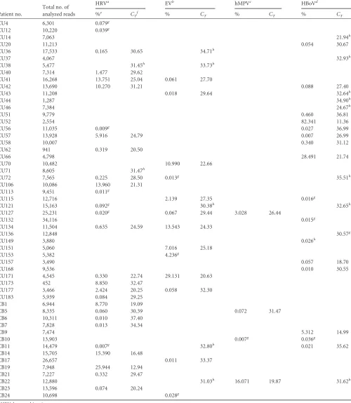
TABLE 2Summary of deep-sequencing and real-time PCR data
Patient no.
Total no. of analyzed reads
HRVa EVb hMPVc HBoVd
%e C
Tf % CT % CT % CT
CU4 6,301 0.079g
CU12 10,220 0.039g
CU14 7,063 21.94h
CU20 11,213 0.054 30.67
CU36 17,533 0.165 30.65 34.71h
CU37 4,067 32.93h
CU38 5,477 31.45h 33.73h
CU40 7,314 1.477 29.62
CU41 16,268 13.751 25.04 0.061 27.70
CU42 13,690 10.270 31.21 0.088 27.40
CU43 11,208 0.018 29.64 32.64h
CU44 1,287 34.90h
CU46 7,384 24.67h
CU51 9,779 0.460 36.81
CU52 2,554 82.341 11.36
CU56 11,035 0.009g 0.027 36.99
CU57 13,928 5.916 24.79 0.007 26.99
CU58 10,007 0.340 31.12
CU62 941 0.319 20.50
CU66 4,798 28.491 21.74
CU70 10,482 10.990 22.66
CU71 8,605 31.47h
CU72 7,565 0.225 28.50 0.013g 35.51h
CU106 10,086 13.960 21.31
CU113 9,451 0.011g
CU115 12,716 2.139 27.35 0.016g
CU121 15,163 0.092g 30.38h 32.65h
CU127 25,231 0.020g 0.067 29.44 3.028 26.44
CU132 34,116 0.015g
CU134 11,504 0.635 24.59 13.543 24.33
CU136 12,848 30.57g
CU149 3,880 0.026h
CU151 5,060 7.016 25.18
CU153 5,382 4.236g
CU157 3,490 0.057 18.70
CU168 9,536 0.010 30.55
CU171 4,545 0.330 22.74 29.131 20.63
CU173 452 8.850 32.47
CU177 3,466 2.424 20.25 0.058 32.30
CU183 5,939 0.084 29.25
CB1 6,944 8.770 19.09
CB5 8,335 0.060 30.39 0.072 31.47
CB6 10,311 0.010 37.40
CB7 7,828 0.013 34.34
CB9 7,474 5.312 14.99
CB10 13,903 0.007g 0.036g
CB11 14,479 0.007g 32.80h 0.021 35.62
CB14 15,705 15.390 16.48
CB17 26,657 0.011 33.37
CB19 7,948 25.944 12.94
CB21 7,227 0.332 29.47
CB22 12,880 31.03h 16.071 19.87 31.62h
CB23 13,596 0.074 20.24
CB24 10,698 0.028g
aHRV, human rhinovirus. b
EV, human enterovirus.
chMPV, human metapneumovirus. d
HBoV, human bocavirus.
ePercentage of virus reads out of the total number of analyzed reads. f
CT, real-time PCR cycle threshold value. gSamples positive only by deep sequencing. h
Samples positive only by real-time PCR.
on May 16, 2020 by guest
http://jcm.asm.org/
tween the 81 analyzed samples. This was the case for one bocavirus
read of 159 bp, which was identical in the samples from patients
CU58 and CU66. These patients, however, had 34 and 1,367
bo-cavirus reads, respectively, and the one identical read does not
change the interpretation of our data. In addition to the exclusion
of all samples with
⬍
10,000 analyzed next-generation sequencing
reads, samples that contained one or only a set of identical HRV,
EV, hMPV, or HBoV reads were assumed to be negative by the
deep-sequencing approach (
Fig. 2F
). The overall strong
correla-tion between the
C
Tvalues and percentages of viral reads
identi-fied by sequencing remained.
An advantage of using a next-generation sequencing approach
to detect viruses in clinical specimens is that it can also be used to
obtain information regarding the virus species and/or type of
vi-rus that was identified, in contrast to the real-time RT-PCR assays
used in this study. Indeed, we obtained virus type information (see
Table S2 in the supplemental material). Near-full-length
rhinovi-rus, enterovirhinovi-rus, human metapneumovirhinovi-rus, and bocavirus
ge-nomes were obtained from 11 different patients, in whom
⬎
10%
of the analyzed reads (mean
C
Tvalue, 20.7; range, 11.4 to 31.2)
were of the described viruses (
Table 2
; see also Table S2). The
genetic relationships of these viruses with representative viral
ge-nomes of the respective viral families were assessed (
Fig. 3A
to
D
).
Rhinoviruses from patients CB14, CB19, and CU106 showed the
highest nucleotide identity (91.9%, 90.6%, and 83.6%) with HRV-A
types 23, 61, and 88, respectively. The rhinoviruses obtained from
patients CU41 and CU42 showed the highest nucleotide identity
(
⬃
97.6% and 67%) with HRV-C types 4 and 9, respectively, in the
FIG 2Correlation between deep-sequencing and real-time RT-PCR assays. The percentages of viral reads of the total number of analyzed reads are displayed against the real-time RT-PCRCTvalues for rhinovirus (A), human metapneumovirus (B), enterovirus (C), and bocavirus (D). The theoretical next-generation
sequencing detection limit (based on an average of 10,000 analyzed reads per sample) of 0.01% is indicated by dashed lines. Red, blue, and green dots indicate samples for which⬍5,000,ⱖ5,000 and⬍10,000, orⱖ10,000 reads, respectively, were analyzed. Samples depicted on thexaxis were negative by next-generation sequencing and positive by real-time RT-PCR. (E) A combination of panels A to D depicting the percentages of viral reads of the total number of analyzed reads displayed against the real-time RT-PCRCTvalues for all samples and viruses analyzed with⬎10,000 analyzed deep sequence reads. (F) A combination of panels
A to D depicting the percentages of viral reads of the total number of analyzed reads displayed against the real-time RT-PCRCTvalues for all samples with ⬎10,000 analyzed deep sequence reads and with samples that contained one or only a set of identical HRV, EV, hMPV, or HBoV reads assumed to be negative by the deep-sequencing approach.
on May 16, 2020 by guest
http://jcm.asm.org/
[image:5.585.112.473.62.455.2]FIG 3Phylogenetic analysis of obtained genome sequences. (A) A phylogenetic tree of the near-complete rhinovirus genomes from patients CU41, CU42, CU106, CB14, and CB19 and representative human rhinoviruses (corresponding to nucleotides [nt] 132 to 6677 of HRV-23) was generated using MEGA6 with the neighbor-joining method with p-distance parameter and 1,000 bootstrap replicates. Bootstrap values are shown. (B) A phylogenetic tree of the near-complete enterovirus genomes from patients CU70, CU134, and CU171 and representative human enteroviruses (corresponding to nt 126 to 7143 of EV-NZ-2010-541) was generated using MEGA6 with the neighbor-joining method with the maximum-likelihood composite parameter and 1,000 bootstrap replicates. Bootstrap values are shown. (C) A phylogenetic tree of the near-complete hMPV genome from patient CB22 and representative human metapneumoviruses (correspond-ing to nt 32 to 13080 of hMPV-gz01) was generated us(correspond-ing MEGA6 with the neighbor-join(correspond-ing method with p-distance parameter and 1,000 bootstrap replicates. Bootstrap values are shown. (D) A phylogenetic tree of the near-complete bocavirus genomes from patients CU52 and CU66 and representative human bocaviruses (corresponding to nt 61 to 5201 of HBoV-CU74) was generated using MEGA6 with the neighbor-joining method with the maximum-likelihood composite parameter and 1,000 bootstrap replicates. Bootstrap values are shown. GenBank accession numbers we used are shown in Table S3 in the supplemental material.
on May 16, 2020 by guest
http://jcm.asm.org/
[image:6.585.45.540.65.599.2]phylogenetic tree (
Fig. 3A
). Rhinovirus CU42 may constitute a new
rhinovirus C type (type 55), as it displayed the highest nucleotide
identity (
⬃
74%) to HRV-51, HRV-26, and HRV-36 (
29
). Three
near-complete enterovirus genomes were obtained from patients
CU70, CU134, and CU171, and these genomes showed the highest
nucleotide identity to enterovirus 68 strains from Japan and New
Zealand (
Fig. 3B
). The human metapneumovirus genome from
pa-tient CB22 was most closely related (
⬃
98% nucleotide identity) to a
genetic group A hMPV from China (
Fig. 3C
). The two bocavirus
strains from patients CU52 and CU66 were most closely related
(
⬃
99% nucleotide identity) to HBoV-1 strains previously identified
in Thailand (
Fig. 3D
). Thus, virus type information was available in
the next-generation sequencing data.
DISCUSSION
In principle, random amplification combined with
next-genera-tion sequencing would be advantageous in clinical viral
diagnos-tics, as there is no need to design specific primers to amplify target
sequences. It eliminates the need for the design and validation of
several tens or hundreds of specific primers/probes targeting
mul-tiple viral pathogens, and it does not require continuous
adapta-tion of the primer sequences with the descripadapta-tion of new variants
and species. Instead, it allows the characterization of hundreds of
different known pathogens simultaneously without
a priori
knowledge of the virus present and provides in-depth virus
spe-cies/type information.
A recent review of the application of next-generation
sequenc-ing technology in clinical diagnostics described several major
drawbacks, among which is the fact that random amplification
results in the amplification of all nucleic acids, including host
nucleic acids, suggesting that an analytical sensitivity similar to
that of diagnostic PCRs cannot be expected, even with the
in-creased depth of sequencing and specific pathogen enrichment
steps applied (
11
). Our data indicate that this may be less of a
problem, as a strong correlation existed between the
C
Tvalues
from diagnostic real-time RT-PCRs and the percentages of viral
reads identified by next-generation sequencing for rhinovirus,
en-terovirus, bocavirus, and human metapneumoviruses, which is in
line with previous observations (
30
). Using a next-generation
se-quencing platform that generated an average of 10,000 reads per
sample, our sequencing approach was at least as sensitive as
diag-nostic real-time RT-PCRs for rhinovirus and human
metapneu-movirus detection. However, the deep-sequencing approach
seemed less sensitive than did real-time RT-PCRs for enterovirus
and bocavirus detection. This possibly reflects differences in the
optimization of real-time RT-PCR assays and detection with
next-generation sequencing. The detection of RNA viruses with the
sequencing approach was generally better than for DNA viruses,
which may be due to genome size differences and/or the nucleic
acid isolation procedure, as the removal of host DNA was more
substantial in the RNA isolation than in the DNA isolation
proce-dure. As shown by Wylie and coworkers (
31
), increasing the
se-quencing depth using other next-generation sese-quencing
plat-forms with increased sequence output may increase the sensitivity
of the next-generation sequencing approach even further. Thus,
achieving an analytical sensitivity comparable to that of diagnostic
real-time RT-PCRs using next-generation sequencing platforms
may actually be possible.
A common belief is that the necessary genetic information
re-quired for accurate virus typing is unpredictable when partial
se-quences are acquired due to insufficient genome coverage (
11
).
This, however, depends on a number of variables, including the
divergence of viruses within the virus families under study and
which part of the genome is sequenced. In our study, typing
in-formation was readily available from next-generation sequencing
data from 11 different patients in whom
⬎
10% of the analyzed
reads (mean
C
Tvalue, 20.7; range, 11.4 to 31.2) were of the
de-scribed viruses. In line with previous observations, virus type
in-formation was obtained even when only a few sequence reads were
available (
31
). For example, most enterovirus-positive samples
contained viral reads that were typed as enterovirus D type 68.
Until recently, reports of respiratory infections due to enterovirus
were rare, but over the past 3 years, outbreaks in Japan, the
Phil-ippines, and the Netherlands, as well as epidemic clusters in the
United Kingdom, have implicated EV68 as an emerging
respira-tory pathogen (
32–36
). Our data confirm previous observations
regarding enterovirus 68 infections in Thailand (
16
). In addition,
we typed most of the rhinovirus-positive samples as being A, B, or
C and obtained near-full-length genome sequences that were
typed as A23, A61, or A88; C4; or a new type that we designated
C55 (
29
). All the hMPV strains were typed as A, the human
boca-viruses were type I, and RSV (64% type A, 27% type B) and human
herpesvirus (HHV) infections (22% HHV-6B, 67% HHV-5, 11%
HHV-4) were typed as well.
The next-generation sequencing approach identified a number
of viruses that are usually not screened for in respiratory infections
using routine diagnostic assays, including
Herpesviridae
. It is of
note that herpesviruses were identified in 12% of the samples
an-alyzed here (mean, 0.24% of anan-alyzed reads; range, 0.007% to 2%)
and that they always occurred in combination with at least one
other viral infection. This may be due to the detection of latent
viruses, but it is also possible that respiratory disease caused by
other viruses results in the reactivation of latent herpesviruses.
However, it cannot be ruled out that human herpesviruses are
involved in respiratory disease. Cytomegalovirus (CMV)
infec-tions have been described to cause pneumonia sporadically, and in
this study, CMV was detected at relatively high levels using
next-generation sequencing (
⬃
2% of the analyzed reads) in an
8-month-old patient (CU188) and seemingly dominated the
si-multaneous RSV infection.
Another prominent observation was the detection of
anellovi-ruses in respiratory samples from more than half of the children
with respiratory illness. Anellovirus infections are commonly
ac-quired during early childhood, during which the virus establishes
a chronic productive infection with long-lasting detectable
viremia (
37
). They are endemic worldwide and can be detected in
blood and various tissues in the body, including cerebrospinal and
bronchoalveolar lavage fluid (
37
). Many investigations have been
carried out to unravel their epidemiological, clinical, and
patho-genic properties, but at present, their causative role in disease is
not considered likely. Previously, we showed that the incidence of
human anelloviruses in vitreous fluid of patients with seasonal
hyperacute panuveitis (SHAPU), a potentially blinding ocular
dis-ease occurring in Nepal that principally affects young children,
was significantly higher than that in non-SHAPU patients. The
data suggested that anelloviruses observed in vitreous fluid
sam-ples of patients with uveitis, but not in patients with retinal
de-tachment, most likely originated from the systemic anellovirus
pool upon inflammation-induced disruption of the blood-ocular
barrier (
38
). Likewise, inflammation in the respiratory tract
on May 16, 2020 by guest
http://jcm.asm.org/
tem, caused by an infection, for example, may cause increased
permeability of the capillary barrier, resulting in edema, an influx
of inflammatory cells, and the concomitant increase in the
amount of anellovirus in the respiratory tract.
One of the major questions is how to interpret next-generation
sequencing data in terms of what is clinically relevant for the
pa-tient. The comparison of the next-generation sequencing data to
real-time RT-PCR
C
Tvalues showed that the interpretation may
not need to be very different than the interpretation of
C
Tvalues,
as there is a very strong correlation between the percentage of viral
reads detected and the
C
Tvalue obtained from the same sample.
However, as the sensitivity of the next-generation sequencing
as-say compared to that of the real-time RT-PCR asas-says is not the
same for each RT-PCR assay, the next-generation sequencing
as-say may need to be validated with each real-time RT-PCR asas-say. In
addition, our data show that multiple viral infections occurred
substantially more often than did single-virus infections (60%
versus 28%, respectively). Even when taking only well-established
respiratory pathogens into account, 25% of the cases were
multi-ple infections. In most cases, one of the detected viruses seemed to
dominate. Although major hurdles for the routine use of
next-generation sequencing approaches do exist, including cost, labor
intensity, and especially turnaround time, the sensitivity of a
next-generation sequencing assay may actually reach that of current
diagnostic routine real-time RT-PCR assays, and it potentially
provides more information regarding virus species/type, thus
re-maining of interest for virus diagnosis in clinical and public health
settings. Although it might not be used for routine diagnostics at
present, a sequence-independent next-generation sequencing
as-say may be applied to samples that remain negative with routine
diagnostics and, as such, may function as an additional diagnostic
tool with the added value of surveillance for (re)emerging viruses.
ACKNOWLEDGMENTS
This work was partially funded by the Virgo Consortium, which is funded by the Dutch government (project number FES0908), the Netherlands Genomics Initiative (NGI) (project number 050-060-452), and ZonMW TOP (project 91213058).
Thai sites included the National Research University Project, Office of Higher Education Commission (HR1155A, WCU-001,007-HR-57); the Center of Excellence in Clinical Virology, Chulalongkorn University; the Centenary Academic Development Project, Integrated Innovation Aca-demic Center; the Chulalongkorn University Centenary AcaAca-demic Devel-opment Project (CU56-HR01); the Ratchadaphiseksomphot Endowment Fund of Chulalongkorn University (RES560530093); and the Outstand-ing Professor and RGJ Ph.D. program of the Thailand Research Fund (DPG5480002, PHD/0114/2551).
A. D. M. E. Osterhaus and S. L. Smits are part-time employees of Viroclinics Biosciences B.V. This does not alter our adherence to all the policies on sharing data and materials.
REFERENCES
1.Chu CM, Lin DY, Yeh CT, Sheen IS, Liaw YF.2001. Epidemiological characteristics, risk factors, and clinical manifestations of acute non-A-E hepatitis. J. Med. Virol.65:296 –300.http://dx.doi.org/10.1002/jmv.2033. 2.Finkbeiner SR, Kirkwood CD, Wang D.2008. Complete genome se-quence of a highly divergent astrovirus isolated from a child with acute diarrhea. Virol. J.5:117.http://dx.doi.org/10.1186/1743-422X-5-117. 3.Granerod J, Crowcroft NS.2007. The epidemiology of acute
enceph-alitis. Neuropsychol. Rehabil.17:406 – 428.http://dx.doi.org/10.1080 /09602010600989620.
4.Juvén T, Mertsola J, Waris M, Leinonen M, Meurman O, Roivainen M, Eskola J, Saikku P, Ruuskanen O.2000. Etiology of community-acquired
pneumonia in 254 hospitalized children. Pediatr. Infect. Dis. J.19:293– 298.http://dx.doi.org/10.1097/00006454-200004000-00006.
5.Saeed M, Zaidi SZ, Naeem A, Masroor M, Sharif S, Shaukat S, Angez M, Khan A.2007. Epidemiology and clinical findings associated with entero-viral acute flaccid paralysis in Pakistan. BMC Infect. Dis.7:6.http://dx.doi .org/10.1186/1471-2334-7-6.
6.Capobianchi MR, Giombini E, Rozera G. 2013. Next-generation se-quencing technology in clinical virology. Clin. Microbiol. Infect.19:15– 22.http://dx.doi.org/10.1111/1469-0691.12056.
7.Lipkin WI.2010. Microbe hunting. Microbiol. Mol. Biol. Rev.74:363– 377.http://dx.doi.org/10.1128/MMBR.00007-10.
8.Mokili JL, Rohwer F, Dutilh BE.2012. Metagenomics and future per-spectives in virus discovery. Curr. Opin. Virol.2:63–77.http://dx.doi.org /10.1016/j.coviro.2011.12.004.
9.Smits SL, Osterhaus AD.8 April 2013. Virus discovery: one step beyond. Curr. Opin. Virol3:1– 6.http://dx.doi.org/10.1016/j.coviro.2013.03.007. 10. van Boheemen S, de Graaf M, Lauber C, Bestebroer TM, Raj VS, Zaki
AM, Osterhaus AD, Haagmans BL, Gorbalenya AE, Snijder EJ, Fouchier RA.2012. Genomic characterization of a newly discovered coro-navirus associated with acute respiratory distress syndrome in humans. mBio3:e00473-12.http://dx.doi.org/10.1128/mBio.00473-12.
11. Lecuit M, Eloit M.2014. The diagnosis of infectious diseases by whole genome next generation sequencing: a new era is opening. Front. Cell Infect. Microbiol.4:25.http://dx.doi.org/10.3389/fcimb.2014.00025. 12. Smits SL, Osterhaus ADME.2012. Emerging viral infections, p 1142–
1154.InGinsburg GS, Willard HF (ed), Genomic and personalized med-icine, vol 1. Academic Press, London, United Kingdom.
13. Suwannakarn K, Payungporn S, Chieochansin T, Samransamruajkit R, Amonsin A, Songserm T, Chaisingh A, Chamnanpood P, Chutinimit-kul S, Theamboonlers A, Poovorawan Y.2008. Typing (A/B) and sub-typing (H1/H3/H5) of influenza A viruses by multiplex real-time RT-PCR assays. J. Virol. Methods152:25–31.http://dx.doi.org/10.1016/j.jviromet .2008.06.002.
14. Auksornkitti V, Kamprasert N, Thongkomplew S, Suwannakarn K, Theamboonlers A, Samransamruajkij R, Poovorawan Y.2014. Molecular characterization of human respiratory syncytial virus, 2010 –2011: identifica-tion of genotype ON1 and a new subgroup B genotype in Thailand. Arch. Virol.159:499 –507.http://dx.doi.org/10.1007/s00705-013-1773-9. 15. Linsuwanon P, Payungporn S, Samransamruajkit R, Posuwan N,
Mak-koch J, Theanboonlers A, Poovorawan Y. 2009. High prevalence of human rhinovirus C infection in Thai children with acute lower respira-tory tract disease. J. Infect.59:115–121.http://dx.doi.org/10.1016/j.jinf .2009.05.009.
16. Linsuwanon P, Puenpa J, Suwannakarn K, Auksornkitti V, Vichiwat-tana P, Korkong S, Theamboonlers A, Poovorawan Y.2012. Molecular epidemiology and evolution of human enterovirus serotype 68 in Thai-land, 2006 –2011. PLoS One7:e35190.http://dx.doi.org/10.1371/journal .pone.0035190.
17.Sriwanna P, Chieochansin T, Vuthitanachot C, Vuthitanachot V, Theamboonlers A, Poovorawan Y.2013. Molecular characterization of human adenovirus infection in Thailand, 2009 –2012. Virol. J.10:193. http://dx.doi.org/10.1186/1743-422X-10-193.
18. Ruampunpong H, Payungporn S, Samransamruajkit R, Prathee-pamornkul T, Theamboonlers A.2014. Human parainfluenza virus in-fection in Thai children with lower respiratory tract inin-fection from 2010 to 2013. Southeast Asian Pac. J. Trop. Med. Public Health45:610 – 621. 19. Bodewes R, van der Giessen J, Haagmans BL, Osterhaus AD, Smits SL.
2013. Identification of multiple novel viruses, including a parvovirus and a hepevirus, in feces of red foxes. J. Virol.87:7758 –7764.http://dx.doi.org /10.1128/JVI.00568-13.
20. van den Brand JM, van Leeuwen M, Schapendonk CM, Simon JH, Haagmans BL, Osterhaus AD, Smits SL.2012. Metagenomic analysis of the viral flora of pine marten and European badger feces. J. Virol.86: 2360 –2365.http://dx.doi.org/10.1128/JVI.06373-11.
21. van Leeuwen M, Williams MM, Koraka P, Simon JH, Smits SL, Oster-haus AD.2010. Human picobirnaviruses identified by molecular screen-ing of diarrhea samples. J. Clin. Microbiol.48:1787–1794.http://dx.doi .org/10.1128/JCM.02452-09.
22. Hoek RA, Paats MS, Pas SD, Bakker M, Hoogsteden HC, Boucher CA, van der Eerden MM.2013. Incidence of viral respiratory pathogens caus-ing exacerbations in adult cystic fibrosis patients. Scand. J. Infect. Dis. 45:65– 69.http://dx.doi.org/10.3109/00365548.2012.708942.
23. Kantola K, Sadeghi M, Antikainen J, Kirveskari J, Delwart E, Hedman
on May 16, 2020 by guest
http://jcm.asm.org/
K, Soderlund-Venermo M.2010. Real-time quantitative PCR detection of four human bocaviruses. J. Clin. Microbiol.48:4044 – 4050.http://dx .doi.org/10.1128/JCM.00686-10.
24. Tamura K, Stecher G, Peterson D, Filipski A, Kumar S.2013. MEGA6: Molecular Evolutionary Genetics Analysis version 6.0. Mol. Biol. Evol. 30:2725–2729.http://dx.doi.org/10.1093/molbev/mst197.
25. Chieochansin T, Simmonds P, Poovorawan Y.2010. Determination and analysis of complete coding sequence regions of new discovered human bocavirus types 2 and 3. Arch. Virol.155:2023–2028.http://dx.doi.org/10 .1007/s00705-010-0781-2.
26. Gaunt ER, Jansen RR, Poovorawan Y, Templeton KE, Toms GL, Sim-monds P.2011. Molecular epidemiology and evolution of human respi-ratory syncytial virus and human metapneumovirus. PLoS One6:e17427. http://dx.doi.org/10.1371/journal.pone.0017427.
27. Palmenberg AC, Spiro D, Kuzmickas R, Wang S, Djikeng A, Rathe JA, Fraser-Liggett CM, Liggett SB. 2009. Sequencing and analyses of all known human rhinovirus genomes reveal structure and evolution. Sci-ence324:55–59.http://dx.doi.org/10.1126/science.1165557.
28. Wisdom A, Kutkowska AE, McWilliam Leitch EC, Gaunt E, Templeton K, Harvala H, Simmonds P.2009. Genetics, recombination and clinical features of human rhinovirus species C (HRV-C) infections; interactions of HRV-C with other respiratory viruses. PLoS One4:e8518.http://dx.doi .org/10.1371/journal.pone.0008518.
29. McIntyre CL, Knowles NJ, Simmonds P.2013. Proposals for the classi-fication of human rhinovirus species A, B and C into genotypically as-signed types. J. Gen. Virol.94:1791–1806.http://dx.doi.org/10.1099/vir.0 .053686-0.
30. de Vries M, Oude Munnink BB, Deijs M, Canuti M, Koekkoek SM, Molenkamp R, Bakker M, Jurriaans S, van Schaik BD, Luyf AC, Ola-barriaga SD, van Kampen AH, van der Hoek L.2012. Performance of VIDISCA-454 in feces-suspensions and serum. Viruses 4:1328 –1334. http://dx.doi.org/10.3390/v4081328.
31.Wylie KM, Mihindukulasuriya KA, Sodergren E, Weinstock GM,
Storch GA.2012. Sequence analysis of the human virome in febrile and afebrile children. PLoS One7:e27735.http://dx.doi.org/10.1371/journal .pone.0027735.
32. Imamura T, Fuji N, Suzuki A, Tamaki R, Saito M, Aniceto R, Galang H, Sombrero L, Lupisan S, Oshitani H.2011. Enterovirus 68 among chil-dren with severe acute respiratory infection, the Philippines. Emerg. In-fect. Dis.17:1430 –1435.http://dx.doi.org/10.3201/eid1708.101328. 33. Jacobson LM, Redd JT, Schneider E, Lu X, Chern SW, Oberste MS,
Erdman DD, Fischer GE, Armstrong GL, Kodani M, Montoya J, Magri JM, Cheek JE.2012. Outbreak of lower respiratory tract illness associated with human enterovirus 68 among American Indian children. Pediatr. Infect. Dis. J.31:309 –312.http://dx.doi.org/10.1097/INF.0b013e3182443eaf.
34. Kaida A, Kubo H, Sekiguchi J, Kohdera U, Togawa M, Shiomi M, Nishigaki T, Iritani N.2011. Enterovirus 68 in children with acute respi-ratory tract infections, Osaka, Japan. Emerg. Infect. Dis.17:1494 –1497. http://dx.doi.org/10.3201/eid1708.110028.
35. Rahamat-Langendoen J, Riezebos-Brilman A, Borger R, van der Heide R, Brandenburg A, Scholvinck E, Niesters HG.2011. Upsurge of human en-terovirus 68 infections in patients with severe respiratory tract infections. J. Clin. Virol.52:103–106.http://dx.doi.org/10.1016/j.jcv.2011.06.019. 36. Xiang Z, Gonzalez R, Wang Z, Ren L, Xiao Y, Li J, Li Y, Vernet G,
Paranhos-Baccala G, Jin Q, Wang J.2012. Coxsackievirus A21, entero-virus 68, and acute respiratory tract infection, China. Emerg. Infect. Dis. 18:821– 824.http://dx.doi.org/10.3201/eid1805.111376.
37. Maggi F, Bendinelli M.2010. Human anelloviruses and the central ner-vous system. Rev. Med. Virol.20:392– 407.http://dx.doi.org/10.1002/rmv .668.
38. Smits SL, Manandhar A, van Loenen FB, van Leeuwen M, Baarsma GS, Dorrestijn N, Osterhaus AD, Margolis TP, Verjans GM.2012. High prevalence of anelloviruses in vitreous fluid of children with seasonal hyperacute panuveitis. J. Infect. Dis.205:1877–1884.http://dx.doi.org/10 .1093/infdis/jis284.

![FIG 3 Phylogenetic analysis of obtained genome sequences. (A) A phylogenetic tree of the near-complete rhinovirus genomes from patients CU41, CU42,CU106, CB14, and CB19 and representative human rhinoviruses (corresponding to nucleotides [nt] 132 to 6677 of](https://thumb-us.123doks.com/thumbv2/123dok_us/8286318.849616/6.585.45.540.65.599/phylogenetic-sequences-phylogenetic-rhinovirus-representative-rhinoviruses-corresponding-nucleotides.webp)
