0095-1137/09/$08.00
⫹
0
doi:10.1128/JCM.01707-08
Copyright © 2009, American Society for Microbiology. All Rights Reserved.
Development and Evaluation of a Liquid Bead Microarray Assay for
Genotyping Genital Human Papillomaviruses
䌤
†
Qinghua Feng,
1* Stephen Cherne,
1Rachel L. Winer,
2Akhila Balasubramanian,
2Shu-Kuang Lee,
3Stephen E. Hawes,
2Nancy B. Kiviat,
1and Laura A. Koutsky
2Department of Pathology, School of Medicine,
1and Department of Epidemiology
2and Department of Biostatistics,
3School of
Public Health and Community Medicine, University of Washington, Seattle, Washington
Received 3 September 2008/Returned for modification 30 October 2008/Accepted 5 January 2009
We developed a liquid bead microarray (LBMA) assay for genotyping genital human papillomaviruses
(HPVs) based on the MY09–MY11–HMB01 PCR system and the reverse line blot (RLB) assay probe
se-quences. Using individual HPV plasmids, we were able to detect as few as 50 copies per reaction. In two
separate retrospective studies, the LBMA assay was compared to the RLB assay and to the Hybrid Capture II
(hc2) assay. Testing was performed without knowledge of other assay results. In the first study, 614 cervical
swab samples (enriched for HPV infection) from 160 young women were tested for HPV DNA, and 360 (74.8%)
type-specific HPV infections were detected by both assays, 71 (14.8%) by the LBMA assay only, and 50 (10.4%)
by the RLB assay only. Type-specific agreement for the two assays was excellent (99.1%; kappa
ⴝ
0.85; 95%
confidence interval [95% CI], 0.82 to 0.88). Samples with discrepant LBMA and RLB test results tended to
have low viral loads by a quantitative type-specific PCR assay. In the second study, cervical swab samples from
452 women (including 54 women with histologically confirmed cervical-intraepithelial neoplasia grade 2 or
worse [
>
CIN2]) were tested initially by the hc2 and subsequently by the LBMA assay. The estimated
sensitivities for
>
CIN2 were similar for the LBMA and hc2 assays (98.4% [95% CI, 95.0 to 100%] and 95.6%
[95% CI, 89.2 to 100%], respectively). The percentages of negative results among 398 women without
>
CIN2
were similar for the LBMA and hc2 assays (45% and 50%, respectively). The repeat test reproducibility for 100
samples was 99.1% (kappa
ⴝ
0.92; 95% CI, 0.90 to 0.95). We conclude that the new LBMA assay will be useful
for clinical and epidemiological research.
Human papillomavirus (HPV) is the central etiological
agent for virtually all cervical cancers, for a substantial
propor-tion of other anogenital tract cancers, and for a smaller
pro-portion of head and neck cancers (3, 20). Currently, more than
100 different HPV types have been identified, and at least 40
types infect the anogenital epithelium. The risk of cancer is not
the same for all HPV types. High-risk HPV types include HPV
type 16 (HPV-16) and HPV-18, -31, -33, -35, -39, -45, -51, -52,
-56, -58, -59, -68, -73, and -82. Low-risk HPV types include
HPV-6, -11, -40, -42, -43, -44, -54, -61, -70, -72, -81, and
-CP6108. Potentially high-risk types include HPV-26, -53, and
-66 (20, 21). HPV DNA testing has been used (i) for triage of
women with a Papanicolaou (Pap) test finding of atypical
squa-mous cells of undetermined significance (ASC-US), (ii) for
monitoring for recurrence of a precancerous cervical lesion or
cancer after treatment, and (iii) as a primary screening method
for cervical cancer in women 30 years old and older (1, 23, 31).
The only FDA-approved HPV assay for clinical testing, the
Hybrid Capture II (hc2) assay, distinguishes high-risk HPVs
from low-risk HPVs but does not provide individual HPV
genotyping information. HPV type-specific assays are likely to
have a role in the clinical management of the neoplastic
dis-eases associated with HPV infection. Although 60 to 70% of
U.S. women become infected with one or more high-risk
gen-ital types of HPV during their lifetime, most infections are
quickly resolved and without consequence (2, 6, 19). Women
who remain persistently positive for the same high-risk HPV
type for extended periods are at increased risk for progression
to cancer (11, 12). HPV genotyping is needed to differentiate
women who are repeatedly positive for the same high-risk
HPV type from those who are simply sequentially infected with
different high-risk types of HPV. As the use of prophylactic
HPV vaccines becomes more widespread, surveillance for
population-level effectiveness will become an increasingly
im-portant activity that is likely to require the use of an HPV
type-specific assay (10, 27). Estimates of the duration of
vac-cine-induced protection and the potential for herd immunity,
for cross-protection, or for replacement (i.e., an increase in the
prevalence of non-vaccine-type cervical lesions despite a
de-crease in the prevalence of vaccine-type lesions) will require
HPV type-specific testing. Further, with increased coverage of
HPV vaccines and the development of new vaccines that are
likely to target more HPV types, the usefulness and methods of
HPV testing and genotyping in cervical cancer prevention
pro-grams will likely need reevaluation and revision (14).
Cur-rently, HPV genotyping is indispensable for epidemiological
and clinical studies of the transmission, natural history, and
pathogenesis of HPV, and it is likely to have a role in the
management of HPV-related precancerous lesions and cancers
in the future.
DNA sequencing is the “gold standard” for HPV
genotyp-ing; however, it is costly, time-consuming, and difficult to apply
* Corresponding author. Mailing address: 815 Mercer Street, UW
Medicine, Box 358050, Department of Pathology, University of
Wash-ington, Seattle, WA 98109. Phone: (206) 1583. Fax: (206)
897-1334. E-mail: [email protected].
† Supplemental material for this article may be found at http://jcm
.asm.org/.
䌤
Published ahead of print on 14 January 2009.
547
on May 16, 2020 by guest
http://jcm.asm.org/
to clinical samples, which frequently have multiple infections
and produce nonspecific PCR products. Currently, the most
widely used multiplex HPV genotyping assays are reverse line
blot (RLB) assays (7, 15). These assays are based on
solid-phase hybridization of amplified HPV sequences to a slot blot
membrane. However, RLB assays are labor-intensive, are not
easily automated, have limited reproducibility (because they
rely on a subjective visual readout) (5), and are increasingly
expensive.
Recent reports support the potential use of the liquid bead
microarray (LBMA) assay based on Luminex technology for
HPV genotyping, using either the existing GP5
⫹
–GP6
⫹
sys-tem or the PGMY PCR syssys-tem (13, 26, 28). This assay format
is sensitive and amenable to high-throughput configuration
and potentially can be automated. However, little is known
about the analytical sensitivity and specificity of this new assay
or about how it performs on clinical specimens in comparison
to the other HPV genotyping assays.
In our present study, we developed an LBMA assay based on
the MY09–MY11–HMB01 PCR system for genotyping
clini-cally important HPV types (11, 18). We determined the
ana-lytical sensitivity and specificity of the LBMA assay using
in-dividual HPV plasmids, compared the genotyping results of
the LBMA assay with those of the RLB assay, and estimated
the clinical performance of the LBMA assay in comparison to
that of the hc2 test using archived cervical swab samples.
MATERIALS AND METHODS
HPV LBMA assay development. (i) Probe selection.The previously reported RLB probe sequences (Roche Molecular Diagnostics, Pleasanton, CA) were used in the development of our assay (7, 24). As in the RLB assay, two probes were used for HPV-16, -18, -31, -33, -35, -39, -42, -45, -51, -52, -53, -54, -55, -58,
-59, -61, -62, -66, -67, -68, -69, -71, -72, -73, -81, -82, -83, -83, -IS39, and -CP6108, while a single probe was used for HPV-6, -11, -26, -40, -56, -64, and -70 (see the table in the supplemental material).
(ii) Probe conjugation to beads.Thirty-seven Luminex bead sets (sets 17 to 21, 24, 25, 33 to 38, 42 to 47, 51 to 56, 61 to 66, and 72 to 77) were obtained (MiraiBio, Alameda, CA), and each bead set (approximately 5⫻106beads/set)
[image:2.585.42.285.77.343.2]containing an intrinsic fluorescent signature was attached to specific HPV probes according to the manufacturer’s oligonucleotide coupling protocol. Briefly, each
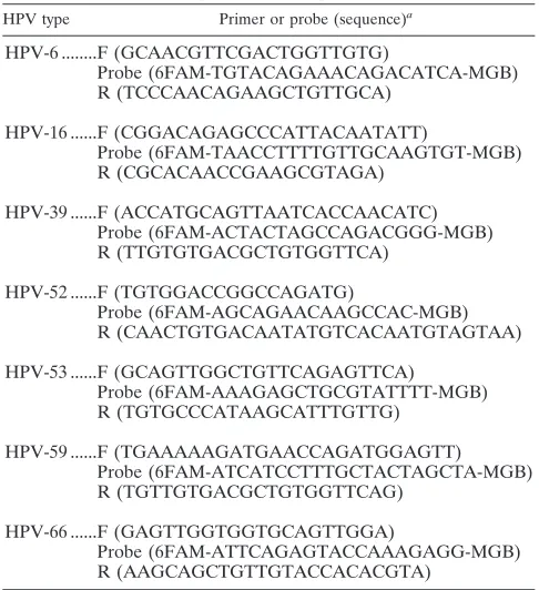
TABLE 1. Primers and probes for the quantification of HPV loads
HPV type Primer or probe (sequence)a
HPV-6 ...F (GCAACGTTCGACTGGTTGTG)
Probe (6FAM-TGTACAGAAACAGACATCA-MGB)
R (TCCCAACAGAAGCTGTTGCA)
HPV-16 ...F (CGGACAGAGCCCATTACAATATT)
Probe (6FAM-TAACCTTTTGTTGCAAGTGT-MGB)
R (CGCACAACCGAAGCGTAGA)
HPV-39 ...F (ACCATGCAGTTAATCACCAACATC)
Probe (6FAM-ACTACTAGCCAGACGGG-MGB)
R (TTGTGTGACGCTGTGGTTCA)
HPV-52 ...F (TGTGGACCGGCCAGATG)
Probe (6FAM-AGCAGAACAAGCCAC-MGB)
R (CAACTGTGACAATATGTCACAATGTAGTAA)
HPV-53 ...F (GCAGTTGGCTGTTCAGAGTTCA)
Probe (6FAM-AAAGAGCTGCGTATTTT-MGB)
R (TGTGCCCATAAGCATTTGTTG)
HPV-59 ...F (TGAAAAAGATGAACCAGATGGAGTT)
Probe (6FAM-ATCATCCTTTGCTACTAGCTA-MGB)
R (TGTTGTGACGCTGTGGTTCAG)
HPV-66 ...F (GAGTTGGTGGTGCAGTTGGA)
Probe (6FAM-ATTCAGAGTACCAAAGAGG-MGB)
R (AAGCAGCTGTTGTACCACACGTA)
[image:2.585.304.539.87.606.2]a6FAM, 6-carboxyfluorescein; MGB, minor groove binder.
TABLE 2. Characteristics of the women in the two
study populations
Population and characteristic Value
Study population 1
No. of subjects...
160
Age range (yr) (mean, SD) ...18–26 (21.9, 1.7)
No. (%) of the following race/ethnicity:
White ...
106 (66.3)
Asian...
30 (19.1)
Hispanic...
5 (2.9)
Other ...
19 (11.7)
No. of cervical swab samples...
614
No. (%) of samples with the following
RLB assay result:
Negative ...
360 (58.6)
Positive ...
254 (41.4)
No. (%) of positive samples with the
following no. of HPV types detected:
1 types...
145 (57.1)
2 types...
76 (29.9)
3 types...
22 (8.7)
4 types...
8 (3.2)
5 types...
3 (1.2)
No. of persistent infections (no. of positive
samples)...
107 (315)
No. of transient infections (no. of positive
samples)...
95 (95)
Study population 2
No. of subjects...
452
Age range (yr) (mean, SD) ...18–47 (24.1, 5.9)
No. (%) of the following race/ethnicity:
White ...
335 (74.1)
Black ...
40 (8.8)
Hispanic...
20 (4.4)
Asian...
12 (2.7)
Other ...
45 (10.0)
No. (%) with the following cervical hc2
assay result:
Negative ...
200 (44.2)
Positive ...
252 (55.8)
No. (%) with the following histologic
diagnosis
a:
Negative ...
85 (39.5)
Atypia ...
40 (18.6)
CIN1 ...
30 (14.0)
CIN1 to CIN2...
4 (1.9)
CIN2 ...
17 (7.9)
CIN2 to CIN3...
8 (3.7)
CIN3 or worse ...
29 (13.5)
Insufficient...
2 (0.9)
No. (%) with the following liquid-based
Pap test result
b:
Negative ...
295 (65.3)
ASC-US...
76 (16.8)
LSIL...
35 (7.7)
HSIL or greater ...
37 (8.2)
Insufficient...
9 (2.0)
aA total of 215 subjects had histologically confirmed diagnoses.bLSIL, low-grade squamous intraepithelial lesion; HSIL, high-grade squa-mous intraepithelial lesion.
on May 16, 2020 by guest
http://jcm.asm.org/
TABLE 3. Detection of individual HPV plasmids by the LBMA assay
Plasmid (no. of copies)
Result (RLU) with specific probes of the following type (cutoff)a:
HPV-6 (358)
HPV-11 (293)
HPV-16 (263)
HPV-18 (393)
HPV-33 (367)
HPV-35 (322)
HPV-39 (473)
HPV-45 (418)
HPV-52 (317)
HPV-59 (405)
HPV-66 (312)
HPV-67 (331)
Negative controls:
dH
2O
17
18
23
26
24
15
17
24
20
28
27
28
K562
27
18
17
35
27
29
44
34
25
36
24
37
HPV-6
95,722
1,560
23
24
31
25
22
28
35
25
29
28
28
100
763
26
26
41
28
29
35
33
19
32
22
37
50
361
32
29
38
29
26
32
32
21
31
27
38
10
735
23
24
31
25
27
29
34
23
34
26
34
HPV-11
348,786
17
1,515
10
18
19
17
17
18
14
23
12
28
100
14
580
10
21
13
14
22
21
11
26
18
28
50
14
532
13
20
20
14
23
25
13
27
17
30
10
19
145
16
27
21
15
21
27
16
27
23
29
HPV-16
106,476
28
31
1,145
38
41
32
32
54
25
40
30
39
100
45
41
1,991
68
45
43
43
56
45
50
44
51
50
39
33
2,004
36
41
38
35
46
35
48
36
47
10
32
27
1,733
32
41
40
41
41
25
44
33
43
HPV-18
1,187,373
24
23
27
2,098
25
32
30
28
27
33
26
33
100
27
26
18
2,359
21
24
34
32
16
38
26
41
50
32
30
21
1,740
24
26
30
35
25
29
25
37
10
30
25
26
41
29
20
31
40
26
34
24
38
HPV-33
25,395
29
35
19
35
2,499
28
29
38
24
39
33
43
100
26
24
28
35
2,608
31
28
33
21
38
30
34
50
26
21
18
26
2,660
25
18
32
18
32
26
32
10
22
17
21
27
2,658
18
27
26
20
34
28
29
HPV-35
568,041
33
19
36
30
27
2,310
27
33
23
39
30
42
100
42
33
31
43
37
2,566
41
52
31
50
34
45
50
35
37
29
47
34
2,158
43
50
32
46
37
55
10
35
36
35
38
35
122
37
46
36
43
34
45
HPV-39
20,766
33
36
32
44
17
28
1,678
36
30
34
31
43
100
15
14
14
22
15
16
449
20
12
22
15
21
50
17
13
11
23
15
16
490
22
13
24
17
21
10
13
12
12
21
12
14
50
21
11
24
17
22
HPV-45
311,229
38
29
30
35
20
24
30
782
32
39
31
38
100
28
26
22
31
29
30
27
2,044
21
32
30
31
50
26
16
19
37
26
25
26
1,991
22
34
28
32
10
20
23
22
29
28
23
29
40
29
33
27
43
HPV-52
476,139
31
26
28
29
35
34
31
43
1,616
38
32
36
100
39
39
34
54
33
28
36
46
1,108
41
36
53
50
45
41
43
55
50
41
46
51
722
51
40
50
10
29
30
26
39
27
22
24
38
27
31
26
36
HPV-59
102,762
44
29
39
45
43
38
36
39
44
1,534
45
46
100
29
25
27
43
32
19
30
40
23
1,099
21
34
50
27
31
24
30
30
18
30
35
28
1,065
21
34
10
15
21
12
23
22
13
26
24
20
210
24
27
HPV-66
107,706
19
23
22
38
28
15
23
33
18
24
2,121
37
100
27
23
16
29
29
29
22
28
23
36
1,655
36
50
24
24
20
25
24
25
32
29
16
34
1,266
32
10
23
21
17
30
25
22
26
25
20
30
1,585
37
HPV-67
248,838
25
15
12
27
134
18
28
26
19
30
21
1,703
100
36
20
11
22
20
18
46
34
16
33
20
1,082
50
22
15
14
23
26
17
37
27
15
31
16
1,259
10
21
14
12
16
22
12
28
26
9
24
14
517
a
Values for positive samples are in boldface. The cutoff for each HPV type is calculated as 10 times the mean RLU for 12 negative-control samples.
on May 16, 2020 by guest
http://jcm.asm.org/
bead set and its specific amine-substituted probes were first incubated with freshly prepared 1-ethyl-3-(3-dimethylaminopropyl)-carbodiimide (EDC) at room temperature for 30 min and then washed and resuspended in Tris-EDTA at 50,000 beads/l. All conjugated beads were pooled in suspension to form the 37-plex HPV-LBMA assay mixture (1,350 beads/set/l).
HPV plasmids.Plasmids of 12 HPV types (HPV-6, -11, -16, -18, -33, -35, -39, -45, -52, -59, -66, and -67) were available for the study. Nine of these (HPV-6, -11, -16, -33, -45, -52, -59, -66, and -67) contain full-length HPV genomes, while the remaining three (HPV-18, -35, and -39) contain a partial L1 open reading frame that encompasses the MY09–MY11–HMB01 amplicon (kindly donated by Denise Galloway). The concentrations of purified plasmids were determined with a UV spectrometer and were converted to copies per microliter. The plasmids were diluted, and 1 to 106
copies were mixed with 30 ng of purified K562 genomic DNA and amplified using MY09–MY11–HMB01 primers.
Amplification of the HPV L1 fragment.MY09–MY11–HMB01 primers were used to amplify the HPV L1 fragment under the following conditions: 95°C for 9 min; 40 cycles of 95°C for 30 s, 55°C for 1 min, and 72°C for 1 min; and a 5-min extension at 72°C. The ramp speed was set at 1°C/s (29). The presence of the correct PCR product was confirmed by gel electrophoresis; then the product was purified with a QIAquick PCR purification column (Qiagen, Valencia, CA) to remove the remaining excess primers.
Generation of a biotin-labeled single-stranded PCR product.Biotin-labeled single-stranded HPV PCR products were generated from the PCR-amplified HPV L1 fragment by using biotin-labeled MY11 in place of MY09–MY11– HMB01 and the cycle conditions described above, except that only 20 cycles were run.
Hybridization assay.One hundred nanograms of biotin-labeled PCR products or one-third of the second round of PCR products was mixed with conjugated beads, denatured at 95°C for 10 min, and hybridized at 55°C in 1.5⫻TMAC (tetramethyl ammonium chloride) solution for 30 min according to the Luminex oligonucleotide hybridization and DNA buffer protocols. After hybridization, the beads were washed twice with 1⫻TMAC using a 96-well filter plate (Qiagen, Valencia, CA) and then incubated with 4 g/ml phycoerythrin-conjugated streptavidin (diluted in 1⫻TMAC; BD Pharmingen) at 55°C for 30 min. Finally, the beads were washed again with 1⫻TMAC and resuspended in 100l 1⫻ TMAC for detection on the LiquiChip system (the Luminex 100 platform) according to the manufacturer’s protocol (Qiagen, Valencia, CA). For each assay run, four HPV-negative controls (genomic DNA isolated from the K562 cell line) were included, and the background RLU was determined as the average for these four negative controls. A sample was considered to be weakly positive or
positive for a specific HPV type if the RLU was greater than 7 or 10 times the background RLU, respectively, for that specific HPV.
HPV DNA load analysis by quantitative PCR.Type-specific TaqMan assays were designed for HPV-6, -16, -39, -52, -53, -59, and -66 based on the HPV E7 gene. Primers and probe sequences are listed in Table 1. The quantitative PCR assays were performed on an ABI Prism 7900 sequence detection system (ABI, Foster City, CA). Relative quantification (RQ) was determined by the 2⫺⌬⌬CT method. For each HPV type, the RQ of the sample with the highest viral load was set at 1.
Clinical samples.Archived cervical swab specimens collected in specimen transport medium (STM) from two different NIH-funded research projects were used in this study. Both sets of specimens were enriched for the detection of high-risk HPV infections. The first set of specimens consisted of 614 cervical swab samples from 160 female university students participating in a longitudinal study of the natural history of HPV infections (30). HPV genotyping by an RLB assay had been performed previously, and all RLB assay-positive cervical spec-imens, all RLB assay-negative cervical specimens for which a corresponding vulvar/vaginal or self-collected vaginal specimen was positive, and a random sample of the negative cervical specimens were included in this analysis. The second group of specimens consisted of cervical swab samples from 452 women enrolled in a cervical cancer screening study that included HPV DNA screening by the hc2 assay (16). The selected group of women was enriched for those with histologically confirmed diagnoses and included 54 women with cervical intra-epithelial neoplasia grade 2 or worse (ⱖCIN2). The protease K-digested ar-chived clinical samples, stored at ⫺20°C, were used for reextraction via a QIAamp column according to the manufacturer’s protocol (Qiagen, Valencia, CA) for the LBMA assay and the type-specific quantitative PCR assay. All LMBA assays were performed without knowledge of prior laboratory or clinical test results. The demographic characteristics of both study populations are listed in Table 2.
[image:4.585.45.542.80.333.2]Statistical analysis.The first group of archived cervical swab samples was used to assess the concordance of type-specific HPV DNA detection between the LBMA and RLB assays by using an unweighted kappa statistic to determine the percentage of agreement beyond that expected by chance. To account for cor-relation within subjects, 95% confidence intervals (95% CI) were computed using percentile bootstrap methods with 1,000 repetitions. By using data from the second group of cervical swab samples, different cervical cancer screening strat-egies were defined based on the use of LBMA, hc2, and/or cytology tests. Because only 13 HPV types (HPV-16, -18, -31, -33, -35, -39, -45, -51, -52, -56, -58, -59, and -68) are included in the high-risk hc2 assay, samples were considered
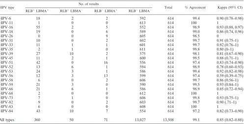
TABLE 4. HPV type-specific agreement on cervical swab samples
HPV type
No. of results
Total % Agreement Kappa (95% CI) RLB⫹LBMA⫹ RLB⫹LBMA⫺ RLB⫺LBMA⫹ RLB⫺LBMA⫺
HPV-6
18
2
2
592
614
99.4
0.90 (0.78–0.98)
HPV-11
1
0
0
613
614
100
1
HPV-16
55
2
5
552
614
98.9
0.93 (0.88, 0.97)
HPV-18
19
0
6
589
614
99.0
0.86 (0.74, 0.96)
HPV-26
0
0
9
605
614
98.5
0
HPV-31
10
0
2
602
614
99.7
0.91 (0.75–1)
HPV-33
11
1
1
601
614
99.7
0.92 (0.76–1)
HPV-35
2
1
0
611
614
99.8
0.80 (0–1)
HPV-39
27
10
2
575
614
98.1
0.81 (0.67–0.90)
HPV-45
11
2
1
600
614
99.5
0.88 (0.71–1)
HPV-51
42
0
16
556
614
97.4
0.83 (0.74–0.90)
HPV-52
13
6
1
594
614
98.9
0.78 (0.60–0.93)
HPV-53
24
3
1
586
614
99.4
0.92 (0.82–0.98)
HPV-56
12
3
13
599
614
97.4
0.59 (0.39–0.75)
HPV-58
6
0
2
606
614
99.7
0.86 (0.56–1)
HPV-59
21
3
0
590
614
99.5
0.93 (0.84–1)
HPV-66
21
6
1
586
614
98.9
0.85 (0.72–0.94)
HPV-68
2
0
0
612
614
100
1
HPV-73
7
1
0
606
614
99.8
0.93 (0.75–1)
HPV-82
9
0
2
603
614
99.7
0.90 (.71–1)
HPV-83
6
0
0
608
614
100
1
HPV-84
43
10
7
554
614
97.2
0.82 (0.73–0.90)
All types
360
50
71
13,027
13,508
99.1
0.85 (0.82–0.88)
on May 16, 2020 by guest
http://jcm.asm.org/
positive for the LBMA assay only if 1 or more of those 13 HPV types were detected. Detection of other HPV types was considered a negative result for HPV for the purposes of this analysis. The strategies were evaluated for their sensitivities in detecting histologically confirmedⱖCIN2. Since this population was enriched for women with histologically confirmed disease, it was not possible to estimate meaningful measures of specificity for the four screening strategies based on the LBMA assay, the hc2 assay, and/or cytology. Instead, an indication of the relative specificities (and of the relative percentages of results that were “false positive” forⱖCIN2) of the LMBA and hc2 assays for a group of women withoutⱖCIN2 was provided by the calculated percentage of this group that is negative for high-risk HPV DNA by the LBMA or hc2 assay. Estimates of sensitivity were corrected for verification bias by using a previously described inverse probability weighting method (16). Ninety-five percent confidence inter-vals were computed using the 2.5th and 97.5th percentiles of the bootstrap distribution of weighted estimates of sensitivity. Weights were applied using the pweights command in Stata (StataCorp LP, TX). All statistical analyses were performed using STATA, version 10.0 (StataCorp LP, TX).
RESULTS
Analytical sensitivity and specificity of the LBMA assay.
We
determined the detection limits of the assay using HPV
plas-mids. As shown in Table 3, the LBMA assay was able to detect
as few as 50 copies of each HPV plasmid mixed with genomic
DNA. The assay had high analytical specificity for each HPV
type when tested with high copy numbers of plasmids (10
5to
10
6copies/reaction).
Agreement of the LBMA assay and the RLB assay.
The
performance of the LBMA assay was evaluated using 614
ar-chived cervical swab samples from 160 subjects. These
speci-mens had previously been genotyped using the RLB assay for
27 HPV types. Because HPV-57 was not included in the
LBMA assay, and there are four low-risk HPV types (HPV-40,
-42, -54, and -55) that appear to be clinically insignificant, we
restricted all comparisons to 22 genotypes (HPV-6, -11, -16,
-18, -26, -31, -33, -35, -39, -45, -51, -52, -53, -56, -58, -59, -66,
-68, -73, -82, -83, and -84). Of the 614 cervical swab samples,
254 (41.4%) were positive for one or more of these types of
HPV by the RLB assay and 254 (41.4%) were positive by the
LBMA assay. By the two assays together, a total of 481
specific HPV infections were detected. Overall, 74.8% of
type-specific HPV infections were detected by both assays, 14.8%
were detected by the LBMA assay only, and 10.4% were
de-tected by the RLB assay only. By pooling across HPV types,
the type-specific percentage of agreement for all HPV types
was 99.1% (kappa
⫽
0.85; 95% CI, 0.82 to 0.88) (Table 4).
In order to further understand the cause of the discrepancy
between the two assays, type-specific quantitative PCR assays
targeting the HPV E7 region were designed. Specifically, we
focused on high-risk or potentially high risk HPV types
(HPV-16, -39, -52, -53, -59, and -66) for which the RLB assay gave
positive results but that were more likely missed by the LBMA
assay. In addition, an HPV-6 type-specific assay was designed.
We analyzed samples that were positive by either the RLB or
the LBMA assay for one or more of these seven HPV types.
Samples showing discordant results between the RLB and the
LBMA assays tended to have lower viral loads than samples
that were positive by both assays (Fig. 1).
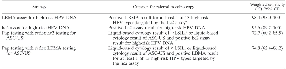
Relative performances of the LBMA and hc2 assays for
detection of histologically confirmed
>
CIN2.
The
perfor-mance of screening strategies defined by results from the
LBMA assay, the hc2 assay, and/or cytology was evaluated for
452 women (Table 5). Among 54 women with histologically
confirmed
ⱖ
CIN2, the estimated weighted sensitivities for the
LBMA and hc2 assays were similar (98.4% [95% CI, 95.0 to
100] and 95.6% [95% CI, 89.2 to 100], respectively). The
[image:5.585.44.284.69.196.2]per-FIG. 1. HPV loads of samples positive by either the LBMA or the
RLB assay. DNA viral loads of samples positive for HPV-6, -16, -39,
-52, -53, -59, or -66 by either the LBMA or the RLB assay were
determined by a quantitative PCR assay based on the HPV E7
se-quence. RQ was determined using the 2
⫺⌬⌬CTmethod. For each HPV
type, the RQ of the sample with the highest viral load was set at 1.
TABLE 5. Sensitivities of four different strategies for detecting histologically confirmed
ⱖ
CIN2
aStrategy Criterion for referral to colposcopy Weighted sensitivity
(%) (95% CI)
LBMA assay for high-risk HPV DNA
Positive LBMA result for at least 1 of 13 high-risk
HPV types targeted by the hc2 assay
b98.4 (95.0–100)
hc2 assay for high-risk HPV DNA
Positive hc2 assay result for high-risk HPV DNA
95.6 (89.2–100)
Pap testing with reflex hc2 testing for
ASC-US
Liquid-based cytology result of
ⱖ
LSIL,
cor liquid-based
cytology result of ASC-US and positive hc2 assay
result for high-risk HPV DNA
72.7 (60.2–85.5)
Pap testing with reflex LBMA testing
for ASC-US
Liquid-based cytology result of
ⱖ
LSIL, or liquid-based
cytology result of ASC-US and positive LBMA result
for at least 1 of 13 high-risk HPV types targeted by
the hc2 assay
74.8 (62.4–86.2)
aInsufficient and inadequate results were included in the “positive screening result” category, and for the strategy involving Pap testing with reflex LBMA testing for ASC-US, missing follow-up hc2 assay results were considered to be negative. Changing these assumptions did not markedly impact the findings.
bHigh-risk HPV types not targeted by the hc2 assay were included in the “negative for HPV DNA” category.
cⱖLSIL, low-grade squamous intraepithelial lesion or worse. Inadequate cytology results were included in theⱖLSIL category, since women with inadequate cytology results are generally asked to return for a follow-up visit.
on May 16, 2020 by guest
http://jcm.asm.org/
[image:5.585.40.543.563.685.2]centages of negative results for 398 women without
histologi-cally confirmed
ⱖ
CIN2 were also similar for the LBMA and
RLB assays (45% and 50%, respectively) (Table 6).
Reproducibility of the LBMA assay.
We repeated the
LBMA assay on 100 randomly selected cervical swab samples.
When each of 37 HPV types was considered separately for the
reproducibility analysis (100
⫻
37
⫽
3,700 comparisons in
total), 207 (5.6%) samples were concordantly positive, 3,460
(93.5%) were concordantly negative, and 33 (0.9%) were
dis-cordant. Therefore the agreement between the two repeated
tests was 99.1% (kappa
⫽
0.92; 95% CI, 0.90 to 0.95).
DISCUSSION
We developed an LBMA assay for genotyping HPVs. The
LBMA assay was able to detect as few as 50 copies of the HPV
genome and displayed high analytical specificity. When tested
on 614 archived cervical samples, the LBMA assay showed
excellent reproducibility and excellent agreement with the
RLB assay for HPV genotyping. Using cervical swab samples
from 452 subjects, we observed that the LBMA assay had an
estimated clinical sensitivity for
ⱖ
CIN2 that was comparable
to that of the hc2 assay. These results indicate that this newly
developed LBMA assay is likely to be a valid and reliable
alternative method for HPV genotyping and a sensitive assay
for identifying high-grade cervical lesions.
Several previous studies reported the feasibility of
establish-ing a Luminex-based HPV genotypestablish-ing assay targetestablish-ing 15 to 45
different HPVs (9, 13, 17, 22, 26, 28). Various primer systems
were used for the development of these Luminex assays,
in-cluding PGMY09–PGMY11 (28), GP5
⫹
–GP6
⫹
(26), MY09–
MY11 (13), type-specific primers (9), and YBT L1–GP6-1 (22).
Using archived clinical samples, these assays reported 74 to
99% agreement with other HPV detection (hc2 assay) or
geno-typing (RLB assay, type-specific PCR, or HPV microarray
as-say) assays (Table 7).
While these previous reports support the LBMA technology
for HPV DNA genotyping, to our knowledge, the present
report is the first to provide results of a large-scale
investiga-tion that compared HPV genotyping results for the LBMA
assay to those of widely used HPV genotyping assays based on
RLB technology. In addition, the results for the LBMA assay
were compared to those of the FDA-approved hc2 test in order
to assess the potential clinical performance of the LBMA assay
for detection of
ⱖ
CIN2. Although many of the samples
in-cluded in the genotyping comparison were positive for more
than 1 of the 22 targeted HPV types (42.9% of 254 RLB
assay-positive samples), type-specific agreement was high,
in-dicating excellent sensitivity and specificity for type-specific
HPV DNA detection, even in the setting of mixed infections.
In the current study, we did not compare the LBMA assay to
the commercially available Roche Linear Array (LA) assay,
because these archived samples had already been genotyped by
the RLB assay. Recently, Castle et al. compared the
perfor-mances of the RLB and LA assays using archived clinical
samples (3). The percentage of agreement for carcinogenic
HPV DNA detection was 88% (kappa
⫽
0.76). Although the
LA assay appeared to be more sensitive than the RLB assay in
detecting HPV DNA, the authors suggested that this was due
largely to the different DNA isolation methods used and the
different amounts of DNA input for PCR amplification. When
equal amounts of DNA extracted by the same method were
used, no difference in overall or type-specific HPV detection
was observed (3). The LBMA assay, like any other bead-based
assay or solid-phase assay with an automated reading, is
pos-sibly more objective than the RLB assay, because unlike the
RLB assay, it does not rely on a subjective visual readout.
Moreover, the LMBA assay is amenable to a high-throughput
configuration, can potentially be automated, can easily be
scaled up to 100 HPV types, and can be combined with
Lumi-nex-based assays for the detection of HPV type-specific
anti-bodies.
Several aspects of the LBMA assay might be improved in the
future. First, a

-globin or other housekeeping gene probe
[image:6.585.43.284.89.233.2]could be incorporated to monitor the sample input. Second,
the assay could be streamlined by using fluorescently labeled
TABLE 6. LBMA and hc2 test results for women with and without
histologically confirmed
ⱖ
CIN2
Histologic categorya
No. (%) positive by the following assay:
LBMAb
hc2
Histologically confirmed (
n
⫽
213)
ⱖ
CIN2 (
n
⫽
54)
53 (98.2)
52 (96.2)
ⱖ
CIN3 (
n
⫽
37)
36 (97.3)
36 (97.3)
CIN2 (
n
⫽
17)
17 (100.0)
16 (94.1)
CIN1 or less (
n
⫽
159)
124 (78.0)
121 (76.1)
No histological confirmation (
n
⫽
239)
Cytologic findings of SIL (
n
⫽
13)
12 (92.3)
13 (100.0)
Cytologic findings of ASC-US (
n
⫽
26)
14 (53.9)
14 (53.9)
Normal cytologic findings (
n
⫽
197)
67 (34.0)
52 (26.4)
Inadequate cytologic findings (
n
⫽
3)
1 (33.3)
0 (0.0)
aSIL, squamous intraepithelial lesion. b
Number (percent) positive for at least 1 of 13 high-risk HPV types targeted by the hc2 assay. High-risk types not targeted by the hc2 assay were included in the “negative for HPV DNA” category.
TABLE 7. Comparison of various reported bead-based HPV genotyping assays
Primer
No. of HPV types
detected
No. of clinical
samples Assay compared
Reported agreement (%)
Reported kappa (95% CI)
Source or reference
PGMY09–PGMY11
45
429
hc2
82
0.68 (0.65, 0.72)
28
GP5
⫹
–GP6
⫹
22
94
RLB
85
0.92
26
MY09–MY11
26
133
Type-specific PCR sequencing
90
13
Type specific
25
109
hc2
95
9
YBT L1–GP6-1
15
53
HPV microarray
74
22
MY09–MY11–HMB01
22
614
RLB
99
0.85 (0.82, 0.88)
Present study
on May 16, 2020 by guest
http://jcm.asm.org/
[image:6.585.43.540.634.724.2]primers in the PCR, to avoid the need for a second
hybridiza-tion with phycoerythrin-conjugated streptavidin. Third, the
as-say sensitivity could be improved by using biotin- or fluorescent
dye-labeled nucleotides in the PCR instead of labeled primers.
Finally, in order to expand the LBMA assay to allow it to
genotype cutaneous HPV types, which are more
heteroge-neous and for which it is difficult to design common degenerate
primers for amplification, signal amplification should be
con-sidered as an alternative to PCR amplification.
In conclusion, our data showed excellent correlation
be-tween the RLB and the LBMA assays when they were used for
genotyping clinical samples, and it also showed comparable
sensitivities for the hc2 and LMBA assays for the detection of
biopsy-confirmed CIN2 or worse. Several recent studies
under-score the importance of standardization of HPV genotyping
assay protocols (3–5, 8, 25). The LBMA assay described here is
amenable to standardization and thus shows promise for use in
large-scale epidemiological studies of HPV pathogenesis, in
surveillance of HPV immunization programs for
population-level effectiveness, and in clinical investigations of new
ap-proaches to the prevention, diagnosis, and management of
HPV-related cancers and precancerous lesions.
ACKNOWLEDGMENTS
This work was supported by grants from the National Institute of
Health/National Cancer Institute (CA34493 and CA105181).
Informed consent was obtained according to procedures approved
by the Human Subjects Committee of the University of Washington.
REFERENCES
1.ASCUS-LSIL Triage Study (ALTS) Group.2003. Results of a randomized trial on the management of cytology interpretations of atypical squamous cells of undetermined significance. Am. J. Obstet. Gynecol.188:1383–1392. 2.Baseman, J. G., and L. A. Koutsky.2005. The epidemiology of human
papillomavirus infections. J. Clin. Virol.32(Suppl. 1):S16–S24.
3.Castle, P. E., P. E. Gravitt, D. Solomon, C. M. Wheeler, and M. Schiffman. 2008. Comparison of linear array and line blot assay for detection of human papillomavirus and diagnosis of cervical precancer and cancer in the Atypical Squamous Cell of Undetermined Significance and Low-Grade Squamous Intraepithelial Lesion Triage Study. J. Clin. Microbiol.46:109–117. 4.Castle, P. E., C. Porras, W. G. Quint, A. C. Rodriguez, M. Schiffman, P. E.
Gravitt, P. Gonzalez, H. A. Katki, S. Silva, E. Freer, L.-J. Van Doorn, S. Jime´nez, R. Herrero, and A. Hildesheim for the CVT Group.2008. Compar-ison of two PCR-based human papillomavirus genotyping methods. J. Clin. Microbiol.46:3437–3445.
5.Dunn, S. T., R. A. Allen, S. Wang, J. Walker, and M. Schiffman.2007. DNA extraction: an understudied and important aspect of HPV genotyping using PCR-based methods. J. Virol. Methods143:45–54.
6.Gravitt, P. E., and R. Jamshidi.2005. Diagnosis and management of onco-genic cervical human papillomavirus infection. Infect. Dis. Clin. N. Am. 19:439–458.
7.Gravitt, P. E., C. L. Peyton, R. J. Apple, and C. M. Wheeler.1998. Geno-typing of 27 human papillomavirus types by using L1 consensus PCR prod-ucts by a single-hybridization, reverse line blot detection method. J. Clin. Microbiol.36:3020–3027.
8.Gravitt, P. E., M. Schiffman, D. Solomon, C. M. Wheeler, and P. E. Castle. 2008. A comparison of linear array and hybrid capture 2 for detection of carcinogenic human papillomavirus and cervical precancer in ASCUS-LSIL triage study. Cancer Epidemiol. Biomarkers Prev.17:1248–1254. 9.Han, J., D. C. Swan, S. J. Smith, S. H. Lum, S. E. Sefers, E. R. Unger, and
Y. W. Tang.2006. Simultaneous amplification and identification of 25 human papillomavirus types with Templex technology. J. Clin. Microbiol.44:4157– 4162.
10.Harper, D. M., E. L. Franco, C. Wheeler, D. G. Ferris, D. Jenkins, A. Schuind, T. Zahaf, B. Innis, P. Naud, N. S. De Carvalho, C. M. Roteli-Martins, J. Teixeira, M. M. Blatter, A. P. Korn, W. Quint, and G. Dubin. 2004. Efficacy of a bivalent L1 virus-like particle vaccine in prevention of infection with human papillomavirus types 16 and 18 in young women: a randomised controlled trial. Lancet364:1757–1765.
11.Hildesheim, A., M. H. Schiffman, P. E. Gravitt, A. G. Glass, C. E. Greer, T. Zhang, D. R. Scott, B. B. Rush, P. Lawler, M. E. Sherman, et al.1994.
Persistence of type-specific human papillomavirus infection among cytolog-ically normal women. J. Infect. Dis.169:235–240.
12.Ho, G. Y., R. Bierman, L. Beardsley, C. J. Chang, and R. D. Burk.1998. Natural history of cervicovaginal papillomavirus infection in young women. N. Engl. J. Med.338:423–428.
13.Jiang, H. L., H. H. Zhu, L. F. Zhou, F. Chen, and Z. Chen.2006. Genotyping of human papillomavirus in cervical lesions by L1 consensus PCR and the Luminex xMAP system. J. Med. Microbiol.55:715–720.
14.Kiviat, N. B., S. E. Hawes, and Q. Feng.2008. Screening for cervical cancer in the era of the HPV vaccine—the urgent need for both new screening guidelines and new biomarkers. J. Natl. Cancer Inst.100:290–291. 15.Kleter, B., L. J. van Doorn, L. Schrauwen, A. Molijn, S. Sastrowijoto, J. ter
Schegget, J. Lindeman, B. ter Harmsel, M. Burger, and W. Quint.1999. Development and clinical evaluation of a highly sensitive PCR-reverse hy-bridization line probe assay for detection and identification of anogenital human papillomavirus. J. Clin. Microbiol.37:2508–2517.
16.Kulasingam, S. L., J. P. Hughes, N. B. Kiviat, C. Mao, N. S. Weiss, J. M. Kuypers, and L. A. Koutsky.2002. Evaluation of human papillomavirus testing in primary screening for cervical abnormalities: comparison of sen-sitivity, specificity, and frequency of referral. JAMA288:1749–1757. 17.Liu, M., C. X. Wang, X. M. Deng, L. S. Wang, J. Zhang, W. Li, G. X. Zheng,
and J. F. Wang.2007. Study on the genotyping of human papillomavirus using a new DNA liquid chip in women of high-risk group of Shandong province. Zhonghua Liu Xing Bing Xue Za Zhi.28:487–490. (In Chinese.) 18.Manos, M. M., Y. Ting, D. K. Wright, A. J. Lewis, T. R. Broker, and S. M. Wolinsky.1989. The use of polymerase chain reaction amplification for the detection of genital human papillomavirus, p. 209–214.InM. Furth and M. Greaves (ed.), Molecular diagnostics of human cancer, vol. 7. Cold Spring Harbor Laboratory Press, Cold Spring Harbor, NY.
19.Monsonego, J., F. X. Bosch, P. Coursaget, J. T. Cox, E. Franco, I. Frazer, R. Sankaranarayanan, J. Schiller, A. Singer, T. C. Wright, Jr., W. Kinney, C. J. Meijer, J. Linder, E. McGoogan, and C. Meijer.2004. Cervical cancer con-trol, priorities and new directions. Int. J. Cancer108:329–333.
20.Mun˜oz, N., F. X. Bosch, S. de Sanjose, R. Herrero, X. Castellsague´, K. V. Shah, P. J. Snijders, and C. J. Meijer.2003. Epidemiologic classification of human papillomavirus types associated with cervical cancer. N. Engl. J. Med. 348:518–527.
21.Mun˜oz, N., X. Castellsague´, A. B. de Gonza´lez, and L. Gissmann.2006. Chapter 1: HPV in the etiology of human cancer. Vaccine24(Suppl. 3):S1– S10.
22.Oh, Y., S. M. Bae, Y. W. Kim, H. S. Choi, G. H. Nam, S. J. Han, C. H. Park, Y. Cho, B. D. Han, and W. S. Ahn.2007. Polymerase chain reaction-based fluorescent Luminex assay to detect the presence of human papillomavirus types. Cancer Sci.98:549–554.
23.Paraskevaidis, E., M. Arbyn, A. Sotiriadis, E. Diakomanolis, P. Martin-Hirsch, G. Koliopoulos, G. Makrydimas, J. Tofoski, and D. H. Roukos.2004. The role of HPV DNA testing in the follow-up period after treatment for CIN: a systematic review of the literature. Cancer Treat. Rev.30:205–211. 24.Peyton, C. L., P. E. Gravitt, W. C. Hunt, R. S. Hundley, M. Zhao, R. J. Apple,
and C. M. Wheeler.2001. Determinants of genital human papillomavirus detection in a US population. J. Infect. Dis.183:1554–1564.
25.Sabol, I., M. Salakova, J. Smahelova, M. Pawlita, M. Schmitt, N. M. Gas-perov, M. Grce, and R. Tachezy.2008. Evaluation of different techniques for identification of human papillomavirus types of low prevalence. J. Clin. Microbiol.46:1606–1613.
26.Schmitt, M., I. G. Bravo, P. J. Snijders, L. Gissmann, M. Pawlita, and T. Waterboer.2006. Bead-based multiplex genotyping of human papillomavi-ruses. J. Clin. Microbiol.44:504–512.
27.Villa, L. L., R. L. Costa, C. A. Petta, R. P. Andrade, K. A. Ault, A. R. Giuliano, C. M. Wheeler, L. A. Koutsky, C. Malm, M. Lehtinen, F. E. Skjeldestad, S. E. Olsson, M. Steinwall, D. R. Brown, R. J. Kurman, B. M. Ronnett, M. H. Stoler, A. Ferenczy, D. M. Harper, G. M. Tamms, J. Yu, L. Lupinacci, R. Railkar, F. J. Taddeo, K. U. Jansen, M. T. Esser, H. L. Sings, A. J. Saah, and E. Barr.2005. Prophylactic quadrivalent human papilloma-virus (types 6, 11, 16, and 18) L1 papilloma-virus-like particle vaccine in young women: a randomised double-blind placebo-controlled multicentre phase II efficacy trial. Lancet Oncol.6:271–278.
28.Wallace, J., B. A. Woda, and G. Pihan.2005. Facile, comprehensive, high-throughput genotyping of human genital papillomaviruses using spectrally addressable liquid bead microarrays. J. Mol. Diagn.7:72–80.
29.Weaver, B. A., Q. Feng, K. K. Holmes, N. Kiviat, S. K. Lee, C. Meyer, M. Stern, and L. A. Koutsky.2004. Evaluation of genital sites and sampling techniques for detection of human papillomavirus DNA in men. J. Infect. Dis.189:677–685.
30.Winer, R. L., J. P. Hughes, Q. Feng, S. O’Reilly, N. B. Kiviat, K. K. Holmes, and L. A. Koutsky.2006. Condom use and the risk of genital human papil-lomavirus infection in young women. N. Engl. J. Med.354:2645–2654. 31.Wright, T. C., Jr., M. Schiffman, D. Solomon, J. T. Cox, F. Garcia, S. Goldie,
K. Hatch, K. L. Noller, N. Roach, C. Runowicz, and D. Saslow.2004. Interim guidance for the use of human papillomavirus DNA testing as an adjunct to cervical cytology for screening. Obstet. Gynecol.103:304–309.