RESEARCH ARTICLE
Retinoid signaling controls spermatogonial differentiation by
regulating expression of replication-dependent core histone genes
Yao Chen1,2, Li Ma3, Cathryn Hogarth4, Gang Wei3, Michael D. Griswold4,* and Ming-Han Tong1,2,*ABSTRACT
Retinoic acid (RA) signaling is crucial for spermatogonial differentiation, which is a key step for spermatogenesis. We explored the mechanisms underlying spermatogonial differentiation by targeting expression of a dominant-negative mutant of retinoic acid receptor α (RARα) specifically to the germ cells of transgenic mice to subvert the activity of endogenous receptors. Here we show that: (1) inhibition of retinoid signaling in germ cells completely blocked spermatogonial differentiation identical to vitamin A-deficient (VAD) mice; (2) the blockage of spermatogonial differentiation by impaired retinoid signaling resulted from an arrest of entry of the undifferentiated spermatogonia into S phase; and (3) retinoid signaling regulated spermatogonial differentiation through controlling expression of its direct target genes, including replication-dependent core histone genes. Taken together, our results demonstrate that the action of retinoid signaling on spermatogonial differentiation in vivo is direct through the spermatogonia itself, and provide the first evidence that this is mediated by regulation of expression of replication-dependent core histone genes.
KEY WORDS: Spermatogonial differentiation, Spermatogenesis, Retinoic acid, Dominant-negative retinoic acid receptor, Testis, Replication-dependent core histone genes
INTRODUCTION
Spermatogenesis is a highly organized and complex process that allows for the continuous production of millions of haploid spermatozoa throughout male adult life and for transferring the intact genome and appropriate epigenome from generation to generation (Clermont, 1972; Oatley and Brinster, 2008). The transition of undifferentiated spermatogonia into A1spermatogonia
(termed spermatogonial differentiation) is an initial and irreversible step of spermatogenesis (de Rooij, 2001). The undifferentiated spermatogonia can be subdivided into Asingle (As), Apaired (Ap)
and Aaligned (Aal) spermatogonia and include spermatogonial
stem cells (SSCs) and progenitor spermatogonia. This cohort of undifferentiated spermatogonia express self-renewal- and proliferation-associated genes such asPou5f1(Pesce et al., 1998), Lin28a(Tong et al., 2011; Zheng et al., 2009),Mir-21(Niu et al., 2011),Mir-17∼92(Mirc1) (Tong et al., 2012),Foxo1(Goertz et al.,
2011),Nanos2 (Sada et al., 2009; Zhou et al., 2015),Neurog 3 (Ngn3) (Nakagawa et al., 2007), Sox3 (Laronda and Jameson, 2011),Taf4b(Falender et al., 2005), andZbtb16(Plzf) (Buaas et al., 2004; Costoya et al., 2004) to maintain the capacity for self-renewal and proliferation. During spermatogonial differentiation, the undifferentiated spermatogonia downregulate these self-renewal associated genes and upregulate genes associated with differentiation such asSohlh1 (Ballow et al., 2006),Sohlh2 (Hao et al., 2008), Stra8 (Endo et al., 2015; Zhou et al., 2008), Kit (Schrans-Stassen et al., 1999), Ccnd2(Beumer et al., 2000) and Sall4(Hobbs et al., 2012; Gely-Pernot et al., 2015). Despite this fact, the molecular mechanisms that govern spermatogonial differentiation remain incomplete.
Retinoic acid (RA), an active derivative of vitamin A, is essential for spermatogonial differentiation as: (1) the transition of the undifferentiated spermatogonia into A1spermatogonia is blocked in
vitamin A deficient (VAD) rodents, and (2) RA administration to VAD animals reinitiates spermatogonial differentiation (Clagett-Dame and Knutson, 2011; Griswold et al., 1989; Huang and Hembree, 1979; Morales and Griswold, 1987; Wilson et al., 1953; Wolbach and Howe, 1925; Wolgemuth and Chung, 2007; van Pelt and de Rooij, 1990). There are 12 stages of the cycle of seminiferous epithelium (hereafter referred to as epithelial stages I-XII) in the mouse (Clermont, 1972; Hogarth and Griswold, 2010; Oakberg, 1956). Although the undifferentiated spermatogonia in epithelial stages II-VIII are competent for spermatogonial differentiation in the adult mouse testis, spermatogonial differentiation occurs only in epithelial stages VII and/or VIII as the RA level reaches its peak (de Rooij, 2001; Endo et al., 2015; Hasegawa and Saga, 2012; Hogarth et al., 2015; Hogarth and Griswold, 2010). Moreover, RA treatment could induce precocious differentiation of the undifferentiated spermatogonia in epithelial stages II-VII into A1 spermatogonia
(Hogarth et al., 2015; Endo et al., 2015). However, the mechanisms underlying RA-induced spermatogonial differentiation remain largely unknown.
The action of RA on expression of target genes is mediated through two families of nuclear hormone receptors; the retinoic acid receptors (RARs) and the retinoid X receptors (RXRs), each with three subtypes,α,β, and γ, which are encoded by distinct genes (Chambon, 1996). RAR and RXR usually function as RAR-RXR heterodimers, which bind to retinoic acid response elements (RAREs) in regulatory regions of the target genes (Bastien and Rochette-Egly, 2004). RAREs are typically composed of two direct repeats of a core hexameric motif, PuG(G/T)TCA, separated by a 5 bp spacer sequence (referred to as DR5) (Bastien and Rochette-Egly, 2004). Several subtypes of RARs and RXRs are expressed in both Sertoli cells and germ cells including spermatogonia and exert redundant functions (Vernet et al., 2006b; Gaemers et al., 1998). Ikami et al. showed that ectopic expression ofRargcould induce the differentiation of RARG-negative undifferentiated spermatogonia by RA (Ikami et al., 2015). Global inactivation of individual RAR Received 28 January 2016; Accepted 2 March 2016
1
State Key Laboratory of Molecular Biology, Institute of Biochemistry and Cell Biology, Shanghai Institute for Biological Sciences, Chinese Academy of
Sciences, Shanghai 200031, China.2Shanghai Key Laboratory of Molecular
Andrology, Institute of Biochemistry and Cell Biology, Shanghai Institute for Biological Sciences, Chinese Academy of Sciences, Shanghai 200031, China.
3
CAS-MPG Partner Institute for Computational Biology, Shanghai Institute for Biological Sciences, Chinese Academy of Sciences, Shanghai 200031, China.
4
School of Molecular Biosciences, Washington State University, Pullman, WA 99164, USA.
*Authors for correspondence ([email protected]; [email protected])
DEVEL
O
genes such as Rara results in male sterility and aberrant spermatogenesis (Lufkin et al., 1993). Several lines of compound mutants lacking multiple RARs or RXRs have been studied in testis, suggesting that retinoid signaling plays a crucial role in spermatogonial differentiation (Gely-Pernot et al., 2012, 2015). However, the RA target genes implicated in spermatogonial differentiation need to be identified.
To address these questions in the current study, we used conditional dominant-negative mouse models to block retinoid signaling specifically in germ cells. We demonstrate that impaired retinoid signaling in germ cells resulted in a complete blockage of spermatogonial differentiation. One of the major biological functions of RA is to inhibit cell proliferation (Bohnsack and Hirschi, 2004; Clagett-Dame and Knutson, 2011); however, RA is capable of stimulating cell proliferation in some type of cells such as neural crest-derived mesenchyme in the forebrain (Schneider et al., 2001) and neonatal germ cells (Busada et al., 2014). We report here that RA-induced entry into S phase of the undifferentiated spermatogonia could be crucial for spermatogonial differentiation. We further show that retinoid signaling could directly control expression of replication-dependent core histone genes that is essential for entry into S phase during spermatogonial differentiation. These findings thus provide novel insights into the molecular mechanisms by which retinoid signaling could regulate expression of replication-dependent core histone genes, and thereby control spermatogonial differentiationin vivo.
RESULTS
Inactivation of retinoid signaling in spermatogonia impaired spermatogenesis
To directly determine whether retinoid signaling in germ cells controls spermatogenesis, we employed a conditional dominant-negative mutant of RARα403 (dnRAR) transgene strategy (Rosselot et al., 2010). RARα403, which is a truncated form of human RARα, retains the ability to dimerize and bind the RARE but lose its transcriptional activation function (Damm et al., 1993). Previous studies have demonstrated that RARα403 can completely block wild-type RAR and RXR function in a dose-dependent manner (Damm et al., 1993). A dnRAR transgene was inserted into the ROSA26Rlocus and was preceded by a floxed STOP sequence that is excised in cells expressing Cre, activating the expression of
dnRAR (Fig. S1A) (Rosselot et al., 2010). We conditionally expresseddnRARin a subset of spermatogonia using aStra8-Cre transgenic line withCreexpression in germ cells starting at∼3 days post-partum (dpp) (Sadate-Ngatchou et al., 2008). Throughout this study, we hereafter referred to the two different genotypes of mice as: germ-cell mutant (dnRARflox/flox, Stra8-Cre), heterozygous
germ-cell mutant (dnRARflox/+, Stra8-Cre).
To test whether the expression of dnRAR can impair retinoid signaling, we used a RARElacZ reporter line containing a RARE-driven lacZ transgene, which allows the distribution of retinoid signaling to be visualized by 5-bromo-4-chloro-3-indolyl-β -D-galactoside (X-gal) staining (Rossant et al., 1991). NolacZ activity was detected in testes from the mice expressing both alleles of the dnRARtransgene (dnRARflox/flox, Stra8-Cre,RARElacZ), whereas
strong lacZ staining was seen in the control testes (Fig. S1B). However, in those animals expressing a single allele of dnRAR (dnRARflox/+, Stra8-Cre, RARElacZ) testes, lacZ activity was
reduced but still detectable (Fig. S1B), suggesting that thednRAR in this line can block endogenous retinoid signaling in a dose-dependent manner as previously shown (Rosselot et al., 2010). Histologically, the seminiferous epithelium in control testes contained lacZ-positive spermatogonia and spermatocytes (Fig. S1C). Significantly, no lacZ-positive cells can be found in testes from the mice expressing both alleles of thednRARtransgene (Fig. S1C), but more than 40% of seminiferous tubes contained lacZ-positive spermatogonia in the testes from the mice expressing a single allele of thednRAR(Fig. S1C).
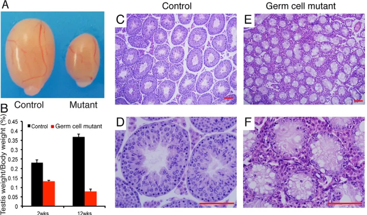
[image:2.612.49.409.525.736.2]Germ-cell mutant males were sterile but they exhibited normal copulating behavior. Germ-cell mutant testes were much smaller than control littermate testes (Fig. 1A,B); at 2 weeks old, control=0.231±0.016 (mean testis weight/body weight×100±s.d.), germ-cell mutants=0.132±0.006,P<0.001,n=12; at 12 weeks old, control=0.368±0.014, germ-cell mutants=0.079±0.012, P<0.001, n=8. Histological examinations of adult germ-cell mutants showed severe defects in spermatogenesis (Fig. 1C-F). In contrast to control seminiferous tubules that contained the full complement of germ cells (Fig. 1C,D), adult germ-cell mutant seminiferous tubules showed a reduced diameter and contained only morphologically normal Sertoli cells and undifferentiated spermatogonia cells (Fig. 1E,F). Moreover, compared with control testes, seminiferous epithelium were also devoid of differentiated germ cells, containing
Fig. 1. Impaired spermatogenesis in germ-cell mutant testes.(A) Gross morphology of representative testes from an 8-week-old control and an age-matched germ-cell mutant. (B) Comparisons of testis weight from 2- or 12-week-old control and mutant mice (n=8-12 for each genotype per data point). Data are expressed as mean±s.d. (C-F) Hematoxylin and Eosin staining of control (C,D) and germ-cell mutant (E,F) testes at 8 weeks old. Scale bars: 20μm.
DEVEL
O
only Sertoli cells and undifferentiated spermatogonia at the basal membrane in 2- or 3-week-old germ-cell mutants (Fig. S2A-D).
Germ-cell mutants exhibit complete blockage of spermatogonial differentiation
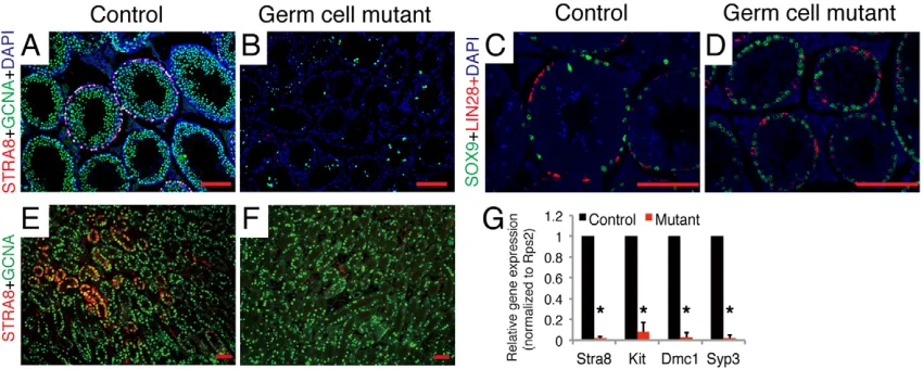
To further characterize the remaining spermatogonia in germ-cell mutants, we employed immunostaining with antibodies to spermatogonial markers. On immunostaining for STRA8, a marker for differentiated spermatogonia, STRA8-positive germ cells were not observed in adult germ-cell mutant testes (Fig. 2B), whereas control testes showed many seminiferous tubules with STRA8-positive germ cells (Fig. 2A), demonstrating that the undifferentiated spermatogonia in adult germ-cell mutants fail to differentiate. Consistent with this, KIT-positive germ cells were rarely seen in germ-cell mutant testes in contrast to control testes that contained many seminiferous tubules with KIT-positive germ cells (Fig. S3A,B). We next examined the undifferentiated spermatogonial markers, LIN28 and PLZF, in both control and germ-cell mutant testes. The LIN28-expressing (Fig. 2C,D) or PLZF-expressing (Fig. S3C,D) undifferentiated spermatogonia in germ-cell mutants were similar to those in controls. Co-immunostaining for SOX9, a marker for Sertoli cells, showed normal Sertoli cell development in adult germ-cell mutant testes (Fig. 2C,D; Fig. S3C,D). These results revealed complete blockage of spermatogonial differentiation in adult germ-cell mutants. Furthermore, immunostaining with spermatogonial markers showed that seminiferous tubules were depleted of STRA8-expressing differentiated spermatogonia (Fig. S2E-H) and had normal LIN28-expressing undifferentiated spermatogonia (Fig. S2I-L) at 2 or 3 weeks old, indicating that inactivation of retinoid signaling in spermatogonia also causes impaired spermatogonial differentiation during the first wave of spermatogenesis. Taken together, the observed defects in germ-cell mutant testes are identical to the abnormalities present in VAD animals. We further found that the defects in spermatogonial differentiation first occurred by 4.5 days old (Fig. 2E-G). We did not observe a significant difference in apoptosis of PLZF-expressing spermatogonia between control and germ-cell mutant testes using a TUNEL assay (Fig. S4A-C). Thus, we conclude that
inactivation of retinoid signaling in germ cells causes complete blockage of spermatogonial differentiation in both the first wave of spermatogenesis and adult spermatogenesis.
Inactivation of retinoid signaling blocks entry into S phase in the undifferentiated spermatogonia
The above data indicated that impaired retinoid signaling in germ cells causes the blockage at the differentiation of undifferentiated spermatogonia into A1 spermatogonia. To pinpoint the
spermatogonial differentiation defects caused by impaired retinoid signaling in germ cells, we administered a short (4 h) pulse of 5-ethynyl-2′-deoxyuridine (EdU), a marker of S-phase cell cycle progression, to control and germ-cell mutant mice. We found that the ratio (EdU+PLZF+/PLZF+) of cells positive for both
EdU and PLZF (EdU+PLZF+) to cells positive for PLZF (PLZF+)
in germ-cell mutant testes was significantly lower than that of controls (Fig. 3A-C), indicating that germ-cell mutants had more undifferentiated spermatogonia in the G0/G1 phase of the cell cycle. Only a subset of undifferentiated spermatogonia, which are arrested in the G0/G1 phase of the cycle, are competent for spermatogonial differentiation (Kluin and de Rooij, 1981; Endo et al., 2015). We thus speculated that inaction of retinoid signaling could result in G1/S phase transition arrest of the undifferentiated spermatogonia, accounting for impaired spermatogonial differentiation observed in germ-cell mutant testes.
To test this hypothesis, we examined cell cycle progression of the undifferentiated spermatogonia (THY1+spermatogonia) in control
[image:3.612.94.519.505.675.2]and germ-cell mutants by fluorescence-activated cell sorting (FACS) analysis. We found that, compared with control testes, impaired retinoid signaling in spermatogonia inhibited cell cycle progression by significantly increasing the G1 population of the undifferentiated spermatogonia (Fig. 3D,E), suggesting that the undifferentiated spermatogonia in the germ-cell mutants underwent an arrest of entry into S phase. To confirm that impaired spermatogonial differentiation results from an arrest of entry of the undifferentiated spermatogonia into S phase, we injected WIN18,466, which chemically inhibits RA synthesis and blocks spermatogonial differentiation (Hogarth et al., 2013), into the
Fig. 2. Complete blockage of spermatogonial differentiation in germ-cell mutants.(A,B) Immunohistochemical staining for STRA8 (red) in sections of 8-week-old control (A) and germ-cell mutant (B) testes, with co-staining for germ-cell marker GCNA (green) and DAPI (blue). (C,D) Immunohistochemical staining for LIN28 (red), SOX9 (green) and DAPI (blue) in sections of 8-week-old control (C) and germ-cell mutant (D) testes. (E,F) Immunohistochemical staining for STRA8 (red) in sections of 4.5-day-old control (E) and germ-cell mutant (F) testes, with co-staining for germ-cell marker GCNA (green). Scale bars: 10μm. (G) qRT-PCR analysis of mRNA levels of marker for spermatogonial differentiation in control and germ-cell mutant testes at 4.5 days old. Data are expressed as
mean±s.d. fold changes compared with controls, normalized toRps2.n=3-4, *P<0.01, Student’st-test.
DEVEL
O
control mice. As predicted, WIN18,466 treatment led to a substantial accumulation of the undifferentiated spermatogonia in G1 phase whereas spermatogonial differentiation is blocked (Fig. 3F). Because injected RA induced differentiation of the undifferentiated spermatogonia into A1 spermatogonia, we
predicted that RA could rescue the G1/S transition arrest by WIN18,466 treatment. We initially found that, after RA injection, the accumulation of cells in the G1 phase following WIN18,466 treatment was significantly reduced (Fig. S5A,B). We then showed that STRA8-positive spermatogonia incorporated EdU in RA-treated mice as previously reported, whereas both mice without RA treatment and germ-cell mutant mice with RA administration did not contain STRA8-positive spermatogonia in the testes (Fig. 3G-I; Fig. S5C-E), indicating newly differentiating spermatogonia enter into mitotic S phase through RA induction.
Collectively, these findings provide strong evidence that impaired spermatogonial differentiation in the germ-cell mutant testes results from an arrest of entry into S phase in the undifferentiated spermatogonia.
Retinoid signaling controls spermatogonial differentiation through expression of the target genes including replication-dependent core histone genes
To investigate alterations in gene expression that result from impaired retinoid signaling, we conducted RNA sequencing to profile the transcriptome of germ-cell mutant and control THY1+
spermatogonia. Gene ontology analysis of the top-ranked genes indicated enrichment in genes associated with roles in reproduction, transcription and spermatogenesis (Fig. 4A). In total, we identified 1633 and 742 transcripts [reads per kilobase of transcript per million mapped reads (RPKM) >1] that were significantly (P<0.05, >1.5-fold difference) down- and upregulated, respectively, in the germ-cell mutants compared with the controls (Table S1). It is of note that
there was a dramatic upregulation of a specific subset of transcripts encoding proteins previously reported to be expressed by the undifferentiated spermatogonia in germ-cell mutants relative to controls (Fig. 4B). By contrast, expression of genes known to be involved in spermatogonial differentiation was significantly downregulated in germ-cell mutants compared with controls (Fig. 4B). This finding suggested that the expression program of spermatogonia in germ-cell mutants was switched to the undifferentiated spermatogonia program.
The above data showed an arrest of entry of the undifferentiated spermatogonia into S phase in germ-cell mutants. Interestingly, we found that the majority of transcripts of the replication-dependent core histone genes, histone cluster 1 (Hist1) (Osley, 1991; Kurat et al., 2014; Marzluff et al., 2002) were downregulated in germ-cell mutants (Fig. 4C; Table S1). In mammals, the genes for the five histones H1, H2A, H2B, H3 and H4 are clustered in two loci, Hist1and Hist2(Osley, 1991; Kurat et al., 2014; Marzluff et al., 2002). Transcription of the Hist1 and Hist2 cluster genes is initiated at the G1/S transition and downregulated soon after completion of genome duplication in the S phase (Kurat et al., 2014; Osley, 1991). The downregulation of individualHist1genes was further validated by qRT-PCR on RNA from isolated germ-cell mutant and control THY1+ spermatogonia. Seven out
of eight Hist1 genes examined showed significant decreases in expression in germ-cell mutants compared with controls (Fig. 4D). Downregulation of the Hist1 cluster genes is consistent with G1/S transition arrest of the undifferentiated spermatogonia in germ-cell mutants. To further investigate whether RA controls the transcription of Hist1 genes, we examined expression of the individual Hist1genes in THY1+spermatogonia from mice with
[image:4.612.48.375.56.361.2]WIN18,466 treatment alone and WIN18,466-treated mice with RA exposure. As shown in Fig. 4E, RA treatment resulted in a significant increase in Hist1 mRNA levels. Taken together, we
Fig. 3. Retinoid signaling regulates G1/S phase transition of the undifferentiated spermatogonia. (A,B) Immunostaining for PLZF (green) and EdU (red) in sections of 4-week-old control (A) and germ-cell mutant testes (B). Arrowheads indicate both PLZF- and EdU-positive spermatogonia (orange).
(C) Quantification of proliferative spermatogonia in control and germ-cell mutant testes at 4 weeks of age. The number of both PLZF- and EdU-positive cells was scored per number of PLZF-positive cells. All seminiferous tubules at each section were counted (n=3-4). Error bars represent s.d., *P<0.05 by Student’s
t-test. (D-F) FACS analysis showing the effect of retinoid signaling on cell cycle progression in spermatogonia. The data shows one of the representative FACS and means±s.d. of four independent analyses, *P<0.01 by Student’st-test. (G-I) Whole-mount immunostaining of seminiferous tubules for STRA8 (green), EdU (red) and DAPI (blue) in WIN18,466-treated controls (G), RA-injected WIN18,466-treated controls (H), and RA-injected germ-cell mutants (I). Arrowheads indicate both STRA8- and EdU-positive representative spermatogonia. Scale bars: 20μm.
DEVEL
O
conclude that retinoid signaling could play anin vivo role in the regulation of the expression of replication-dependent core histone genes located in theHist1cluster.
In addition, we found that expression ofE2fandCcnd2, which are crucial for regulation of cell cycle, was significantly suppressed in the undifferentiated spermatogonia of germ-cell mutants, whereas, in VAD mice, RA administration stimulated Ccnd2 expression in spermatogonia (Fig. S6), as previously reported (Beumer et al., 2000).
Retinoid signaling regulates expression of replication-dependent core histone genes
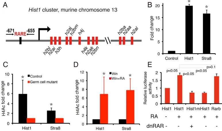
The mouse Hist1 cluster is located on chromosome 13 and the histone genes in theHist1cluster are arranged in three subclusters (Marzluff et al., 2002) (Fig. 5A). Given that most of Hist1gene transcripts are coordinately regulated in THY1+spermatogonia by
retinoid signaling, we assume that expression of Hist1 genes is directly controlled by retinoid signaling.
To test this hypothesis, we found a putative RARE upstream of the histone-encoding genes of the Hist1 cluster. The RARE is located between positions −671 and −655 relative to the transcription start site of Hist1h2bl, the first Hist1 gene (Fig. 5A). Notably, chromatin immunoprecipitation assays (ChIPs) performed on mouse testis at day 5 using an anti-RARG antibody revealed that RARG is present at the RARE region upstream of the Hist1 cluster, at similar levels to the Stra8
promoter (Fig. 5B). Compared with controls, there was a significant decrease in acetylated H4 (H4Ac) levels at the regulatory regions of both Hist1 and Stra8 in the germ-cell mutants (Fig. 5C). In addition, RA administration to WIN18,466-treated mice induces a striking upregulation in H4Ac at the regulatory regions of bothHist1andStra8(Fig. 5D). To further examine whether the−671 to−655 region is a functional RARE, we inserted∼1.5 kb of the upstream regulatory region of theHist1 cluster containing this putative RARE into a luciferase reporter vector and transfected the reporter into RAR/RXR-expressing P19 cells (Kruyt et al., 1991; Schoorlemmer et al., 1995). As shown in Fig. 5E, luciferase activity was significantly induced by RA treatment compared with vehicle-treated controls; however, RA-induced luciferase activity was significantly inhibited by co-transfection withdnRARvector. Furthermore, a mutation of the RARE in the reporter significantly disrupted the RA-induced luciferase activity (Fig. 5E). Collectively, these results reveal that a functional RARE is present upstream of theHist1cluster and that the Hist1 cluster genes could be direct targets for retinoid signaling.
DISCUSSION
Retinoid signaling directly controls spermatogonial differentiation
[image:5.612.89.523.58.352.2]Retinoid signaling is central to spermatogonial differentiation. We and others have demonstrated that testicular RA mainly originates Fig. 4. Alterations of the mRNA transcriptome in germ-cell mutant spermatogonia.(A) Gene ontology term enrichment analyses of retinoid signaling-regulated genes. The top 10 most enriched biological processes based on theirP-values are shown. (B) Differential expression between controls and germ-cell mutants of genes reported previously to be expressed by undifferentiated or differentiated spermatogonia. (C) A heat map showing expression of 36Hist1cluster genes in control and germ-cell mutant spermatogonia. (D) qRT-PCR analysis of mRNA levels ofHist1cluster genes in control and germ-cell mutant spermatogonia. (E) qRT-PCR analysis of mRNA levels ofHist1cluster genes in WIN18,466-treated and RA-injected WIN18,466-treated mouse spermatogonia. Data in D,E are expressed as fold differences compared with controls (D) or WIN18,466-treated (E), respectively, normalized toRps2. Data are expressed as mean±s.d.,n=3, *P<0.05 by Student’st-test.
DEVEL
O
from Sertoli cells especially in puberty; however, it remains elusive whether the action of RA on spermatogonial differentiation is through germ cells or Sertoli cells, or both Sertoli and germ cells, as its receptors, RARs and RXRs, are expressed in both Sertoli cells and the undifferentiated spermatogonia (Gely-Pernot et al., 2012; Tong et al., 2013; Vernet et al., 2006b; Gaemers et al., 1998; DeFalco et al., 2015). For instance, genetic ablation ofRarg in germ cells resulted in only mild spermatogonial differentiation defects in mutants younger than one year old, indicating that either retinoid signaling in Sertoli cells is involved in spermatogonial differentiation, or that other subtypes of RARs in spermatogonia compensate for loss of RARγfunction, as it is the case for many other developmental process (Gely-Pernot et al., 2012; Mark et al., 2009). Thus, to better understand whether the modulation of spermatogonial differentiation by retinoid signaling occurs directly via the germ cells, it is necessary to completely inactivate retinoid signaling, specifically in germ cells. In this study, we used a germ-cell-specific line ofdnRARfl/fl transgenic mice expressingCre to
impair retinoid signaling in germ cells. We found that if there was no retinoid signaling in germ cells, spermatogonial differentiation was completely blocked during the first wave of spermatogenesis and in adult spermatogenesis. Therefore, our findings presented in this study provide strong functional evidence that retinoid signaling directly controls spermatogonial differentiation through the spermatogonia themselves.
Gely-Pernot et al. have recently analyzed mice simultaneously lacking all RARs or all RXRs specifically in undifferentiated spermatogonia and found that testicular defects of both mutants are not identical to those of VAD mice (Gely-Pernot et al., 2015). The
[image:6.612.111.497.60.289.2]phenotype we see in our study is more severe than that reported by Gely-Pernot et al. They reported that excision of genes for all three RXRs resulted in an age-dependent testicular degeneration but spermatogonial differentiation proceeded normally and some cells entered meiosis. In contrast to our results they showed that ablation of allRxror allRargenes in spermatogonia did not alter the first wave of spermatogenesis. This difference might be because the dnRARused in our studies could bind all availableRxrs andRars. Because the dnRAR would sequester all Rxrs it is possible that some of the defects seen in our mutant testes could result from the lack of action of other RXR binding partners such as vitamin D receptor (VDR), thyroid receptor, or the PPAR receptor. However, we feel this result is unlikely because the knockout of theVdror Pparstill results in the completion of spermatogenesis although with a reduced sperm count (Blomberg Jensen et al., 2013; Yao et al., 2015). In addition, male mice lacking individual thyroid receptor genes or bothTRalpha1andTRbeta1(also known asThra and Thrb, respectively) are still fertile, and inactivation of TRalpha1actually results in a larger testis with enhanced Sertoli and germ cell numbers (Cooke, 1991; Gao et al., 2014; Gothe et al., 1999; Holsberger et al., 2005). Therefore, the observations from Gely-Pernot et al. and our findings that germ-cell mutants exhibit complete defects in spermatogonial differentiation identical to VAD mice reveal that RARs could act both with and without an RXR, or that RXRs could act both with and without an RAR, in germ cells. Indeed, it has been demonstrated that RAR functions in Sertoli cells independently of RXRs (Vernet et al., 2006a). Thus, the transgenic model used in this study should be a powerful tool for exploring the molecular mechanisms of retinoid Fig. 5. Functional RARE occurs upstream of theHist1cluster.(A) The histone genes in the mouseHist1cluster and the position of putative RARE are shown. (B) Native ChIP with anti-RARG or IgG antibodies followed by qPCR with primers encompassing RARE upstream ofHist1,Stra8( positive controls) or the control site (2 kb upstream ofHist1RARE) revealing the presence of RARG at the upstream RARE ofHist1andStra8. Mean fold enrichment of three independent experiments at theHist1andStra8site is relative to the amount of DNA at the control site. Error bars represent s.d., *P<0.001. (C) The level of acetylated histone H4 (H4Ac), a mark for open and actively transcribed chromatin, at RARE upstream of bothHist1andStra8is significantly higher in control spermatogonia than in germ-cell mutant spermatogonia. Mean±s.d.,n=3, *P<0.01 by Student’st-test. (D) The H4Ac level at RARE upstream of bothHist1andStra8is significantly increased in RA-injected WIN18,466-treated mouse spermatogonia compared with WIN18,466-treated mouse spermatogonia. Mean±s.d.,n=3, *P<0.01 by Student’st-test. (E) Quantitative evaluation of the putative RARE at upstream of theHist1cluster. Relative firefly luciferase activity was normalized toRenillafor individual conditions. Data shown in graph represent the mean±s.d. fold change from control media (without RA treatment) of three independent experiments. *P<0.01. Hist1, a luciferase reporter containing the putative RARE at upstream ofHist1cluster; mHist1, a luciferase reporter containing mutant putative RARE as described in Materials and methods; Rarb, a luciferase reporter containing classic RARE at theRarbgene promoter.
DEVEL
O
signaling in spermatogonial differentiation, meiotic initiation and spermiogenesis.
RA-induced entry of the undifferentiated spermatogonia into S phase are crucial to spermatogonial differentiation
RA, a potent regulator of cell growth, exerts pleiotropic effects in regulating cellular proliferation and differentiation, depending on the cell types present during embryogenesis and in adult tissues (Bohnsack and Hirschi, 2004; Chambon, 1996; Clagett-Dame and Knutson, 2011). We here provide functional evidence that in the testis, retinoid signaling might control entry of the undifferentiated spermatogonia into S phase, and then promote spermatogonial differentiation. First, impaired retinoid signaling in germ cells causes the arrest of the undifferentiated spermatogonia into S phase, accounting for the blockage of spermatogonial differentiation, as evidenced by: (1) a significant accumulation of the undifferentiated spermatogonia in the G0/G1 phase occurred in germ-cell mutants whereas the differentiation of the undifferentiated spermatogonia into A1spermatogonia is blocked; (2) inhibition of RA synthesis by
WIN18,466 resulted in a blockade of entry into S phase in the undifferentiated spermatogonia as previously suggested by van Pelt et al. (van Pelt et al., 1995; van Pelt and de Rooij, 1990); (3) expression of a burst of replication-dependent core histone genes, whose expression is induced right before and during S phase (Kurat et al., 2014), was downregulated in the undifferentiated spermatogonia of germ-cell mutants; and (4) expression ofCcnd2 andE2f, key regulators for the progression from G1 to S phase (Bohnsack and Hirschi, 2004), was reduced in the germ-cell mutants. Second, during reinitiation of spermatogonial differentiation in WIN18,466-treated mice upon RA administration, early differentiating spermatogonia (STRA8-positive spermatogonia) enter into the S phase. Consistent with this, transcription ofHist1cluster genes andCcnd2 is upregulated in spermatogonia during RA-induced spermatogonial differentiation. Finally, this hypothesis is also supported by previously published data. For instance, during RA-induced precocious spermatogonial differentiation in vivo, the undifferentiated spermatogonia in epithelial stages II-VI, which are arrested in G0/G1 phase of the cell cycle, are released from the G1 block and subsequently enter into mitotic S phase (Endo et al., 2015; Kluin and de Rooij, 1981). Furthermore, undifferentiated spermatogonia in epithelial stage VII-VIII have been shown to enter mitotic S phase during spermatogonial differentiation (Endo et al., 2015).
Retinoid signaling can regulate expression of replication-dependent core histone genes in the undifferentiated spermatogonia
The regulation of cell cycle progression is tightly controlled within cells. At the G1/S phase transition, the cell duplicates the genome, a process that requires the initiation of DNA replication coupled with activation of core histone gene expression, whereas outside of S phase, core histone synthesis is suppressed to avoid overproduction (Osley, 1991). Expression of the core histone genes that encode proteins for packaging the newly synthesized genome is exquisitely controlled at transcriptional and posttranscriptional levels within cells (Osley, 1991; Eliassen et al., 1998). Our studies showed that impaired RA signaling in germ cells inhibited the transcription of replication-dependent core histone genes in the undifferentiated spermatogonia. By contrast, RA treatment upregulated expression of core histone gene expression in spermatogonia. Collectively, these data indicate that RA-dependent signaling in spermatogonia controls replication-dependent core histone gene expression. Because the expression of many histone genes such as Hist1 cluster genes is coordinately activated upon entry into S phase, it is possible that a common pathway (e.g. transcriptional factors) contributes to their simultaneous regulation. Using bioinformatic analyses, ChIP assays and luciferase reporter experiments, we identified a functional RARE present in the upstream regulatory region of the Hist1 cluster. Thus, we demonstrate that retinoid signaling directly controls the transcription ofHist1cluster genes in spermatogonia.
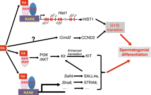
This, together with a wealth of evidence that CCND2 plays crucial roles in the mitotic G1/S transition (Beumer et al., 2000) leads us to conclude that retinoid signaling directly functions on the undifferentiated spermatogonia to upregulate the expression of Ccnd2for initiating DNA synthesis and to activate the transcription of replication-dependent histone genes for packaging the new genome. This results in the induction of entry of the undifferentiated spermatogonia into S phase (Fig. 6). It will be of interest in the future to study whether retinoid signaling could promote the premeiotic G1/S transition.
[image:7.612.52.354.545.735.2]Recent studies reveal that several RA target genes have also been implicated in spermatogonial differentiation. Endo et al. demonstrated that RA target gene, Stra8, could promote spermatogonial differentiation (Endo et al., 2015). Gely-Pernot et al. indicated that Sall4, a RA target gene, could control spermatogonial differentiation through upregulation of Kit
Fig. 6. A proposed model for retinoid signaling-induced spermatogonial differentiation.RA signaling induces spermatogonial differentiation through activating expression of genes (e.g.Hist1,Ccnd2) required for G1/S phase transition and stimulating other gene expression such as
Stra8,Sall4andKitin both genomic and non-genomic pathways.
DEVEL
O
expression (Gely-Pernot et al., 2015). Busada et al. showed that RA could also utilize non-genomic pathways via PI3K/AKT/mTOR signaling to stimulate translation of mRNA for Kit during spermatogonial differentiation (Busada et al., 2014). Based on these results, we postulate that retinoid signaling might induce the entry of the undifferentiated spermatogonia into S phase through upregulation of expression of replication-dependent histone genes and Ccnd2, and subsequently promote the differentiation of undifferentiated spermatogonia into A1 spermatogonia with
RA-induced expression ofStra8(Endo et al., 2015; Zhou et al., 2008), Sall4 (Gely-Pernot et al., 2015) and Kit (Schrans-Stassen et al., 1999; Busada et al., 2014) (Fig. 6).
MATERIALS AND METHODS Mice
The dnRARflox/flox mice were a generous gift from the Mendelsohn
laboratory (Rosselot et al., 2010; Lécureuil et al., 2002). TheStra8-Cre line was generated in the Braun laboratory (Sadate-Ngatchou et al., 2008). The RARElacZ reporter line was created in the Rossant laboratory (Rossant et al., 1991). All animal experiments were approved by the Animal Care and Use Committee at Institute of Biochemistry and Cell Biology, Shanghai Institutes for Biological Sciences.
Histological and immunohistochemical analyses
Testes were fixed in Bouins solution or 4% paraformaldehyde (PFA), embedded in paraffin and sectioned. Sections were deparaffinized, rehydrated, and stained with Hematoxylin and Eosin. For immunohistochemical studies, slides were boiled in 10 mM sodium citrate buffer, pH 6.0, for 15 min, brought to room temperature, washed in PBS with 0.1% Triton X-100, and then incubated for 60 min at room temperature with blocking buffer (10% donkey serum, 1% BSA, 0.1% Triton X-100 in PBS). The sections were then incubated with a 1:50 dilution of rat anti-GCNA IgM (kindly provided by Dr G. Enders, University of Kansas, USA) and rabbit anti-STRA8 IgG or rabbit anti-PLZF (SC-22839, Santa Cruz Biotechnology) or rabbit anti-SOX9 IgG (AB5535, Millipore) or 1:50 dilution of rat anti-KIT (MABF510, Millipore) overnight at 4°C and rabbit anti-STRA8 IgG or rabbit anti-Alexa Fluor 488-and 594-conjugated donkey secondary antibody (711545152 488-and 711585152, Jackson ImmunoResearch Laboratories) at 1:500 dilution were added. After 60 min at room temperature, the sections were washed in PBS, rinsed quickly in pure ethanol, mounted in Prolong Gold Antifade medium with DAPI (Molecular Probes), and then analyzed by a fluorescence microscope (Olympus).
Isolation of mouse THY1+spermatogonia and flow cytometry
THY1+spermatogonia were isolated using magnetic activated cell sorting (MACS) with magnetic microbeads conjugated to anti-THY1+(Miltenyi Biotech) as described previously (Tong et al., 2012). To determine cell cycle progression, THY1+ spermatogonia were fixed in 75% ethanol at 4°C overnight, washed with PBS, and incubated with 25 µg/ml RNase A (Sigma) and 50 µg/ml propidium iodide (Sigma) in PBS at 37°C for 30 min, followed by FACS analysis on a BD LSRII SORP (BD Biosciences).
RNA sequencing analyses
Total RNA was extracted from THY1+spermatogonia isolated from control and germ-cell mutant mice using Trizol reagent (Invitrogen). Libraries of cDNA were constructed by the Omics Core of CAS-MPG Partner Institute for Computational Biology at Shanghai Institutes for Biological Sciences using the TrueSeq Stranded Total RNA Library Prep Kit (Illumina) following manufacturer’s instructions. Libraries were sequenced using single reads (100 nt) on an Illumina HiSeq 2000. Sequencing reads were mapped to the ENSEMBL mouse reference genome (GENCODE vM6/ GRCm38) using TopHat (Johns Hopkins University Center for Computational Biology) with standard setting. Confidently aligned reads for each sample were analyzed with the Cuffdiff2 program (Johns Hopkins University Center for Computational Biology) to determine differential expression between control and germ-cell mutant samples. Genes with a fold change≥1.5 were selected as differential genes.
Quantitative RT-PCR assays
Total RNA was extracted using Trizol reagent, and treated with DNaseI (Ambion). Total RNA was reverse transcribed using an iScript cDNA Synthesis Kit (Bio-Rad). Quantitative PCR was performed with Fast SYBR Green PCR mastermix (Applied Biosystems) on the Applied Biosystems 7500 Fast system. Relative expression of genes was analyzed by the comparative CT method with use of ribosomal protein S2 (Rps2) as a normalized control. RT-PCR primer sequences are listed in Table S2.
ChIP assay
As previously described (Tong et al., 2011), mouse spermatogonia were cross-linked with 1% formaldehyde/PBS for 10 min at room temperature and quenched by adding glycine to a final concentration of 0.25 M for 5 min. Cells were collected and washed twice with cold PBS containing 1× protease inhibitor cocktail (Roche). Cell pellets were lysed in 1 ml of lysis buffer (1% SDS, 50 mM Tris-HCl pH 8.0, 10 mM EDTA and 1× protease inhibitor cocktail) for 10 min on ice, and sonicated on ice to obtain a chromatin size of 200-500 bp. After pre-clearing with protein A/G agarose beads (Upstate), an equivalent amount of sheared chromatin was immunoprecipitated with antibody overnight at 4°C followed by an incubation with protein A/G for 1 h. Antibodies used in ChIP assays were anti-RARG (1:250; sc-7387, Santa Cruz), anti-acetylated histone H4 (H4Ac) (1:500; 06-866, Upstate), and IgG control (1:1000; sc-66931, Santa Cruz). Protein-DNA complexes were eluted in fresh 1% SDS/0.1 M NaHCO3. Crosslinking was reversed and protein was removed. DNA was recovered and purified for PCR using specific primers listed in Table S3.
Luciferase assay
The pGL3-Hist1-luciferase reporter vector was constructed by replacing the cytomegalovirus (CMV) promoter sequence of the pGL3-luciferase reporter plasmid with a 1.5 kb sequence upstream of theHist1cluster containing a putative RARE region (−671 to−655: AGGCCAAGGGGAAGTGA). The mutations were introduced into the RARE region of the pGL3-Hist1-luciferase construct to generate the pGL3-mHist1-pGL3-Hist1-luciferase reporter. The pGL3-Rarb-luciferase reporter was generated by replacing the CMV promoter of the pGL3-luciferase plasmid with aRarbpromoter sequence, which contains classic RARE. The pGL3-Hist1-luciferase, pGL3-mHist1-luciferase, or pGL3-Rarb-luciferase was co-transfected with aRenillavector into P19 cells using Lipofectamine 2000 transfection regent (Invitrogen) according to the manufacturer’s instruction. At 24 h post-transfection, the cells were treated with or without 10−6mol/l RA for another 24 h before harvesting cells for dual luciferase assay according to the manufacturer’s protocol (Promega). The relative luciferase activity was normalized to
Renillaluciferase activity.
WIN18,466, RA administration and EdU labeling
For WIN18,466 treatment (Hogarth et al., 2013), 2-day-old animals were pipette-fed with WIN18,466 (100μg/g body weight) suspended in 1% gum tragacanth for seven consecutive days. For RA injection experiments, mice received an intraperitoneal (i.p.) injection of all-trans-RA (Sigma; 400μg/ injection) in 10μl of DMSO for 24 h.
For EdU labeling, as described previously (Tong et al., 2013), mice were i.p. injected with EdU (Invitrogen) (50μg/g body weight) in PBS. The mice were euthanized 4 h later and testes were fixed in 4% PFA/PBS solution, embedded in paraffin and sectioned. The sections were immunostained with antibody against PLZF or STRA8 first and the EdU incorporation was then detected by Click-It EdU Alexa Fluor 594 Imaging Kit according to the manufacturer’s protocol (Invitrogen).
X-gal staining
dnRARfl/fl, Stra8-Cre+ (female) mice were crossed with a RARElacZ
reporter line harboring RARE-Hspa1b-lacZ alleles to obtain dnRARfl/+,
Stra8-Cre+, RARElacZand dnRARfl/+,RARElacZmice. ThednRARfl/+,
Stra8-Cre+,RARElacZ(female) anddnRARfl/+,RARElacZmice were used
to produce dnRARfl/fl, Stra8-Cre+, RARElacZ, dnRARfl/+, Stra8-Cre+,
RARElacZanddnRARfl/fl,RARElacZmice. Testes, epididymis and kidneys
from animals bearing RARE-Hspa1b-lacZ alleles were fixed in 4%
DEVEL
O
paraformaldehyde (PFA) in PBS for 2 h at room temperature, washed, stained in bromo-chloro-indolyl-galactopyranoside (X-gal) at 37°C overnight, washed and then photographed. The stained testes were then processed, embedded in paraffin and sectioned. Sections were counterstained with Fast Red.
Acknowledgements
We thank Dr G. Enders for providing anti-GCNA antibody and Dr C. Mendelsohn for providingdnRARmice.
Competing interests
The authors declare no competing or financial interests.
Author contributions
Y.C. carried out most experiments and data analysis; L.M. and G.W. performed RNA sequencing data analysis; C.H., M.D.G. and M.-H.T. analyzed data and wrote the manuscript. All the authors were involved in the discussion on the manuscript.
Funding
This work supported by the National Natural Science Foundation of China [grant 31471401 to M.-H.T.] and the Ministry of Science and Technology of the People’s Republic of China [grant 2014CB943101 to M.-H.T.] and National Institutes of Health [grants HD10808 to M.D.G. and HD06777 to M.-H.T. and M.D.G]. Deposited in PMC for release after 12 months.
Data availability
RNA sequencing data have been deposited at Gene Expression Omnibus with accession number GSE79863.
Supplementary information
Supplementary information available online at
http://dev.biologists.org/lookup/suppl/doi:10.1242/dev.135939/-/DC1
References
Ballow, D., Meistrich, M. L., Matzuk, M. and Rajkovic, A.(2006). Sohlh1 is essential for spermatogonial differentiation.Dev. Biol.294, 161-167.
Bastien, J. and Rochette-Egly, C.(2004). Nuclear retinoid receptors and the transcription of retinoid-target genes.Gene328, 1-16.
Beumer, T. L., Roepers-Gajadien, H. L., Gademan, I. S., Kal, H. B. and de Rooij, D. G.(2000). Involvement of the D-type cyclins in germ cell proliferation and differentiation in the mouse.Biol. Reprod.63, 1893-1898.
Blomberg Jensen, M., Lieben, L., Nielsen, J. E., Willems, A., Jørgensen, A., Juul, A., Toppari, J., Carmeliet, G. and Rajpert-De Meyts, E. (2013). Characterization of the testicular, epididymal and endocrine phenotypes in the Leuven Vdr-deficient mouse model: targeting estrogen signalling. Mol. Cell Endocrinol.377, 93-102.
Bohnsack, B. L. and Hirschi, K. K. (2004). Nutrient regulation of cell cycle progression.Annu. Rev. Nutr.24, 433-453.
Buaas, F. W., Kirsh, A. L., Sharma, M., McLean, D. J., Morris, J. L., Griswold, M. D., de Rooij, D. G. and Braun, R. E.(2004). Plzf is required in adult male germ cells for stem cell self-renewal.Nat. Genet.36, 647-652.
Busada, J. T., Kaye, E. P., Renegar, R. H. and Geyer, C. B.(2014). Retinoic acid induces multiple hallmarks of the prospermatogonia-to-spermatogonia transition in the neonatal mouse.Biol. Reprod.90, 64.
Chambon, P.(1996). A decade of molecular biology of retinoic acid receptors. FASEB J.10, 940-954.
Clagett-Dame, M. and Knutson, D. (2011). Vitamin A in reproduction and development.Nutrients3, 385-428.
Clermont, Y. (1972). Kinetics of spermatogenesis in mammals: seminiferous epithelium cycle and spermatogonial renewal.Physiol. Rev.52, 198-236. Cooke, P. S.(1991). Thyroid hormones and testis development: a model system for
increasing testis growth and sperm production. Ann. N. Y. Acad. Sci. 637, 122-132.
Costoya, J. A., Hobbs, R. M., Barna, M., Cattoretti, G., Manova, K., Sukhwani, M., Orwig, K. E., Wolgemuth, D. J. and Pandolfi, P. P.(2004). Essential role of Plzf in maintenance of spermatogonial stem cells.Nat. Genet.36, 653-659. Damm, K., Heyman, R. A., Umesono, K. and Evans, R. M.(1993). Functional
inhibition of retinoic acid response by dominant negative retinoic acid receptor mutants.Proc. Natl. Acad. Sci. USA90, 2989-2993.
de Rooij, D. G.(2001). Proliferation and differentiation of spermatogonial stem cells. Reproduction121, 347-354.
DeFalco, T., Potter, S. J., Williams, A. V., Waller, B., Kan, M. J. and Capel, B. (2015). Macrophages contribute to the spermatogonial niche in the adult testis. Cell Rep.12, 1107-1119.
Eliassen, K. A., Baldwin, A., Sikorski, E. M. and Hurt, M. M.(1998). Role for a YY1-binding element in replication-dependent mouse histone gene expression. Mol. Cell. Biol.18, 7106-7118.
Endo, T., Romer, K. A., Anderson, E. L., Baltus, A. E., de Rooij, D. G. and Page, D. C.(2015). Periodic retinoic acid–STRA8 signaling intersects with periodic germ-cell competencies to regulate spermatogenesis.Proc. Natl. Acad. Sci. USA
112, E2347-E2356.
Falender, A. E., Freiman, R. N., Geles, K. G., Lo, K. C., Hwang, K., Lamb, D. J., Morris, P. L., Tjian, R. and Richards, J. S. (2005). Maintenance of spermatogenesis requires TAF4b, a gonad-specific subunit of TFIID.Genes Dev.19, 794-803.
Gaemers, I. C., van Pelt, A. M., van der Saag, P. T., Hoogerbrugge, J. W., Themmen, A. P. and de Rooij, D. G.(1998). Differential expression pattern of retinoid X receptors in adult murine testicular cells implies varying roles for these receptors in spermatogenesis.Biol. Reprod.58, 1351-1356.
Gao, Y., Lee, W. M. and Cheng, C. Y.(2014). Thyroid hormone function in the rat testis.Front. Endocrinol.5, 188.
Gely-Pernot, A., Raverdeau, M., Célébi, C., Dennefeld, C., Feret, B., Klopfenstein, M., Yoshida, S., Ghyselinck, N. B. and Mark, M. (2012).
Spermatogonia differentiation requires retinoic acid receptor gamma.
Endocrinology153, 438-449.
Gely-Pernot, A., Raverdeau, M., Teletin, M., Vernet, N., Féret, B., Klopfenstein, M., Dennefeld, C., Davidson, I., Benoit, G., Mark, M. et al.(2015). Retinoic acid receptors control spermatogonia cell-fate and induce expression of the SALL4A transcription factor.PLoS Genet.11, e1005501.
Goertz, M. J., Wu, Z., Gallardo, T. D., Hamra, F. K. and Castrillon, D. H.(2011). Foxo1 is required in mouse spermatogonial stem cells for their maintenance and the initiation of spermatogenesis.J. Clin. Invest.121, 3456-3466.
Gothe, S., Wang, Z., Ng, L., Kindblom, J. M., Barros, A. C., Ohlsson, C., Vennstrom, B. and Forrest, D.(1999). Mice devoid of all known thyroid hormone receptors are viable but exhibit disorders of the pituitary-thyroid axis, growth, and bone maturation.Genes Dev.13, 1329-1341.
Griswold, M. D., Bishop, P. D., Kim, K.-H., Ping, R., Siiteri, J. E. and Morales, C. (1989). Function of vitamin A in normal and synchronized seminiferous tubules. Ann. N. Y. Acad. Sci.564, 154-172.
Hao, J., Yamamoto, M., Richardson, T. E., Chapman, K. M., Denard, B. S., Hammer, R. E., Zhao, G. Q. and Hamra, F. K.(2008). Sohlh2 knockout mice are male-sterile because of degeneration of differentiating type A spermatogonia. Stem Cells26, 1587-1597.
Hasegawa, K. and Saga, Y.(2012). Retinoic acid signaling in Sertoli cells regulates organization of the blood-testis barrier through cyclical changes in gene expression.Development139, 4347-4355.
Hobbs, R. M., Fagoonee, S., Papa, A., Webster, K., Altruda, F., Nishinakamura, R., Chai, L. and Pandolfi, P. P.(2012). Functional antagonism between Sall4 and Plzf defines germline progenitors.Cell Stem Cell10, 284-298.
Hogarth, C. A. and Griswold, M. D. (2010). The key role of vitamin A in spermatogenesis.J. Clin. Invest.120, 956-962.
Hogarth, C. A., Evanoff, R., Mitchell, D., Kent, T., Small, C., Amory, J. K. and Griswold, M. D. (2013). Turning a spermatogenic wave into a tsunami: synchronizing murine spermatogenesis using WIN 18,446.Biol. Reprod.88, 40. Hogarth, C. A., Arnold, S., Kent, T., Mitchell, D., Isoherranen, N. and Griswold, M. D.(2015). Processive pulses of retinoic acid propel asynchronous and continuous murine sperm production.Biol. Reprod.92, 37.
Holsberger, D. R., Kiesewetter, S. E. and Cooke, P. S.(2005). Regulation of neonatal Sertoli cell development by thyroid hormone receptor alpha1.Biol. Reprod.73, 396-403.
Huang, H. F. S. and Hembree, W. C.(1979). Spermatogenic response to vitamin A in vitamin A deficient rats.Biol. Reprod.21, 891-904.
Ikami, K., Tokue, M., Sugimoto, R., Noda, C., Kobayashi, S., Hara, K. and Yoshida, S. (2015). Hierarchical differentiation competence in response to retinoic acid ensures stem cell maintenance during mouse spermatogenesis. Development142, 1582-1592.
Kluin, P. M. and De Rooij, D. G.(1981). A comparison between the morphology and cell kinetics of gonocytes and adult type undifferentiated spermatogonia in the mouse.Int. J. Androl.4, 475-493.
Kruyt, F. A. E., van den Brink, C. E., Defize, L. H. K., Donath, M.-J., Kastner, P., Kruijer, W., Chambon, P. and van der Saag, P. T.(1991). Transcriptional regulation of retinoic acid receptor beta in retinoic acid-sensitive and -resistant P19 embryocarcinoma cells.Mech. Dev.33, 171-178.
Kurat, C. F., Recht, J., Radovani, E., Durbic, T., Andrews, B. and Fillingham, J. (2014). Regulation of histone gene transcription in yeast.Cell. Mol. Life Sci.71, 599-613.
Laronda, M. M. and Jameson, J. L.(2011). Sox3 functions in a cell-autonomous manner to regulate spermatogonial differentiation in mice.Endocrinology152, 1606-1615.
Lécureuil, C., Fontaine, I., Crepieux, P. and Guillou, F.(2002). Sertoli and granulosa cell-specific Cre recombinase activity in transgenic mice.Genesis33, 114-118.
Lufkin, T., Lohnes, D., Mark, M., Dierich, A., Gorry, P., Gaub, M. P., LeMeur, M. and Chambon, P.(1993). High postnatal lethality and testis degeneration in
DEVEL
O
retinoic acid receptor alpha mutant mice. Proc. Natl. Acad. Sci. USA 90, 7225-7229.
Mark, M., Ghyselinck, N. B. and Chambon, P.(2009). Function of retinoic acid receptors during embryonic development.Nucl. Recept. Signal.7, e002. Marzluff, W. F., Gongidi, P., Woods, K. R., Jin, J. and Maltais, L. J.(2002). The
human and mouse replication-dependent histone genes.Genomics80, 487-498. Morales, C. and Griswold, M. D.(1987). Retinol-induced stage synchronization in
seminiferous tubules of the rat.Endocrinology121, 432-434.
Nakagawa, T., Nabeshima, Y.-i. and Yoshida, S.(2007). Functional identification of the actual and potential stem cell compartments in mouse spermatogenesis. Dev. Cell12, 195-206.
Niu, Z., Goodyear, S. M., Rao, S., Wu, X., Tobias, J. W., Avarbock, M. R. and Brinster, R. L. (2011). MicroRNA-21 regulates the self-renewal of mouse spermatogonial stem cells.Proc. Natl. Acad. Sci. USA108, 12740-12745. Oakberg, E. F.(1956). A description of spermiogenesis in the mouse and its use in
analysis of the cycle of the seminiferous epithelium and germ cell renewal. Am. J. Anat.99, 391-413.
Oatley, J. M. and Brinster, R. L.(2008). Regulation of spermatogonial stem cell self-renewal in mammals.Annu. Rev. Cell Dev. Biol.24, 263-286.
Osley, M. A.(1991). The regulation of histone synthesis in the cell cycle.Annu. Rev. Biochem.60, 827-861.
Pesce, M., Wang, X., Wolgemuth, D. J. and Schöler, H. (1998). Differential expression of the Oct-4 transcription factor during mouse germ cell differentiation. Mech. Dev.71, 89-98.
Rossant, J., Zirngibl, R., Cado, D., Shago, M. and Giguere, V.(1991). Expression of a retinoic acid response element-hsplacZ transgene defines specific domains of transcriptional activity during mouse embryogenesis. Genes Dev. 5, 1333-1344.
Rosselot, C., Spraggon, L., Chia, I., Batourina, E., Riccio, P., Lu, B., Niederreither, K., Dolle, P., Duester, G., Chambon, P. et al.(2010). Non-cell-autonomous retinoid signaling is crucial for renal development.Development137, 283-292.
Sada, A., Suzuki, A., Suzuki, H. and Saga, Y.(2009). The RNA-binding protein NANOS2 is required to maintain murine spermatogonial stem cells.Science325, 1394-1398.
Sadate-Ngatchou, P. I., Payne, C. J., Dearth, A. T. and Braun, R. E.(2008). Cre recombinase activity specific to postnatal, premeiotic male germ cells in transgenic mice.Genesis46, 738-742.
Schneider, R. A., Hu, D., Rubenstein, J. L., Maden, M. and Helms, J. A.(2001). Local retinoid signaling coordinates forebrain and facial morphogenesis by maintaining FGF8 and SHH.Development128, 2755-2767.
Schoorlemmer, J., Jonk, L., Shen, S., van Puijenbroek, A., Feijen, A. and Kruijer, W.(1995). Regulation of Oct-4 gene expression during differentiation of EC cells.Mol. Biol. Rep.21, 129-140.
Schrans-Stassen, B. H. G. J., van de Kant, H. J. G., de Rooij, D. G. and van Pelt, A. M. M.(1999). Differential expression of c-kit in mouse undifferentiated and differentiating type A spermatogonia.Endocrinology140, 5894-5900.
Tong, M.-H., Mitchell, D., Evanoff, R. and Griswold, M. D.(2011). Expression of Mirlet7 family microRNAs in response to retinoic acid-induced spermatogonial differentiation in mice.Biol. Reprod.85, 189-197.
Tong, M.-H., Mitchell, D. A., Mcgowan, S. D., Evanoff, R. and Griswold, M. D. (2012). Two miRNA clusters, Mir-17-92 (Mirc1) and Mir-106b-25 (Mirc3), are involved in the regulation of spermatogonial differentiation in mice.Biol. Reprod.
86, 72.
Tong, M.-H., Yang, Q.-E., Davis, J. C. and Griswold, M. D. (2013). Retinol dehydrogenase 10 is indispensible for spermatogenesis in juvenile males.Proc. Natl. Acad. Sci. USA110, 543-548.
van Pelt, A. M. and de Rooij, D. G.(1990). Synchronization of the seminiferous epithelium after vitamin A replacement in vitamin A-deficient mice.Biol. Reprod.
43, 363-367.
van Pelt, A. M., van Dissel-Emiliani, F. M., Gaemers, I. C., van der Burg, M. J., Tanke, H. J. and de Rooij, D. G.(1995). Characteristics of A spermatogonia and preleptotene spermatocytes in the vitamin A-deficient rat testis.Biol. Reprod.53, 570-578.
Vernet, N., Dennefeld, C., Guillou, F., Chambon, P., Ghyselinck, N. B. and Mark, M.(2006a). Prepubertal testis development relies on retinoic acid but not rexinoid receptors in Sertoli cells.EMBO J.25, 5816-5825.
Vernet, N., Dennefeld, C., Rochette-Egly, C., Oulad-Abdelghani, M., Chambon, P., Ghyselinck, N. B. and Mark, M.(2006b). Retinoic acid metabolism and signaling pathways in the adult and developing mouse testis.Endocrinology147, 96-110.
Wilson, J. G., Roth, C. B. and Warkany, J.(1953). An analysis of the syndrome of malformations induced by maternal vitamin A deficiency. Effects of restoration of vitamin A at various times during gestation.Am. J. Anat.92, 189-217. Wolbach, S. B. and Howe, P. R.(1925). Tissue changes following deprivation of
fat-soluble a vitamin.J. Exp. Med.42, 753-777.
Wolgemuth, D. J. and Chung, S. S. (2007). Retinoid signaling during spermatogenesis as revealed by genetic and metabolic manipulations of retinoic acid receptor alpha.Soc. Reprod. Fertil. Suppl.63, 11-23.
Yao, P.-L., Chen, L., Hess, R. A., Müller, R., Gonzalez, F. J. and Peters, J. M. (2015). Peroxisome Proliferator-activated Receptor-D (PPARD) Coordinates Mouse Spermatogenesis by Modulating Extracellular Signal-regulated Kinase (ERK)-dependent Signaling.J. Biol. Chem.290, 23416-23431.
Zheng, K., Wu, X., Kaestner, K. H. and Wang, P. J.(2009). The pluripotency factor LIN28 marks undifferentiated spermatogonia in mouse.BMC Dev. Biol.9, 38. Zhou, Q., Li, Y., Nie, R., Friel, P., Mitchell, D., Evanoff, R. M., Pouchnik, D.,
Banasik, B., McCarrey, J. R., Small, C. et al.(2008). Expression of stimulated by retinoic acid gene 8 (Stra8) and maturation of murine gonocytes and spermatogonia induced by retinoic acid in vitro.Biol. Reprod.78, 537-545. Zhou, Z., Shirakawa, T., Ohbo, K., Sada, A., Wu, Q., Hasegawa, K., Saba, R. and
Saga, Y.(2015). RNA binding protein Nanos2 organizes post-transcriptional buffering system to retain primitive state of mouse spermatogonial stem cells.Dev. Cell34, 96-107.