Available online on www.ijpsr.com 1
IJPSR (2010), Vol. 1, Issue 3 (Review Article)
Received 4 January, 2010; received in revised form 23 February, 2010; accepted 26 February, 2010 OCULAR DRUG DELIVERY: A REVIEW
Palani S*1, Nisha Mary Joseph2, Goda C. C.1, Anish Zachariah3 and Zelalem Ayenew2
Faculty of Pharmacy, 7th October University1, Misurata, Libya
School Of Pharmacy, College of Health Sciences, Mekelle University2, Mekelle, Ethiopia School Of Studies in Biotechnology, Jiwaji University3, Gwalior, Madhya Pradesh, India
Keywords: ophthalmic,
ocular, microspheres, nanoparticles,
liposomes
*Correspondence for Author Dr. S. Palani
Faculty of Pharmacy,
7th October University, Misurata, Libya
ABSTRACT
The route of choice for the treatment of ophthalmic diseases is by the topical route because of the blood ocular barrier. The most commonly utilized conventional preparations of ophthalmic dosage forms are the solutions, suspensions and ointments which are relatively inefficient as therapeutic systems. Following administration, a large proportion of the topically applied drug is immediately diluted in the tear film and excess fluid spills over the lid margin and the remainder is rapidly drained into the nasolacrimal duct so required amount of drug is not available for immediate therapeutic action since it binds to the surrounding extra orbital tissues. In view of these losses, frequent topical administration is necessary to maintain adequate drug levels. Systemic administration of a drug to treat ocular disease would require a high concentration of circulating drug in the plasma to achieve therapeutic levels. By using prolonged drug delivery, the duration of drug action can be remarkably prolonged and also the frequency of drug administration can be reduced. Such a drug delivery can be achieved by designing formulations such as microspheres, nanoparticles, liposomes which can act as
ISSN: 0975 - 8232
Available online on www.ijpsr.com 2
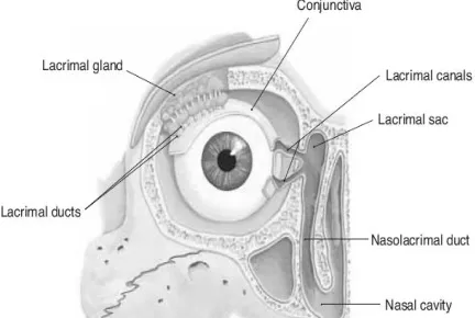
[image:2.612.90.302.327.472.2]INTRODUCTION: The eye is a unique organ, both anatomically (Figure- 1), and physiologically, containing several widely varied structures with independent physiological functions. The complexity of the eye provides unique challenges to drug delivery strategies. Ocular drug delivery is one of the most challenging tasks faced by the Pharmaceutical researchers. One of the major barriers of ocular medication is to obtain and maintain a therapeutic level at the site of action for prolonged period of time. Pharmaceutical treatment and drug delivery methods for treating eye diseases and disorders vary considerably depending on the nature and extent of the disease or disorder 1-2.
FIGURE 1: CROSS SECTIONAL VIEW OF EYE
The bioavailability of traditional ocular drug delivery systems such as eye drops is very poor because eye is protected by a series of complex defense mechanisms that make it difficult to achieve an effective drug concentration within the target area of the eye. The anatomy, physiology of the eye is one of the most complex and unique systems in the human body. lachrymation (figure-2), effective drainage by the nasolacrimal system, the inner and outer blood-retinal barrier, the impermeability of the cornea, and inability of absorption by other
non-corneal structures cause the eye to be exceedingly impervious to foreign substances. While these innate barriers are advantageous for hindering the invasion of undesired molecules, pathogens, and particulates, they pose significant challenges to delivering ocular drugs3-4. Many approaches have been developed to solve the problem in recent decades. Drainage of an administered drug dose by the nasolachrymal system can occur when the volume of fluid in the eye exceeds the normal lachrymal volume of about 7–10μl.
In contrast, the application of one to two drops of a drug medication applied by an eye-dropper as the drug delivery device represents roughly 50–100μl. Much of this dose is wasted or rapidly drained. The remaining applied drug solution is diluted by induced increased lachrymation and physiological tear turnover produced by the applied drug solution. In addition, any remaining drug is subject to non-selective transcorneal adsorption. All these factors taken together can result in a loss of drug from that applied to the eye that can be 500–700 times greater than the rate of absorption of the drug into the anterior chamber.
[image:2.612.324.540.537.682.2]Available online on www.ijpsr.com 3
Three important factors have to be considered when attempting drug delivery to the eye-
1. How the blood-eye barrier (systemic to ocular) or cornea (external to ocular) is crossed by the drug to reach the site of action
2. How to localize the pharmacodynamic action at the eye and minimize drug action on other tissues
3. How to prolong the duration of drug action such that the frequency of drug administration can be reduced
Advanced technology based on the use of nano- carriers (nanoparticles, liposomes, and microspheres) has been investigated recently ocular drug delivery. These systems are claimed to provide a prolonged residence time at the ocular surface, minimizing the effect of natural eye clearance systems. It should be possible with controlled drug delivery to provide drug therapeutic levels for a prolonged time at the site of action.
THE EYE: The eyelids contain skeletal muscle that enables the eyelids to close and cover the front of the eyeball. Eyelashes along the border of each eyelid help keep dust out of the eyes. The eyelids are lined with a thin membrane called the conjunctiva, which is also folded over the white of the eye and merges with the corneal epithelium. Inflammation of this membrane, called conjunctivitis, may be caused by allergies or by certain bacteria or viruses, and makes the eyes red, itchy, and watery. Tears are produced by the lachrymal glands, located at the upper, outer corner of the eyeball, within the orbit. Secretion of tears occurs constantly,
but is increased by the presence of irritating chemicals or dust, and in certain emotional situations. Small ducts take tears to the anterior of the eyeball, and blinking spreads the tears and washes the surface of the eye. Tears are mostly water, with about 1% sodium chloride, similar to other body fluids. Tears also contain lysozyme, an enzyme that inhibits the growth of most bacteria on the wet, warm surface of the eye. At the medial corner of the eyelids are two small openings into the superior and inferior lachrymal canals. These ducts take tears to the lachrymal sac (in the lachrymal bone), which leads to the nasolachrymal duct, which empties tears into the nasal cavity. This is why crying often makes the nose run5.
BARRIERS TO DRUG PERMEATION: The human eye has a spherical shape with a diameter of 23mm. The structural components of the eyeball are divided into three layers: the outermost coat comprises the clear, transparent cornea and the white, opaque sclera; the middle layer comprises the iris anteriorly, the choroid posteriorly, and the ciliary body; and the inner layer is the retina, which is an extension of the central nervous system.
International Journal of Pharmaceutical Sciences and Research ISSN: 0975-8232
Available online on www.ijpsr.com 4
layers of the corneal squamous epithelial cells form a barrier for intercellular drug penetration7.
2. Ocular Wall Barriers: The skeleton of the eye globe consists of the rigid scleral collagenous shell that is lined internally by the uveal tract. The sclera covers the posterior 80% of the eye globe except for a small posterior opening occupied by the optic nerve head, whereas the rest of the globe is covered anteriorly by the cornea. The scleral stroma is composed of bundles of collagen, fibroblasts, and a moderate amount of ground substance. This tissue is essentially a vascular but is lined superficially by the vascular episclera. A large number of channels penetrate the sclera to allow the passage of vessels and nerves to the choroid side.
In humans, the scleral thickness is in the range of 0.3 to 1.0 mm with the posterior pole being the thickest8. The choroid is a highly vascularized tissue, with one of the highest blood flow rates among body tissues. This layer thickness averages 0.25 mm and consists of an innermost layer of fenestrated chorio- capillaris, middle medium and outer large vessels (both nonfenestrated)9.
3. Retinal Barriers: The retina consists of 10 layers: (a) the Retinal Pigment Epithelium, (b) photoreceptor outer segments, (c) external limiting membrane, (d) outer nuclear layer, (e) outer plexiform layer, (f) inner nuclear layer,(g) inner plexiform layer,(h) ganglion cell layer, (i) nerve fiber layer, (j) internal limiting membrane10.
Routes of Ocular drug administration:
1. Topical administration: It is the most
commonly used route of drug
administration for the treatment of anterior segment complications. Posterior segment drug delivery via topical route suffers from drug loss in the precorneal area and anterior segment, drug elimination from the anterior chamber by the canal of Schlemn or via absorption through iris-ciliary body. Enzymatic metabolism in the anterior chamber limits the entry of intact drug into the posterior segment tissues. Limited success has been achieved with topical administration in the area of posterior segment drug delivery.
2. Intra- vitreal administration: In recent advancement in the surgical procedures, intra- vitreal administration of therapeutic agents by direct injection into the mid-vitreous region and sustain and controlled released intra- vitreal implants have become a mainstay treatment option of posterior segment diseases. Longer retention time and higher vitreous concentration of drugs was obtained following this route of administration.
Available online on www.ijpsr.com 5
3. Scleral administration: Due to its large surface area, easy accessibility and relatively high permeability to macromolecules, the sclera recently has become a potential vector for posterior segment drug delivery. Scleral drug delivery has been attempted by different ways, such as scleral plugs and implants, sun conjunctival injection, subtenon injection. Trans-scleral administration of drugs offers a promising therapeutic approach for the treatment of various posterior segment diseases.
4. Systemic administration: Due to the presence of blood retinal barrier, systemic administration has achieved a limited success to deliver drugs to the vitreo-retinal tissues. Only 1-2% of plasma drug concentration is achieved in the vitreous humor and therefore requires frequent administration to maintain therapeutic drug level. This route of administration may also result in non-specific binding of drug to other tissues and cause systemic cytotoxicity.
Drug delivery system for eye: Sustained
ocular drug delivery through
nanoparticles, microspheres and liposomes has been attempted for improvement of various ocular diseases.
1. Microspheres: The major advantages of this formulation prepared by dispersing drug loaded PLGA microspheres are:
Ease of administration
Biocompatibility and biodegradability
Modulation of drug release rates and durations by carefully manipulating the type of microspheres used in the dispersion
Minimal particle migration in vitreous
Frequent re-administrations possible without the need for removal of previous implants.
Various polymers such as ethyl cellulose, chitosan, albumin, gelatin, have been studied for preparation of sustained drug delivery11. Poly (D, L- lactide-co-glycolide) (PLGA) polymers are biocompatible and biodegradable nature, the most studied polymer for the ocular drug delivery. A wide variety of these polymers are available in the wide range of molecular weight and lactide: glycolide ratio. Controlled delivery of drugs via PLGA polymers microspheres has gained wide acceptance. Custom development of a sustained release formulation becomes recent interest of pharmaceutical industry to obtain required duration of drug action in various pathological conditions.
An ideal controlled release formulation should release the entrapped drugs in a continuous manner over desired time periods. Release modifying agents such as ethylene-glycols, isopropyl myristate and surfactants have been studied to achieve constant release of
hydrophilic drugs from PLGA
International Journal of Pharmaceutical Sciences and Research ISSN: 0975-8232
Available online on www.ijpsr.com 6
Amongst the various triblock co-polymers currently available, PLGA-PEG-PLGA is an attractive candidate for use in ocular drug delivery14. The development of thermo- gelling injection outside the eye is of great interest among pharmaceutical scientists. Recent advancement in polymeric ocular delivery seems to provide a great opportunity to develop biocompatible and biodegradable devices in the treatment various vitreo- retinal pathologies. A composite collagen hydro gel containing protein encapsulated alginate microspheres was developed for ocular applications. Bovine serum albumin (BSA) served as a drug model. The composite hydro-gel retained optical clarity and mechanical robustness of control hydrogels without microspheres.
A sustained release of BSA was achieved during an 11-day period in neutral phosphate buffer. The composite hydro-gel supported human corneal epithelial cell growth and had adequate mechanical strength and excellent optical clarity for possible use as therapeutic lens for drug delivery and/or use as corneal substitute for transplantation into patients
who have corneal diseases15. Micro
particulates, such as microspheres, provide an alternative to multiple injections to obtain sustained release of the drug with a single administration. The polymers used to make the injectable micro-particles, the most commonly used are poly (lactic acid), poly (glycolic acid) and copolymers of lactic and glycolic acids because they are biocompatible and degrade to metabolic products that are easily eliminated from the body. This biodegradable polymeric microsphere loaded with drugs, which have been investigated for delivery by intra- vitreous
injection to treat diverse vitreo- retinal diseases16. In-vitro/in-vivo investigation on poly (lactide- co- glycolide) microspheres as carriers for the topical ocular delivery of a peptide drug, vancomycin. The microspheres were able to modulate the in vitro drug release of vancomycin with
behaviour dependent on their
composition. In- vivo studies were carried out by assessing the pharmacokinetic profile of vancomycin in the aqueous humor of rabbits after topical administration of aqueous suspensions of microspheres. High and prolonged vancomycin concentrations and increased area under the curve values (2- fold) with respect to an aqueous solution of the drug were observed17.
Micro-particles smaller than 10 µm in diameter were fabricated by emulsification with poly (lactic-co-glycolic acid) as a core material and, in some cases, poly (ethylene glycol)as a muco- adhesion promoter. Mucoadhesive microdiscs adhered better to the simulated ocular surface than did other types of micro-particles. When a dry tablet embedded with muco-adhesive micro-discs was administered in the cul-de-sac of the rabbit eye in vivo, these micro-discs exhibited longer retention than the other formulations tested in this study. More than 40% and 17% of muco-adhesive micro-discs remained on the pre- ocular surface at 10 minutes and 30 minutesafter administration, respectively18.
2. Nanoparticles: Nano- particulate delivery systems have potential applications for ocular drug delivery. Particulate carriers meet the basic needs of advanced ocular drug carriers, being
Available online on www.ijpsr.com 7
biocompatible, uniform, and
biodegradable in a predictable pace. In addition, they have the ability to provide protection for the delivered molecules while interacting with the ocular surface. Extensive research efforts are underway in the development of ophthalmic particulate delivery systems. The adhesive properties of these polymeric systems contributed to the enhancement of corneal penetration of the drug as a result of the increased residence time of the drug in the pre- corneal environment19.
Furthermore, surface modifications of nanoparticles with a sterically stabilizing layer can modulate their in- vivo biodistribution and lower their tendency to aggregate with bio molecules. Longer residence times on the ocular surface can be achieved if the nanoparticles are coated with a muco-adhesive or charged polymer. In general, there is a drive for target-specific surface modifications based on the notion that both non coated and pegylated particulate systems are nonspecific in their interaction with cells and macromolecules.
PLA nano- sphere colloidal suspensions containing acyclovir provided a marked sustained drug release in the aqueous humor and significantly higher levels of acyclovir compared to the free drug formulation. When loaded in PLA nano-spheres, acyclovir aqueous humor area under the curve was seven times higher compared to the free drug formulation area under the curve, whereas the pegylation of the nano-spheres almost doubled this augmentation20. The potential of chitosan nanoparticles for ocular drug delivery by investigating their interaction with the ocular mucosa in- vivo
and also their toxicity in conjunctival cell cultures. Chitosan nanoparticles were stable upon incubation with lysozyme and did not affect the viscosity of mucin dispersion. In- vivo studies showed that the amounts of Chitosan nanoparticles in cornea and conjunctiva were significantly higher for Chitosan nanoparticles than for a control Chitosan nanoparticles solution, these amounts being fairly constant for up to 24 hrs.
Confocal studies suggest that nanoparticles penetrate into the corneal and conjunctival epithelia. Cell survival at 24 h after incubation with chitosan nanoparticles was high and the viability of the recovered cells was near 100%. Chitosan nanoparticles are promising vehicles for ocular drug delivery21. A modified Emulsion-diffusion-evaporation technique used to prepare biodegradable and biocompatible poly (lactide-co-glycolide) nano-reservoir systems, stabilized by polyvinyl alcohol and chitosan.
The nano-spheres were
International Journal of Pharmaceutical Sciences and Research ISSN: 0975-8232
Available online on www.ijpsr.com 8
increased penetration through corneal epithelium22.
3. Liposomes: Liposomes are microscopic lipid vesicles designed to entrap drugs.
Liposomes composed of natural lipids are biodegradable, biologically inert, weakly immunogenic, produce no antigenic reactions and possess limited intrinsic toxicity. Therefore, drugs encapsulated in liposomes are expected to be transported without rapid poly (lactide-co-glycolide)
degradation and minimum side effects to the recipients.
Moreover, efforts have been made to assess the specificity of drug carriers to the target organs, cells or compartments within the cells. They have been used locally as well as systemically for targeting of drugs to specific organs or for prolonging drug effect. The encapsulation of drugs in liposomes has been shown to reduce the toxicity, provide solubility in plasma, and enhance permeability through tissue barriers. The main drawbacks associated with liposomes are their short shelf life and difficulty in storage, limited drug loading capacity and instability on sterilization and finally, transient blurring of vision after an intra- vitreal injection.
A method has been developed to target drugs locally in the eye via a light based mechanism. The method, called laser-targeted delivery23-24 consists of encapsulating a drug in heat-sensitive liposomes, injecting them intravenously, and releasing their content at the site of choice by non-invasively warming up the targeted tissue with a laser pulse directed through the pupil of the eye. The specific temperature needed for the phase transition is 41oC (105.8 F), which causes the liposomes to release their contents in
the blood in less than 0.1 second. Ciprofloxacin containing therapeutic systems were developed using gel and liposome-based formulations to minimize tear-driven dilution in the conjunctival sac,
a long-pursued objective in
ophthalmology. For gel preparation, the bio-adhesive poly (vinyl alcohol) and polymethacrylic acid derivatives were applied in various concentrations. The polymer hydrogels used in our preparations ensured a steady and prolonged active ingredient release25. Development and optimization of reverse phase evaporation ciprofloxacin hydrochloride liposomes for ocular drug delivery was carried out using a 25 full factorial design based on five independent variables. The effects of the studied
parameters on drug entrapment
efficiency, particle size, and percentage of drug released after 1 and 10 h were investigated.
The results obtained pointed out that the molar concentration of cholesterol was the predominant factor that increased the entrapment efficiency percentage of the drug and the particle size responses. The percentage of drug released after 1 h was significantly controlled by the initial ciprofloxacin concentration while that after 10 h was controlled by molar cholesterol concentration. The designed liposomes had average particle sizes that ranged from 2.5 to 7.23 μm. In addition, liposomes revealed a fast release during the first hour followed by a more gradual drug release during the 24-h period according to Higuchi diffusion model26.
The prepared acetazolamide
Available online on www.ijpsr.com 9
stability study indicated that approximately 89% of acetazolamide retained in liposomal formulations up to a period of three months at 4oC. The intraocular pressure-lowering activity of selected acetazolamide liposomal formulations was determined and compared with that of plain liposomes and acetazolamide solution. Multilamellar acetazolamide liposomes revealed more prolonged effect. The positively charged and neutral liposomes exhibited greater lowering in intraocular pressure and a more prolonged effect than the negatively charged ones. The positive multilamellar liposomes composed of PC:CH:SA (7:4:1) molar ratio showed the maximal response, which reached a value of –7.8 ± 1.04 mmHg after 3 hours of topical administration27.
The conjunctival epithelial cell line was exposed to several concentrations of three different liposome-chitosan nanoparticles complex to determine the cytotoxicity. The uptake of liposome-chitosan nanoparticles by the conjunctival cell line and by primary cultured conjunctival epithelial cells was examined by confocal microscopy. Eyeball and lid
tissues from liposome-chitosan
nanoparticles -treated rabbits were evaluated for the in vivo uptake and acute tolerance of the nanosystems. The in vitro toxicity of liposome- chitosan nanoparticles in the cells was very low. Liposome-chitosan nanoparticles were identified inside cells after 15 min and inside primary cultures of conjunctival epithelial cells after 30 min.
Distribution within the cells had different patterns depending on the formulation. Fluorescence microscopy of
the conjunctiva revealed strong cellular uptake of liposome-chitosan nanoparticles in vivo and less intensive uptake by the corneal epithelium. No alteration was macroscopically observed in vivo after ocular surface exposure to liposome-chitosan nanoparticles. Taken together, these data demonstrate that liposome-chitosan nanoparticles are potentially useful as drug carriers for the ocular surface28.
CONCLUSION: Increasing the residence time of an ophthalmic formulation on the corneal surface increases the drug bioavailability and therefore reduces frequency of administration. Although recent advances have been made in ocular drug delivery systems, eye drops are still the most commonly used formulations as they are the least expensive preparations, easy to use and do not interfere with vision. However, frequent administration is necessary. Efficient and safe delivery of therapeutic agents to the ocular tissues, mainly posterior segment tissues, is a major challenge for the formulation scientists due to the presence of various physiological barriers.
International Journal of Pharmaceutical Sciences and Research ISSN: 0975-8232
Available online on www.ijpsr.com 10
REFERENCES:
1. Mitra AK: Ophthalmic Drug Delivery Systems, 2003; 704.
2. Reddu IK: Ocular therapeutics and drug delivery: CRC Press, 1995.
3. Saettone MF: Progress and Problems in Ophthalmic Drug Delivery. Business Briefing: Pharmatech. 2002:167-171.
4. Qi H, Wenwen C, Chunyan H, Li L, Chuming C, Wenmin L and Chunjie W: Development of a poloxamer analogs/carbopol-based in situ gelling and Mucoadhesive ophthalmic delivery system for puerarin. Int. J. Pharm. 2007; 337:178– 187.
5. Valerie CS, Tina S, Essentials of anatomy and physiology, 5th edition,1999:201
6. Karesh, JW: Topographic anatomy of the eye. Foundations of Clinical Ophthalmology, 2003; 1: 1-16.
7. Smolek MK and Klyce SD: Cornea of Clinical Ophthalmology, 2003; 1: 1-10.
8. De la Maza MS and Foster CS : Sclera. Foundations of Clinical Ophthalmology, vol. 1, eds. W. Tasman and E.A. Jaeger. Philadelphia: Lippincott Williams & Wilkins, chap. 1.
9. Buggage RR, Torczynski E and Grossniklaus HE: Choroid and suprachoroid.Foundations of Clinical Ophthalmology 2003; 1: 1-10.
10.Park SS, Sigelman J and Gragoudas ES: The anatomy and cell biology of the retina. Foundations of Clinical Ophthalmology, 2002; 1: 1-10.
11.Colthurst MJ, Williams RL, Hiscott PS and Grierson I: Biomaterials used in the posterior segment of the eye. Biomaterials, 2000, 21, 649-665.
12.Herrero-Vanrell R, Ramirez L, Fernandez-Carballido A and Refojo MF: Biodegradable PLGA microspheres loaded with ganciclovir for intraocular administration. Encapsulation technique, in vitro release profiles, and sterilization process. Pharm. Res., 2000; 17: 1323-1328.
13.Martinez-Sancho C, Herrero-Vanrell R and Negro S: Optimisation of acyclovir poly (D,L-lactide-co-glycolide) microspheres for intravitreal administration using a factorial design study. Int. J. Pharm., 2004; 273: 45-56.
14.Duvvuri S, Janoria KG and Mitra AK: Development of a novel formulation containing poly (d,l-lactide-co-glycolide) microspheres dispersed in PLGA-PEG-PLGA gel for sustained delivery of ganciclovir. J. Control. Release, 2005; 108: 282-293.
15.Wenguang L, May G and Fengfu L: Alginate microsphere-collagen composite hydrogel for ocular drug delivery and implantation. Journal of Materials Science: Materials in Medicine 2008 ; 19: 11
16.Rocio HV and Miguel FR: Biodegradable microspheres for vitreoretinal drug delivery. Advanced Drug Delivery Reviews 2001; 52: 5-16.
17.Gavini E, Chetoni P, Cossu M, Alvarez MG, Saettone MF and Giunchedi P: PLGA microspheres for the ocular delivery of a peptide drug, vancomycin using emulsification/spray-drying as the preparation method: in vitro/in vivo studies. Eur J Pharm Biopharm. 2004; 57(2):207-12.
18.Young BC,Jung HP, Bernard EM, Henry FE, and Mark RP: Mucoadhesive Microdiscs Engineered for Ophthalmic Drug Delivery: Effect of Particle Geometry and Formulation on Preocular Residence Time. Investigative Ophthalmology and Visual Science. 2008; 49: 4808-4815.
19.Marchal-Heussler L, et al : Antiglaucomatous activity of betaxolol chlorhydrate sorbed onto different isobutylcyanoacrylate nanoparticle preparations. Int J Pharm, 1990, 58:115.
20.Giannavola, C et al: Influence of preparation conditions on acyclovir-loaded poly-D, L-lactic acid nanospheres and effect of PEG coating on ocular drug bioavailability. Pharm Res, 2003, 20:584.
21.De campus AM, Diebold Y, Carvalho E, Sanchez A and Alonso MJ: chitosan nanoparticles as new ocular drug delivery systems: in vitro stability, in vivo fate, and cellular toxicity. Pharmaceutical research 2004; 21: 803-810.
22.Motwani SK, Ahmad FJ, Iqbal Z, Talegaonkar S and Khar RK: Gatifloxacin Nanoparticles for Ophthalmic Delivery. www.nsti.org.
23.Zeimer R, Khoobehi B, Peyman G, Niesman MR and Magin RL: Feasibility of blood flow measurement by externally controlled dye delivery. Invest Ophthalmol Vis Sci 1989; 30:660–667.
Available online on www.ijpsr.com 11
25.Buda L, Hajdu M, Budai M: Gels and liposomes in optimized ocular drug delivery: Studies on ciprofloxacin formulations. International Journal of Pharmaceutics 2007; 343, (1-2):34-40.
26.Mehanna, Mohammed, Elmaradny, Hoda, Samaha, Magda: Ciprofloxacin Liposomes as Vesicular Reservoirs for Ocular Delivery: Formulation, Optimization, and In Vitro Characterization. Drug Development and Industrial Pharmacy 2009; 35 (5), 583-593.
27.Rania MH, Samar M, Nahed DM and Ahmed SG: Liposomes as an Ocular Delivery System for Acetazolamide: In Vitro and In Vivo Studies. AAPS Pharm Sci Tech. 2007; 8(1): 83-85