1449
Introduction
Formation of a tree-like structure via the ramification of epithelial tubules during embryogenesis is known as branching morphogenesis. This process is fundamental to the development of a number of organ systems that share similar tissue architecture, such as the kidney, lung, breast and salivary gland. In the mammalian kidney, branching morphogenesis leads to the formation of the urinary collecting system, and a number of key developmental pathways have now been identified through the study of model systems. These have provided important insights into the mechanisms of disease that result from alteration of the branching program. Defective branching may result in clinical phenotypes that range from complete renal agenesis to diseases, such as hypertension, that exist in the setting of grossly normal appearing kidneys.
This review focuses on the intersection between human kidney disease with in vitro model systems and in vivo mutation data with the aim of correlating established molecular events in kidney development, particularly in the context of branching morphogenesis, with final renal architecture, function and pathology. These events are reviewed in the context of the morphological steps that occur during kidney development, from ureteric bud outgrowth to the cessation of branching.
Kidney organogenesis
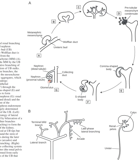
The mammalian kidney, or metanephros, arises at approximately day 35 of human gestation. The kidney originates from two mesenchymally derived components: the Wolffian duct and the metanephric mesenchyme (MM). Directed by inductive signals emanating from the MM, the Wolffian duct forms an epithelial out-pouching called the ureteric bud (UB), which invades the adjacent MM cell cluster. A remarkable series of events follow; reciprocal induction between the UB and MM leads to multiple iterations of branching and elongation of the UB to form the collecting system (where final production of a concentrated urine occurs), while the mesenchyme is induced to condense and epithelialize around the branched tips. Known as the
mesenchymal-to-epithelial transformation, these pre-tubular mesenchymal aggregates then proceed through several morphological stages, including comma- and S-shaped bodies, forming metanephric tubules that eventually mature into the parts of the nephron responsible for regulation of ion and organic molecule transport (proximal and distal tubules), as well as the epithelial glomerular (filtering) components (Fig. 1A).
Iterative branching of the UB occurs ~15 times during human development, normally generating between 300,000 and 1 million nephrons per human kidney (Nyengaard and Bendtsen, 1992). The formation of lateral branches (i.e. branches that grow out from a main stem) is followed by terminal bifid branching (bifurcation of one stem into two daughter branches). As each ureteric tip connects to a nephron, it effectively is removed effectively from further branching events. This type of branching appears to induce the majority of nephrons formed initially. The remaining fraction develops through the poorly understood process of arcade formation and late-phase branching (al-Awqati and Goldberg, 1998; Bush et. al, 2004b). The formation of the renal calyces and renal pelvis – the part of the urinary collecting system that leads to the ureter – occurs through the growth and dilation of early branching segments (Fig. 1B). There is great variability in the number of nephrons per kidney between animal species and currently it is unknown whether similar modes of branching occur across species (Moritz et al., 2003).
Ureteric bud outgrowth
In vitro studies have shown that glial cell-derived neurotrophic factor (GDNF), produced in the MM, is a primary inducer of ureteric budding (Sainio et al., 1997). GDNF interacts with a number of different molecules to play a crucial role in the initiation of downstream branching events. In mice, knockout of GDNF, or factors that affect GDNF expression or signaling, can result in phenotypes ranging from renal agenesis to rudimentary UB systems and kidneys. These factors have been comprehensively reviewed (Steer et al., 2003; Bush et al., 2004b) and include Wilms tumor-suppressor gene 1 (Wt1), Lim1, Paired-box gene 2 (Pax2), Eyes absent 1 (Eya1), Six1, Branching morphogenesis in the kidney is a tightly
regulated, complex process and its disruption potentially can lead to a broad spectrum of diseases, ranging from rare hereditary syndromes to common conditions such as hypertension and chronic kidney failure. This review synthesizes data on branching during kidney development
derived from in vitro and in vivo rodent studies and to apply them to human diseases. It discusses how the broad organization of molecular interactions during kidney development might provide a mechanistic framework for understanding disorders related to aberrant branching.
Summary
Branching morphogenesis and kidney disease
Mita M. Shah*, Rosemary V. Sampogna*, Hiroyuki Sakurai, Kevin T. Bush and Sanjay K. Nigam†
Departments of Pediatrics, Medicine, and Cellular and Molecular Medicine, University of California, San Diego, CA 92093-0693, USA
*These authors contributed equally to this review †Author for correspondence (e-mail: [email protected])
Development 131, 1449-1462
Published by The Company of Biologists 2004 doi:10.1242/dev.01089
Sal-like 1 (Sall1) and the Hox11 homeobox gene paralogous group (Davis et al., 1995; Kreidberg et al., 1993; Laclef et al., 2003; Nishinakamura et al., 2001; Shawlot and Behringer, 1995; Torres et al., 1995; Xu et al., 1999) (see Table 1 for details). In addition, knockout of GFRα1 and Ret, members of the GDNF receptor signaling complex, can also lead to the failure of UB outgrowth and result in renal agenesis (Cacalano et al., 1998; Schuchardt et al., 1996). In many of these knockouts, penetrance is variable; a certain percentage of mutants have rudimentary ureteric bud systems and kidneys (Table 2), indicating that there are compensatory mechanisms that allow kidney development to proceed despite disruption of
the GDNF pathway. Conversely, the depletion of factors that limit the expression of GDNF in the MM, such as Foxc1 (Mf1), or that antagonize inductive signals from the MM, such as BMP4, leads to supernumerary budding and duplication of the collecting system (Kume et al., 2000; Miyazaki et al., 2000). GDNF and BMP4 both require heparan sulfate proteoglycans (HSPG) to promote and suppress, respectively, UB outgrowth along the Wolffian duct (Barnett et al., 2002; Paine-Saunders et al., 2000). HSPGs are glycoproteins that are found on the cell surface and within the extracellular matrix. They are molecules with two distinct components: a core protein and a covalently linked glycosaminoglycan (GAG) side A
B
C
D
E
F G
Metanephric mesenchyme
Ureteric bud
Wolffian duct
Collecting duct Nephron
(distal tubule)
Nephron (proximal tubule)
Glomerulus
S-shaped body
Comma-shaped body
Pre-tubular mesenchymal condensate
A
B
Terminal bifid branch
Arcade Late-phase lateral branching
Renal pelvis
Ureter
Calyx
Lateral branch Fig. 1. (A) Stages of renal branching
morphogenesis and nephron formation. Ureteric bud (UB) outgrowth from the Wolffian duct is induced by signals from the
metanephric mesenchyme (MM) (A). (B,C) Invasion of the MM by the UB is followed by iterative branching of the UB and elongation of UB stalks. (D) At the tips of the branches, the epithelium induces the mesenchyme to form pre-tubular aggregates, which are stimulated to undergo
mesenchymal to epithelial transformation (E,F) through the formation of comma-shaped (E) and S-shaped (F) bodies to form
components of the nephron (G): renal tubules (proximal and distal) and the epithelial component of the
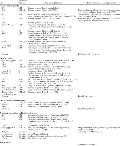
[image:2.612.111.554.71.583.2]Table 1. Known expression patterns of genes expressed in kidney development, and corresponding branching defects in human disease
Location of
Gene expression Mutant mouse phenotype Human (naturally occurring mutation)
Ureteric bud outgrowth
Wt1 MM Bilateral agenesis (Kreidberg et al., 1993)
Pax2 MM, UB Bilateral agenesis (Torres et al., 1995) Renal coloboma syndrome (renal agenesis/hypoplasia, optic nerve coloboma) (Sanyanusin et al., 1995) Sall1 MM Agenesis (32%); single hypoplastic kidney (29%); Townes-Brock syndrome (anal, renal, limb, ear anomalies)
bilateral remnant kidneys (39%) (Nishinakamura et al., 2001) (Kohlhase et al., 1998)
Eya1 MM Bilateral agenesis (100%) (Xu et al., 1999) Brancho-oto-renal syndrome (branchial fistulae, deafness, renal agenesis) (Abdelhak et al., 1997)
Six1 Bilateral agenesis (Xu et al., 2003)
Hoxa11/Hoxd11 MM Variable severity: agenesis to dysplasia. Abnormal branching with elongated primary branches (Davis et al., 1995)
Lim1 UB Bilateral agenesis (Shawlot and Behringer, 1995) Gdnf MM Agenesis caused by lack of UB (Pichel et al., 1996) Ret UB Bilateral agenesis (58%); unilateral agenesis (31%);
decreased branching and undifferentiated MM (11%) (Schuchardt et al., 1996)
Gfra1 MM, UB Bilateral agenesis (76%); unilateral rudiment (23%) (Cacalano et al., 1998)
Foxc1 (Mf1) MM Duplicated collecting system (Kume et al., 2000) Bmp4 MM Homozygotes are lethal at E6.5; no mesoderm formation.
(Winnier et al., 1995). Heterozygotes have a duplicated collecting system (Dunn et al., 1997)
Unknown Duplicated collecting system
Early branching
Gdnf heterozygotes MM Dysplasia; 30% fewer nephrons (Cullen-McEwen et al., 2001) Fgf7 S Thirty percent fewer nephrons (Qiao et al., 1999b)
Fgf10 MM Hypoplasia (Ohuchi et al., 2000)
Emx2 MM, UB Bilateral agenesis secondary to failure of UB branching. Outgrowth normal (Miyamoto et al., 1997)
Foxd1 (BF2) S Hypoplasia (Hatini et al., 1996)
Rara/Rarb2 S Fourfold decrease in the number of UB tips; 4-5 versus 8-9 branching iterations when compared with wild type (Mendelsohn et al., 1999)
Pod1 MM Hypoplasia and 61% decreased branching (Quaggin et al., 1999) Wnt4 MM Small, dysgenic kidneys; undifferentiated MM interspersed
with UB branches (Stark et al., 1994)
Wnt11 UB Thirty-six percent fewer nephrons (Majumdar et al., 2003) Itga8 (integrin α8) MM Bilateral dysgenesis (Muller et al., 1997)
Kama5 (laminin α5) MM Dysgenesis; decreased UB branching (Miner and Li, 2000) Heparan sulfate MM, UB Bilateral agenesis (Bullock et al., 1998)
2-sulfotransferase
Unknown Possibly hypertension
Late branching and maturation
Tgfb1 MM, UB Perinatal lethal; kidneys normal (Kulkarni et al., 1993) Activin MM Perinatal lethal; kidneys normal (Jhaveri et al., 1998) Bmp2 MM Embryonic lethal (Zhang and Bradley, 1996)
Unknown Possibly hypertension
Branching termination and tubule maintenance
Hgf MM Overexpression leads to cysts (Takayama et al., 1997) Tgfa MM Overexpression leads to cysts (Jhappan et al., 1990) Egfr MM Overexpression leads to cysts (Orellana et al., 1995),
knockout leads to dilated collecting ducts (Threadgill et al., 1995)
Tgfb2 MM Cysts (Sanford et al., 1997)
Gpc3 (glypican 3) MM, UB Enhanced branching, medulla degenerates by E15.5; cysts Simpson-Golabi-Behmel syndrome (cystic overgrowth) (Cano-Gauci et al., 1999) (Pilia et al., 1996)
Pkd1, Pkd2 UB (cilia) ADPKD (Wu and Somlo, 2000) Invs (inversin) UB (cilia) Cysts (Morgan et al., 2002)
Kif3a (kinesin) UB (cilia) Conditional knockout leads to cysts (Lin et al., 2003) Polaris UB (cilia) Cysts (Yoder et al., 2002)
Regeneration
Acute tubular necrosis Acquired cystic kidney disease
chain, heparan sulfate (HS). Many growth factors bind to the HS side chains of HSPGs, and the specificity of these interactions is often based on the location and clustering of sulfate modifications along segments of the HS chain (e.g. the degree of 2-O-sulfation versus 6-O-sulfation) and the core protein family (e.g. glypican versus syndecan) (Box 1). For example, GDNF activity is dependent on its binding to HS (Barnett et al., 2002). BMP4 activity, instead, is dependent upon its binding to the cell surface HSPG glypican 3 (Paine-Saunders et al., 2000). These studies indicate that HSPGs influence budding via the specificity of their interactions with morphogenic molecules, a theme that recurs throughout kidney branching morphogenesis.
Defects during initial ureteric bud outgrowth
Renal agenesis is a relatively frequent congenital defect in humans. An estimate of congenital absence of the kidney is 0.48 to 0.58 per 1000 live births (unilateral agenesis occurs in 1/200 births and bilateral agenesis in 1/5000-1/10000 births) (Pohl et al., 2002). Current data indicate that ureteric budding requires the presence of GDNF as a primary inducer. In humans, renal agenesis arises from mutation of single genes known to affect GDNF expression or signaling, for example, in diseases such as Townes-Brock syndrome (SALL1), Renal-coloboma syndrome (PAX2) or Branchio-Oto-Renal syndrome (EYA1) (Abdelhak et al., 1997; Kohlhase et al., 1998; Sanyanusin et al., 1995) (Table 1). These genes are known to interact with GDNF expression or signaling in the mouse, and the clinical phenotype of these gene mutations supports the view that regulation of GDNF expression is a key event in ureteric outgrowth in humans. Collecting system duplication (resulting in bifid ureter and duplex kidney) is another common renal malformation; however, the gene mutations leading to ectopic budding in humans have not yet been identified. A better understanding of congenital renal abnormalities in humans will, in part, depend on the creation of detailed large-scale databases of kidney and lower urinary tract developmental disorders.
Early ureteric bud branching
Initial UB outgrowth is followed by iterative branching of the UB, and this depends upon reciprocal epithelial-mesenchymal interactions (Fig. 1A). Gradients of soluble factors from the
MM are likely to be essential for appropriate spatiotemporal regulation of the arborization pattern (Santos and Nigam, 1993). In vitro, both in UB cell and isolated bud culture, conditions can be optimized to sustain iterative branching in the absence of direct cell-cell contact between epithelia and mesenchyme (Qiao et al., 1999a; Sakurai et al., 1997a). Soluble factors derived from the mesenchyme have been found to support branching and growth so robust that structures eventually occupy much of the matrix, and a major limitation seems to be one of cell nutrition. Whether the reciprocal epithelial-mesenchymal interactions between the UB and the MM are analogous to the positive feedback loops that coordinate growth and patterning in vertebrate limb (Laufer et al., 1994; Sun et al., 2000) needs to be determined.
Multiple factors regulate early branching
In vitro data indicates that GDNF continues to play a prominent role during early branching (Qiao et al., 1999a; Vega et al., 1996). Transcriptional modulators of GDNF expression also influence ureteric branching. For example, Emx2, a homeobox gene expressed in UB epithelia during early branching, maintains expression of GDNF in the MM. Murine Emx2 knockouts exhibit normal UB budding, but the absence of subsequent branching events leads to rapid mesenchymal apoptosis and renal agenesis (Miyamoto et al., 1997). Similarly, maintenance of localized Ret expression in the ureteric bud tips via expression of RARα, RARβ2, Foxd1 and Pod1 appears to be crucial for proper branching morphogenesis (Hatini et al., 1996; Mendelsohn et al., 1999; Quaggin et al., 1999). Homozygous null mutation of Rara, Rarb2, Foxd1 and Pod1 results in decreased ureteric branching within the embryonic kidney (Table 1).
Although GDNF plays an important role in continued branching morphogenesis, in vitro studies indicate that, while necessary, it is not sufficient to induce robust branching in an isolated UB culture model; other soluble factors such as pleiotrophin are also important (Sakurai, 2001). Cell growth continues to be prominent during this stage; however, sculpting of the UB also occurs. Some members of the fibroblast growth factor (FGF) family have been implicated in the early patterning of the UB. FGF1, FGF2, FGF7 and FGF10 all have different effects on branching morphogenesis of the isolated UB (Qiao et al., 2001). Other factors may also operate during early branching. In vitro, hepatocyte growth factor (HGF) induces branched tubule formation in cells embedded within a 3D extracellular matrix (Montesano et al., 1991; Santos and Nigam, 1993), as do the epidermal growth factor (EGF) receptor ligands EGF, TGFα and heparin binding EGF (HB-EGF), as well as amphiregulin (Barros et al., 1995; Sakurai et al., 1997b). Thus, a plethora of soluble mesenchymally derived factors induce the isolated UB or mature collecting system cells to branch. A single key factor has yet to be identified and current studies suggest that multiple factors, perhaps acting in combination, are required during early branching in vivo (Perantoni et al., 1995; Sakurai et al., 1997a; Bush et al., 2004a).
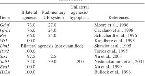
[image:4.612.43.294.91.222.2]HSPGs again feature prominently during early branching morphogenesis. GDNF, pleiotrophin, HGF and various FGFs all bind to HS side chains of HSPGs, and HSPGs appear to differentially modulate the activity of these growth factors (Box 1) (Barnett et al., 2002; Iseki et al., 2002; Rickard et al., Table 2. Knockout phenotypes of early branching
molecules* Unilateral Bilateral Rudimentary agenesis/
Gene agenesis UB system hypoplasia References
Gdnf 73.0 27.0 Moore et al., 1996 Gfra1 76.0 24.0 Cacalano et al., 1998 Ret 66.0 24.0 Schuchardt et al., 1996 Wt1 100.0 Kreidberg et al., 1993 Lim1 Bilateral agenesis (not quantified) Shawlot et al., 1995 Pax2 100.0 Torres et al., 1995 Six1 97.5 2.5 Xu et al., 2003
Sall1 32.0 39.0 29.0 Nishinakamura et al., 2001
Eya1 100.0 Xu et al., 1999
2003; Rubin et al., 2001; Pye et al., 1998). In addition, mice with a gene trap mutation for heparan sulfate 2-O-sulfotransferase (Hs2st), an HS biosynthetic enzyme, die perinatally because of renal agenesis (Bullock et al., 1998). Mice with this mutation appear to have normal UB outgrowth, but mesenchymal condensation and subsequent UB branching is absent. Hs2st–/– mice display a very consistent phenotype, indicating that 2-O-sulfated HS plays an important role during early branching (Table 2). It is important to note that Hs2st, may also function at later stages of kidney development, but termination of development at the early ureteric bud branching stage precludes analysis of the role of Hs2st during late branching morphogenesis.
The binding of growth factors to their receptors ultimately
induces cellular proliferation, shape changes and migration, all of which are required for branching to occur. In UB branching, the ECM may serve several purposes in this regard: providing a substrate for cell migration (Pohl et al., 2000b; Santos and Nigam, 1993), acting as a reservoir for morphogenetic molecules (which exist in gradients) (Pohl et al., 2000a) and transmitting environmental information to the cell via adhesion molecules such as integrins (Kanwar et al., 1997; Zent et al., 2001). Many of the ECM molecules that carry out these functions have been shown to regulate branching morphogenesis in vitro (Pohl et al., 2000a). For example, an appropriate balance of matrix metalloproteinases (MMPs) and their inhibitors – the tissue inhibitors of matrix proteinases (TIMPs) – at tips and leading edges may result in
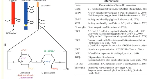
Box 1. Heparan sulfate proteoglycans: multifunctional molecules
HSPGs are modular structures composed of a glycosaminoglycan side chain – heparan sulfate (HS) – covalently linked to a specific core protein. Two major families of plasma membrane-bound HSPGs are known as glypicans (A, red), of which there are six isoforms, and syndecans (A, blue), of which there are four isoforms. Each of the core protein families has distinct functions and tissue specific expression. Minor members of the HSPG superfamily include collagen XVIII (A, white), perlecan and agrin.
Heparan sulfate chain synthesis occurs in the Golgi compartment and is mediated by a variety of biosynthetic enzymes. HS chains are composed of alternating uronic acid and D-glucosamine residues (B) of varying lengths. Following chain polymerization, a series of enzymes modify the HS chain through variable sulfation and C-5 epimerization (of D-glucuronic acid to L-iduronic acid). Variable sulfation can occur at the HNAc- residue (red residue in B) (mediated by the N-deacetylase/N-sulfotransferase enzymes), or at the 6-O (green), 2-O (orange) or 3-2-O (blue) positions (mediated by the respective 2-O-sulfotransferase enzymes) (Esko and Lindahl, 2001). In addition, cells can alter the structure of the HS chain in response to the environment through the action of sulfatases (Ai et al., 2003). Thus, HSPGs are highly diverse and are capable of multifunctional regulation.
HSPGs play multiple roles by interacting with growth factors: they bind growth factors in proximity to high affinity receptors, act as co-receptors in the growth-factor-receptor complex and protect growth factors from proteolytic degradation (reviewed by Bernfield et al., 1999). A feature of many of the factors involved in kidney branching morphogenesis is their interaction with HSPGs (Table I). The interaction of a growth factor with an HSPG is very specific, based on the length of the glycosaminoglycan side chain (Turnbull et al., 1992) and the fine sulfation pattern (Nakato and Kimata, 2002).
O
OH O
O
O
O
x A
B
COO–
COO–
OSO3– CH2OSO3–
HNSO3– OSO3–
D-glucosamine Uronic acid
(D-glucuronic acid
L-iduronic acid)
Glypican Syndecan Collagen XVIII
Endostatin
[image:5.612.46.552.113.385.2]GPI anchor
Table I. Morphogen and heparan sulfate proteoglycan interactions
Factor Characteristics of factor-HS interaction
GDNF 2-O-sulfation required for binding to GFRα1 (Rickard et al., 2003) BMP4 Activity modulated by glypican 3 (Paine-Saunders et al., 2000)
BMP4 antagonist, Noggin, binds HS (Paine-Saunders et al., 2002)
BMP2 Activity modulated by glypican 3 (Grisaru et al., 2001)
WNT Activity simulated by desulfation at 6-O position (Ai et al., 2003)
Pleiotrophin Binds to syndecan (Mitsiadis et al., 1995)
FGF1 2-O- and 6-O-sulfation required for binding (Pye et al., 1998) Cell-bound HS mediates receptor activity (Wu et al., 2003) Highly sulfated HS potentiates mitogenesis (Lundin et al., 2000)
FGF2 Pentasaccharide with N-sulfation and 2-O-sulfation sufficient for binding (Pye et al., 1998)
6-O-sulfation required for activation of FGFR1 (Pye et al., 1998)
FGF7 Heparin abrogates activation of FGFR2IIIb (Ye et al., 2001)
HGF 6-O-sulfation important for binding (Lyon et al., 1994)
TGFβ1 HS potentiates dimerization
Requires high level of N-sulfation for binding (Lyon et al., 1997)
HB-EGF Cell-surface HSPG optimizes activity (Higashiyama et al., 1993)
Endostatin Proteolytic cleavage product of collagen XVIII
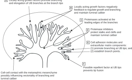
matrix degradation that promotes both branching and elongation (Stuart et al., 1995; Sakurai and Nigam, 1998) (Fig. 2).
Defects during early branching
Aside from complete renal agenesis, very few knockout mutations of genes that operate during early branching result in moderate to severe branching defects (e.g. of the order of 5-10 branching generations) (Table 1). Instead, these phenotypes tend to be slight or undetectable. Fgf7-null mice are characterized by mildly affected phenotypes, having only 30% fewer nephrons than wild type (equivalent to loss of less than one branching generation), although the overall branching architecture is maintained (Qiao et al., 1999b). Wnt11-null mice have a similar defect; these kidneys are characterized by 36% fewer nephrons than wild type with no apparent abnormality in branching pattern (Majumdar et al., 2003).
The lack of moderate to severe branching defects may arise from a requirement for a crucial number of nephrons for organ survival. For example, defects in early branching processes can result in malformed (dysplastic) kidneys that can involute eventually (Hiraoka et al., 2002). In addition, mechanisms such as redundancy may allow for activation of compensatory processes that are able to buffer the effects of genetic mutations (Melton, 1994; Wagner, 2000). For example, although
individual disruption of Hoxa11 or Hoxd11 produces no apparent kidney defects (Small and Potter, 1993; Davis and Capecchi, 1994; Davis et al., 1995), double mutants (Hoxa11/Hoxd11) demonstrate variable kidney hypoplasia (Davis et al., 1995; Patterson et al., 2001) and removal of the last Hox11 paralogous member, Hoxc11, results in renal agenesis (Wellik et al., 2002). Similarly, null mutations of a number of Fgf genes (e.g. Fgf1, Fgf2, Fgf3, Fgf5, Fgf6, Fgf9), another set of growth factors important for in vitro UB branching, result in kidneys that, with respect to growth and branching, appear normal, raising the possibility that substitution of function occurs between these proteins (Kanwar et al., 1997; Mahmood et al., 1995; Okada-Ban et al., 2000; Qiao et al., 2001). Moreover, in vitro data indicate that certain combinations of growth factors may also result in phenotypically equivalent branching; the addition of TGFβand FGF7 to UB culture results in a tree that appears similar to one cultured in the presence of FGF1 alone (Bush et al., 2004a).
Finally, gene dose may also play a role in the lack of moderate to severe branching defects. In other words, expression of the gene product must reach a certain threshold for normal branching to occur; below the threshold abnormalities in renal branching may result. For example, although Gdnf null mice lack kidneys and die, heterozygotes show an array of renal phenotypes ranging from small kidneys A
B
C
D
E
F
G Cell-cell contact with the metanephric mesenchyme possibly influencing vectoriality of branching and luminal caliber
Locally acting growth factors promote branching and elongation of UB branches at the branch tips
Locally acting growth factors negatively feedback to regulate growth and branching and maintain luminal caliber
Proteinases activated at the leading edges of the branches
Proteinase inhibitors protect stalks and clefts and maintain luminal caliber
Cell adhesion molecules and extracellular matrix components: (1) promote branching at UB tips; and (2) establish branch points.
Possible repellent factor at UB tips prevents tip fusion
Fig. 2. The interactions between soluble morphogens, metanephric mesenchyme and matrix molecules during the branching process of the
bilaterally (with approximately a 30% decrease in nephron number) to unilateral agenesis with no intermediate phenotypes (Cullen-McEwen et al., 2001). A similar requirement for adequate gene dose may be operative in human urinary tract development, as highlighted in distal 10q monosomy. In this condition, individuals carrying a single copy of the genes present on the distal 10q chromosome display urinary anomalies such as small under-developed kidneys and defects in the posterior urethral valves. The gene(s) responsible for the observed phenotypes have not yet been identified. However, in some of the cases, haploinsufficiency was not associated with the urinary abnormality, perhaps owing to variable penetrance and/or a dose-dependent gene-phenotype relationship (Ogata et al., 2000). In total, these studies indicate that functional redundancy and/or dose effects exist during early branching.
Late branching and maturation
At some point during development, branching slows down, presumably owing to negative feedback (i.e. corrective information) that serves to dampen the intrinsic branching signals of the UB (slowing growth and proliferation) and promote nephron patterning. There is evidence for these types of signals in other model systems. For example, during the development of respiratory appendages in the Drosophila embryo, the integration of positive and negative feedback systems allows for proper spatiotemporal expression of signals, and hence appropriate patterning (Freeman, 2000). A similar mechanism may operate when differentially expressed modulatory molecules, such as TGFβ and BMPs, act as branching inhibitors and sculptors in the developing kidney (Bush et al., 2004a).
Factors involved in negative feedback
Data obtained from whole organ culture suggest that members of the TGFβ superfamily (BMP2, BMP4, activin and TGFβ1) have direct inhibitory effects on the overall growth and development of the embryonic kidney and cell lines, particularly on UB-derived components (Rogers et al., 1993; Piscione et al., 1997; Raatikainen-Ahokas et al., 2000; Sakurai and Nigam, 1997; Bush et al., 2004a). The in vivo role of these factors has more been difficult to elucidate, as knockout of these genes results in phenotypes that range from early embryonic lethality (Bmp2, Bmp4) (Zhang and Bradley, 1996) to no overt renal phenotype (TGFβ1, activin) (Jhaveri et al., 1998; Kulkarni et al., 1993). The generation of conditional knockouts will help to further clarify these functions.
Negative feedback signals may arise from the MM, perhaps timed to a specific point in MM-derived tubule formation. Fusion between a lateral ureteric branch and metanephric tubule (Fig. 1A, stage F) effectively removes the ureteric branch from further divisions (Oliver, 1968). The ‘stop signal’ may even be present earlier during MM-derived tubule formation, occurring in the earliest branching generations and/or as soon as comma- or S-shaped bodies appear (Fig. 1A) to prevent the intertwining and fusion of branches. The observed shift in expression of the branch-inhibitory factors, BMP2 and BMP7 (Dudley and Robertson, 1997) and the slit proteins (which function in axon repulsive guidance) (Piper et al., 2000) from the condensed mesenchyme to the comma- and S-shaped bodies is consistent with this hypothesis.
Patterning during kidney development also requires structural remodeling by the ECM scaffolding (Fig. 2). Specific ECM proteins can either enhance or inhibit branching in cultured cells (Santos and Nigam, 1993; Sakurai and Nigam, 1997). In addition, differential expression of MMPs and TIMPs at stalks and/or non-branching areas may provide feedback information that prevents further branching and growth (Nigam, 1995; Ota et al., 1998; Pohl et al., 2000a).
As in previous stages, regulatory molecules during late branching and maturation require HSPGs for receptor binding and signaling. Signaling by BMP4 and BMP2 is modulated by glypican 3 (Paine-Saunders et al., 2000; Takada et al., 2003), while the activity of TGFβ appears to be modulated by its binding to HSPGs with N-sulfated HS chains (Lyon et al., 1997). HSPGs may also function to establish the formation of growth factor gradients that modulate patterning of the nephric tree. HSPGs regulate the cellular distribution of Noggin, a BMP antagonist, and may therefore regulate cellular responsiveness to BMPs in vivo through the establishment of a BMP activity gradient (Paine-Saunders et al., 2002).
Derivatives of HSPGs can also influence UB branching directly. For example, endostatin, a proteolytic cleavage product of the HSPG collagen XVIII, inhibits branching in the isolated UB and in cell culture (Karihaloo et al., 2001). Although collagen XVIII is initially present throughout the ureteric epithelium, as development progresses, its expression is lost at the ureteric tips and becomes restricted to the stalk (Lin et al., 2001). Endostatin may suppress further branching specifically at the stalk as a result of this differential spatial expression, which is evident as early as embryonic day 11.5 in the mouse.
Disorders of reduced nephron number
2003). Thus, it is conceivable that defects during early and late branching processes may lead to the development of two very common clinical syndromes: hypertension and progressive renal failure. Importantly, reduced nephron number reflects only one of many possible pathogenic mechanisms that may lead to these diseases.
The number of factors involved in the determination of nephron number is likely to be even more complex than those listed here. It is conceivable that genetic variants (the existence of two or more alleles at significant frequencies in a population) that interact differently with modifiers, suppressors and enhancers can cause subtle changes in nephron number that eventually lead to disease. This could also explain the apparently continuous distribution of nephron number in humans, ranging from approximately 230,000 to 1,800,000 (Nyengaard and Bendtsen, 1992). Variant alleles, identified by single-nucleotide polymorphisms (SNPs), are a source of genetic diversity and are expected to be neutral (or nearly so) with respect to fitness (Fay et al., 2001). During early and late branching, SNPs present in genes governing the branching process may not affect the viability or gross architecture of the kidney, but might predispose to hypertension or chronic kidney disease later in life. This idea may be supported by the recent identification of a SNP in the MMP9 gene, which appears to increase end-stage renal disease susceptibility in humans (Hirakawa et al., 2003). During branching, SNPs might be phenotypically subtle or even silent, consistent with the observation that the branching program seems relatively resistant to single mutations. Because of this resilience, it can be postulated that multiple adverse SNPs in branching genes would be required for a clinically significant decrease in nephron number. Identifying the factors that are involved in these stages of branching may lead to candidate genes for hypertension research and may even be useful in devising therapies.
Branching termination and tubule maintenance Branching termination remains one of the biggest unresolved problems in epithelial organogenesis. Developmental mechanisms must be ‘turned off’ when the structure reaches a certain size and maturity. In the developing kidney, branching must cease and the caliber of the tubule lumen must be regulated. Although clear stop signals have not yet been identified, in vitro studies have suggested several possible candidates. During later stages of branching, increased expression levels of negative regulators (e.g. TGFβ) may overwhelm positive growth-promoting factors (FGFs) and thereby act as stop signals (Santos and Nigam, 1993; Stuart et al., 2003; Bush et al., 2004a). For example, branching of the murine inner medullary collecting duct (mIMCD) cells is inhibited by the addition of TGFβ, leading to the formation of straight, non-branched tubular structures (Sakurai and Nigam, 1997).
Stop/maturation signals also may be correlated with the differentiation of the MM. Recombination assays between cultured UB and freshly isolated MM indicate that the MM regulates tubule lumen caliber and provide signals that induce UB branch elongation (Qiao et al., 1999a; Steer et al., 2003). However, branching does not extend beyond the confines of the MM, which may provide specific stop signals during nephron formation and maturation. Such stop signals could be
short-range secreted molecules or cell-surface proteins (Pohl et al., 2000c). In other systems, membrane-bound ephrins act as stop and maturation signals of epithelial morphogenesis. In C. elegans, ephrins are thought to act as a repulsive signal that prevents the migration of epidermal cells (George et al., 1998). Interestingly, media conditioned by embryonic murine MM cells upregulates expression of ephrin A5 and ephrin B2 in UB cells (Pavlova et al., 1999) suggesting a role for these proteins in UB branching morphogenesis.
Finally, sulfated proteoglycans may also function as stop signals of branching, similar to their roles in other developmental systems. For example, chondroitin sulfate proteoglycans (CSPGs) inhibit axon growth in vitro – perhaps forming a protective ‘jacket’ that masks the target stimulatory molecule(s) (Bradbury et al., 2002; Morgenstern et al., 2002). It also is possible that changes in the proteoglycan environment lead to either enhanced binding of inhibitory factors or diminished activity of stimulatory factors; in effect creating gradients of activity. This appears to be the case in Drosophila wing development, where the expression of the HSPG dally regulates both the cellular response to Dpp (the Drosophila TGFβ/BMP protein homolog) and the distribution of Dpp morphogen in tissues (Fujise et al., 2003).
Disorders of branching termination
As yet, a clear clinical manifestation of disordered stop signals for branching, e.g. a kidney with 4 million nephrons, has not been reported. In fact, clinical studies that have measured gross kidney parameters have failed to find large variation in normal kidney size (kidneys tend to be between 11-12 cm in length in the general population (Brenner and Rector, 2000)). It is possible that a high level of functional redundancy exists among the stop signals. It also is feasible that nutritional or external factors (retroperitoneal space constraints and/or restriction by the kidney capsule) prohibit the generation of significantly over-branched organs.
al., 2001). In Gpc3-deficient mice, there is altered regulation of FGF7, BMP7 and BMP2 that then correlates with increased epithelial cell turnover (i.e. upregulated proliferation and apoptosis), analogous to cyst-forming mechanisms found in human disease (Lanoix et al., 1996).
Mechanisms of cyst formation
Gradients of stimulatory and inhibitory growth factors and other secreted morphogenetic molecules are thought to be normally responsible for maintaining tubule architecture (Pohl et al., 2000c). Gradient disruption and/or alteration of regulatory information presumably leads to an imbalance in the relative concentration of growth and survival signals received by cells (Sakurai and Nigam, 1998), and may play a role in cyst development. Overexpression or upregulation of several branch-promoting factors including HGF, TGFαand EGFR are associated with cystic phenotypes in mice (Takayama et al., 1997). A similar outcome is seen following the disruption of factors that inhibit branching. For example, cystic dilation of tubules occurs in heterozygous mutants of Bmp4 (Dunn et al., 1997) and null mutants of Tgfb2 (Sanford et al., 1997). Similar mechanisms may contribute to human cystic disease as well; autosomal dominant polycystic kidney disease (ADPKD), the most common form of human polycystic kidney disease, is associated with abnormal upregulation of TGFα and EGFR (Lee et al., 1998).
The ECM is another component involved in tubule maintenance and cyst formation. Its influence on epithelial cell behavior is highlighted by cell culture models in which differences in ECM composition can result in phenotypes ranging from branching structures to cysts. For example, immortalized UB cells grown in a matrix of 80% type I collagen/20% Matrigel (an ECM extracted from mouse EHS sarcoma composed of laminin, collagen IV, and HSPGs) form branched tubules; however, when grown in 100% Matrigel matrix, these cells form cysts, even in the presence of tubulogenic growth factors (Sakurai et al., 1997a). Likewise, when Madin-Darby canine kidney (MDCK) cells, which have the properties of the kidney distal tubule and collecting duct cells, are grown in type I collagen alone, they form branched tubules in the presence of HGF, a potent tubulogenic factor for this cell line. However, MDCK cells grown in collagen mixed with Matrigel form cysts even with the addition of HGF (Santos and Nigam, 1993). ECM remodeling by MMPs and their inhibitors (TIMPs) is also important in the formation and maintenance of tubule structure (Pohl et al., 2000a). In the rat model of ADPKD, upregulation of MMP RNA occurs predominantly in cyst lining epithelia. Treatment with the MMP inhibitor called batimastat results in a significant reduction of cyst number (Obermuller et al., 2001). This mechanism may also apply to Von-Hippel-Lindau (VHL) disease, an autosomal dominant tumor syndrome associated with cystic kidney disease. In cell culture, VHL inactivation leads to decreased TIMP and increased MMP levels, suggesting abnormal regulation by these cells (Koochekpour et al., 1999). This re-emphasizes the idea that the relative balance between MMPs and TIMPs is important in proliferation versus stopping and, in this case, cyst versus tubule formation.
Cilia
Although an appropriate balance of stimulatory and inhibitory
growth factors and ECM constraints normally maintains a steady state of proliferation and apoptosis, another cellular feature is emerging as a prominent regulator of tubular architecture: cilia. Cilia are organelles present on the apical surface of tubule-lining epithelial cells that are thought to function as environmental sensors, evident by the presence of many chemo- or mechano-sensing receptors (Calvet, 2003). Loss of ciliary structure and function appears to induce overgrowth of the epithelial cells, thereby leading to cyst formation, although the mechanism for this remains undefined (Bhunia et al., 2002; Lin et al., 2003; Morgan et al., 2002; Hou et al., 2002; Yoder et al., 2002). In humans, cilia gene mutations disrupt regulation of tubule epithelial cell proliferation, thereby resulting in cyst formation. Mutation of PKD1 and PKD2, genes that code for ciliary structural proteins, underlie most forms of ADPKD, whereas mutations in OFD1, a gene involved in cilia generation and structure, and are associated with X-linked dominant polycystic kidney disease (Romio et al., 2003). These studies underscore the importance of cilia function in the regulation of tubule lumen diameter. However, the interactions between cilia, growth factors and ECM proteins in the regulation of branching morphogenesis remain undefined.
Injury/regeneration
Although branching does not occur following acute tubule injury, regeneration employs a combination of mechanisms that overlap with those that are involved in branching morphogenesis (Nigam and Lieberthal, 2000). Unlike glomerular or vascular injury, damage to the nephron tubule can be repaired through regeneration. The most common injury response is acute tubular necrosis (ATN), a sloughing of a large population of epithelial cells occurring in response to oxygen deprivation or chemical insult. Although the proximal tubule is the primary site of injury, more distal injury occurs as well. Recovery from ATN is possible if the adjacent blood supply and basement membrane remain intact, thereby guiding reconstitution of the polarized epithelia. Several mechanisms of cellular repair are activated in the recovery process, including mitosis of neighboring cells, recruitment, differentiation and proliferation of stem cells, and basement membrane remodeling.
culture (Stuart et al., 1995). Cells also may adopt a strategy similar to the one that occurs during Drosophila tracheobronchial development, whereby, after proliferation, extreme cell shape changes primarily account for tube formation (Samakovlis et al., 1996). The end result is a restored, intact epithelial tubule without scar formation.
The process of tubule repair, although ultimately beneficial in the setting of injury, may also go awry and lead to pathology such as in acquired cystic kidney disease (ACKD). ACKD, which is found in adults with end-stage renal disease, is characterized by a bilateral cystic change occurs in response to profound tissue loss. ACKD has been associated with overexpression of EGFR ligands and MMP abnormalities (Concolino et al., 1993; Ito et al., 2000).
Summary/future directions
We have reviewed the molecular basis of kidney epithelial branching; molecules thought to provide key points of control have been parsed within the framework of stages that describe branching morphogenesis from ureteric bud outgrowth to branching termination (Stuart et al., 2003; Nigam, 2003; Bush et al., 2004a). Applied to in vitro systems developed to date, this description may enable characterization of the independent morphogenetic mechanisms functioning during stages of kidney development and shed insight on disease.
Whereas secreted peptides, ECM molecules, growth and transcription factors comprise the mainstay of signals operative during branching, HSPGs have been found to interact with the majority of growth factors at each stage. As seen in other developmental systems, HSPGs may be instrumental in establishing growth factor gradients, as well as acting as morphogenetic modulators via their ability to regulate ‘positive’ and ‘negative’ growth factor activity. Further studies are needed to in order to understand how the integration of tremendous HSPG diversity with growth factor pathways at each branching step drive and/or regulate UB morphogenesis. Such studies may provide key information into the mechanisms underlying branching defects and disease.
As we have discussed, disruption at key points throughout kidney development may lead to disease, including renal agenesis. However, mutagenesis and gene deletion in animal models, including many genes thought to be important in branching based on in vitro studies, often results in kidneys that appear normal. Thus, as judged by these reports, relatively few phenotypes lie between these two extremes. A
decentralized network of molecular interactions that comprise the developmental program, with regulation at many levels, may account for this distribution (Nigam, 2003). Loss of a centrally acting molecule that interacts with or is connected to multiple other molecules (e.g. GDNF) can produce irreparable downstream effects on kidney development. However, the phenotypic consequences of knocking out other genes (e.g. Hgf) may be buffered by the inhomogeneous wiring of complex systems. In certain cases, the plasticity of the system allows development to proceed via a bypass route and/or through alternative molecular mechanisms that are phenotypically equivalent (Barabási et al., 2002; Venter et al., 2001; Nigam, 2003). Thus, a major task of future studies will be to unravel this complex network of molecular interactions, in order to decipher and predict the pathological consequences of developmental errors. Application of microarray analysis to kidney development and model systems is likely to provide essential information about the (non)hierarchical structure of gene networks and key points of regulation (Stuart et al., 2001; Stuart et al., 2003; Schwab et al., 2003).
This review is dedicated to the memory of Robert Oden Stuart II, MD (1962-2004), who made major contributions to the field of kidney development and epithelial biology.
References
Abdelhak, S., Kalatzis, V., Heilig, R., Compain, S., Samson, D., Vincent, C., Weil, D., Cruaud, C., Sahly, I., Leibovici, M. et al. (1997). A human
homologue of the Drosophila eyes absent gene underlies branchio-oto-renal (BOR) syndrome and identifies a novel gene family. Nat. Genet. 15, 157-164.
Ai, X., Do, A. T., Lozynska, O., Kusche-Gullberg, M., Lindahl, U. and Emerson, C. P., Jr (2003). QSulf1 remodels the 6-O sulfation states of cell
surface heparan sulfate proteoglycans to promote Wnt signaling. J. Cell Biol. 162, 341-351.
al-Awqati, Q. and Goldberg, M. R. (1998). Architectural patterns in
branching morphogenesis in the kidney. Kidney Int. 54, 1832-1842.
Barabási, A-L. (2002). Linked: The New Science of Networks. Cambridge,
MA: Perseus.
Barnett, M. W., Fisher, C. E., Perona-Wright, G. and Davies, J. A. (2002).
Signalling by glial cell line-derived neurotrophic factor (GDNF) requires heparan sulphate glycosaminoglycan. J. Cell Sci. 115, 4495-4503.
Barros, E. J., Santos, O. F., Matsumoto, K., Nakamura, T. and Nigam, S. K. (1995). Differential tubulogenic and branching morphogenetic activities
of growth factors: implications for epithelial tissue development. Proc. Natl. Acad. Sci. USA 92, 4412-4416.
Basile, D. P., Rovak, J. M., Martin, D. R. and Hammerman, M. R. (1996).
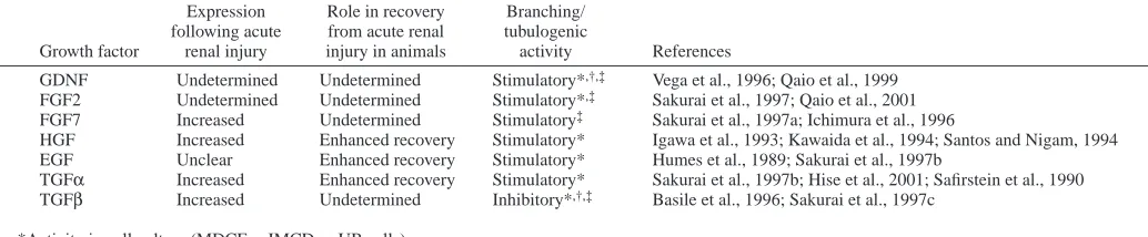
[image:10.612.43.560.87.194.2]Increased transforming growth factor-beta 1 expression in regenerating rat renal tubules following ischemic injury. Am. J. Physiol. 270, F500-F509. Table 3. Growth factors in development and renal recovery
Expression Role in recovery Branching/ following acute from acute renal tubulogenic
Growth factor renal injury injury in animals activity References
GDNF Undetermined Undetermined Stimulatory*,†,‡ Vega et al., 1996; Qaio et al., 1999 FGF2 Undetermined Undetermined Stimulatory*,‡ Sakurai et al., 1997; Qaio et al., 2001 FGF7 Increased Undetermined Stimulatory‡ Sakurai et al., 1997a; Ichimura et al., 1996
HGF Increased Enhanced recovery Stimulatory* Igawa et al., 1993; Kawaida et al., 1994; Santos and Nigam, 1994 EGF Unclear Enhanced recovery Stimulatory* Humes et al., 1989; Sakurai et al., 1997b
TGFα Increased Enhanced recovery Stimulatory* Sakurai et al., 1997b; Hise et al., 2001; Safirstein et al., 1990 TGFβ Increased Undetermined Inhibitory*,†,‡ Basile et al., 1996; Sakurai et al., 1997c
*Activity in cell culture (MDCF, mIMCD or UB cells). †Activity in whole organ culture.
‡Activity in isolated UB culture.
Bernfield, M., Gotte, M., Park, P. W., Reizes, O., Fitzgerald, M. L., Lincecum, J. and Zako, M. (1999). Functions of cell surface heparan
sulfate proteoglycans. Annu. Rev. Biochem. 68, 729-777.
Bhunia, A. K., Piontek, K., Boletta, A., Liu, L., Qian, F., Xu, P. N., Germino, F. J. and Germino, G. G. (2002). PKD1 induces p21(waf1) and
regulation of the cell cycle via direct activation of the JAK-STAT signaling pathway in a process requiring PKD2. Cell 109, 157-168.
Bradbury, E. J., Moon, L. D., Popat, R. J., King, V. R., Bennett, G. S., Patel, P. N., Fawcett, J. W. and McMahon, S. B. (2002). Chondroitinase
ABC promotes functional recovery after spinal cord injury. Nature 416, 636-640.
Brenner, B. M., Garcia, D. L. and Anderson, S. (1988). Glomeruli and blood
pressure. Less of one, more the other? Am. J. Hypertens. 1, 335-347.
Brenner, B. M. and Rector, F. C. (2000). Brenner and Rector’s The Kidney,
6th edn (ed. B. M. Brenner), Philadelphia: WB Saunders.
Bullock, S. L., Fletcher, J. M., Beddington, R. S. and Wilson, V. A. (1998).
Renal agenesis in mice homozygous for a gene trap mutation in the gene encoding heparan sulfate 2-sulfotransferase. Genes Dev. 12, 1894-1906.
Bush, K. T., Sakurai, H., Tsukamoto, T., and Nigam S. K. (1999). Acute
renal failure: cellular features of injury and repair. In Atlas of Diseases of the Kidney (ed. R. W. Schrier), Vol. 1, Chapter 16, pp. 16.1-16.15. Philadelphia: Current Medicine.
Bush, K. T., Sakurai, H., Steer, D. L., Leonard, M. O., Sampogna, R. V., Meyer, T. N., Schwesinger, C., Qiao, J., and Nigam, S. K. (2004a). TGF-β
superfamily members modulate growth, branching, shaping, and patterning of the ureteric bud. Dev. Biol. 266, 285-298.
Bush, K. T., Stuart R. O. and Nigam, S. K. (2004b). Developmental biology
of the kidney. In Brenner and Rector’s The Kidney, 7th edn (ed. B. M. Brenner), Chapter 2. Philadelphia: WB Saunders.
Cacalano, G., Farinas, I., Wang, L. C., Hagler, K., Forgie, A., Moore, M., Armanini, M., Phillips, H., Ryan, A. M., Reichardt, L. F. et al. (1998).
GFRalpha1 is an essential receptor component for GDNF in the developing nervous system and kidney. Neuron 21, 53-62.
Calvet, J. P. (2003). New insights into ciliary function: kidney cysts and
photoreceptors. Proc. Natl. Acad. Sci. USA 100, 5583-5585.
Cano-Gauci, D. F., Song, H. H., Yang, H., McKerlie, C., Choo, B., Shi, W., Pullano, R., Piscione, T. D., Grisaru, S., Soon, S. et al. (1999).
Glypican-3-deficient mice exhibit developmental overgrowth and some of the abnormalities typical of Simpson-Golabi-Behmel syndrome. J. Cell Biol.
146, 255-264.
Concolino, G., Lubrano, C., Ombres, M., Santonati, A., Flammia, G. P. and di Silverio, F. (1993). Acquired cystic kidney disease: the hormonal
hypothesis. Urology 41, 170-175.
Cullen-McEwen, L. A., Drago, J. and Bertram, J. F. (2001). Nephron
endowment in glial cell line-derived neurotrophic factor (GDNF) heterozygous mice. Kidney Int. 60, 31-36.
Cullen-McEwen, L. A., Kett, M. M., Dowling, J., Anderson, W. P. and Bertram, J. F. (2003). Nephron number, renal function, and arterial pressure
in aged GDNF heterozygous mice. Hypertension 41, 335-340.
Davis, A. P. and Capecchi, M. R. (1994). Axial homeosis and appendicular
skeleton defects in mice with a targeted disruption of hoxd-11. Development
120, 2187-2198.
Davis, A. P., Witte, D. P., Hsieh-Li, H. M., Potter, S. S. and Capecchi, M. R. (1995). Absence of radius and ulna in mice lacking hoxa-11 and
hoxd-11. Nature 375, 791-795.
Dudley, A. T. and Robertson, E. J. (1997). Overlapping expression domains
of bone morphogenetic protein family members potentially account for limited tissue defects in BMP7 deficient embryos. Dev. Dyn. 208, 349-362.
Dunn, N. R., Winnier, G. E., Hargett, L. K., Schrick, J. J., Fogo, A. B. and Hogan, B. L. (1997). Haploinsufficient phenotypes in Bmp4 heterozygous
null mice and modification by mutations in Gli3 and Alx4. Dev. Biol. 188, 235-247.
Esko, J. D. and Lindahl, U. (2001). Molecular diversity of heparan sulfate.
J. Clin. Invest. 108, 169-173.
Fay, J. C., Wyckoff, G. J. and Wu, C. I. (2001). Positive and negative
selection on the human genome. Genetics 158, 1227-1234.
Freeman, M. (2000). Feedback control of intercellular signalling in
development. Nature 408, 313-319.
Fujise, M., Takeo, S., Kamimura, K., Matsuo, T., Aigaki, T., Izumi, S. and Nakato, H. (2003). Dally regulates Dpp morphogen gradient formation in
the Drosophila wing. Development 130, 1515-1522.
George, S. E., Simokat, K., Hardin, J. and Chisholm, A. D. (1998). The
VAB-1 Eph receptor tyrosine kinase functions in neural and epithelial morphogenesis in C. elegans. Cell 92, 633-643.
Grisaru, S., Cano-Gauci, D., Tee, J., Filmus, J. and Rosenblum, N. D.
(2001). Glypican-3 modulates BMP- and FGF-mediated effects during renal branching morphogenesis. Dev. Biol. 231, 31-46.
Hatini, V., Huh, S. O., Herzlinger, D., Soares, V. C. and Lai, E. (1996).
Essential role of stromal mesenchyme in kidney morphogenesis revealed by targeted disruption of Winged Helix transcription factor BF-2. Genes Dev.
10, 1467-1478.
Hermiston, M. L., Xu, Z. and Weiss, A. (2003). CD45: a critical regulator
of signaling thresholds in immune cells. Annu. Rev. Immunol. 21, 107-137.
Higashiyama, S., Abraham, J. A. and Klagsbrun, M. (1993).
Heparin-binding EGF-like growth factor stimulation of smooth muscle cell migration: dependence on interactions with cell surface heparan sulfate. J. Cell Biol. 122, 933-940.
Hiraoka, M., Tsukahara, H., Ohshima, Y., Kasuga, K., Ishihara, Y. and Mayumi, M. (2002). Renal aplasia is the predominant cause of congenital
solitary kidneys. Kidney Int. 61, 1840-1844.
Hirakawa, S., Lange, E. M., Colicigno, C. J., Freedman, B. I., Rich, S. S. and Bowden, D. W. (2003). Evaluation of genetic variation and association
in the matrix metalloproteinase 9 (MMP9) gene in ESRD patients. Am. J. Kid. Dis. 42, 133-142.
Hise, M. K., Salmanullah, M., Liu, L., Drachenberg, C. I., Papadimitriou, J. C. and Rohan, R. M. (2001). Control of the epidermal growth factor
receptor and its ligands during renal injury. Nephron 88, 71-79.
Hou, X., Mrug, M., Yoder, B. K., Lefkowitz, E. J., Kremmidiotis, G., D’Eustachio, P., Beier, D. R. and Guay-Woodford, L. M. (2002). Cystin,
a novel cilia-associated protein, is disrupted in the cpk mouse model of polycystic kidney disease. J. Clin. Invest. 109, 533-540.
Hughson, M., III, Douglas-Denton, R., Hoy, W. E. and Bertram, J. F.
(2003). Glomerular number and size in autopsy kidneys: The relationship to birth weight. Kidney Int. 63, 2113-2122.
Humes, H. D., Cieslinski, D. A., Coimbra, T. M., Messana, J. M. and Galvao, C. (1989). Epidermal growth factor enhances renal tubule cell
regeneration and repair and accelerates the recovery of renal function in postischemic acute renal failure. J. Clin. Invest. 84, 1757-1761.
Ichimura, T., Finch, P. W., Zhang, G., Kan, M. and Stevens, J. L. (1996).
Induction of FGF-7 after kidney damage: a possible paracrine mechanism for tubule repair. Am. J. Physiol. 271, F967-F976.
Igawa, T., Matsumoto, K., Kanda, S., Saito, Y. and Nakamura, T. (1993).
Hepatocyte growth factor may function as a renotropic factor for regeneration in rats with acute renal injury. Am. J. Physiol. 265, F61-F69.
Iseki, K., Hagino, S., Mori, T., Zhang, Y., Yokoya, S., Takaki, H., Tase, C., Murakawa, M. and Wanaka, A. (2002). Increased syndecan expression by
pleiotrophin and FGF receptor-expressing astrocytes in injured brain tissue. Glia 39, 1-9.
Ito, F., Nakazawa, H., Ryoji, O., Okuda, H. and Toma, H. (2000). Cytokines
accumulated in acquired renal cysts in long-term hemodialysis patients. Urol. Int. 65, 21-27.
Jhappan, C., Stahle, C., Harkins, R. N., Fausto, N., Smith, G. H. and Merlino, G. T. (1990). TGF alpha overexpression in transgenic mice
induces liver neoplasia and abnormal development of the mammary gland and pancreas. Cell 61, 1137-1146.
Jhaveri, S., Erzurumlu, R. S., Chiaia, N., Kumar, T. R. and Matzuk, M. M. (1998). Defective whisker follicles and altered brainstem patterns in
activin and follistatin knockout mice. Mol. Cell. Neurosci. 12, 206-219.
Kanwar, Y. S., Carone, F. A., Kumar, A., Wada, J., Ota, K. and Wallner, E. I. (1997). Role of extracellular matrix, growth factors and
proto-oncogenes in metanephric development. Kidney Int. 52, 589-606.
Karihaloo, A., Karumanchi, S. A., Barasch, J., Jha, V., Nickel, C. H., Yang, J., Grisaru, S., Bush, K. T., Nigam, S., Rosenblum, N. D. et al. (2001).
Endostatin regulates branching morphogenesis of renal epithelial cells and ureteric bud. Proc. Natl. Acad. Sci. USA 98, 12509-12514.
Kawaida, K., Matsumoto, K., Shimazu, H. and Nakamura, T. (1994).
Hepatocyte growth factor prevents acute renal failure and accelerates renal regeneration in mice. Proc. Natl. Acad. Sci. USA 91, 4357-4361.
Keller, G., Zimmer, G., Mall, G., Ritz, E. and Amann, K. (2003).
Nephron Number in Patients with Primary Hypertension. New Engl. J. Med. 348, 101.
Kohlhase, J., Wischermann, A., Reichenbach, H., Froster, U. and Engel, W. (1998). Mutations in the SALL1 putative transcription factor gene cause
Townes-Brocks syndrome. Nat. Genet. 18, 81-83.
Koochekpour, S., Jeffers, M., Wang, P. H., Gong, C., Taylor, G. A., Roessler, L. M., Stearman, R., Vasselli, J. R., Stetler-Stevenson, W. G., Kaelin, W. G., Jr et al. (1999). The von Hippel-Lindau tumor suppressor
branching morphogenesis in renal carcinoma cells. Mol. Cell Biol. 19, 5902-5912.
Kreidberg, J. A., Sariola, H., Loring, J. M., Maeda, M., Pelletier, J., Housman, D. and Jaenisch, R. (1993). WT-1 is required for early kidney
development. Cell 74, 679-691.
Kulkarni, A., Huh, C., Becker, D., Geiser, A., Lyght, M., Flanders, K., Roberts, A., Sporn, M., Ward, J. and Karlsson, S. (1993). Transforming
growth factor beta1 null mutation in mice causes excessive inflammatory response and early death. Proc. Natl. Acad. Sci. USA 90, 770.
Kume, T., Deng, K. and Hogan, B. L. (2000). Murine forkhead/winged helix
genes Foxc1 (Mf1) and Foxc2 (Mfh1) are required for the early organogenesis of the kidney and urinary tract. Development 127, 1387-1395.
Laclef, C., Hamard, G., Demignon, J., Souil, E., Houbron, C. and Maire, P. (2003). Altered myogenesis in Six1-deficient mice. Development 130,
2239-2252.
Lanoix, J., D’Agati, V., Szabolcs, M. and Trudel, M. (1996). Dysregulation
of cellular proliferation and apoptosis mediates human autosomal dominant polycystic kidney disease (ADPKD). Oncogene 13, 1153-1160.
Laufer, E., Nelson, C. E., Johnson, R. L., Morgan, B. A. and Tabin, C.
(1994). Sonic hedgehog and Fgf-4 act through a signaling cascade and feedback loop to integrate growth and patterning of the developing limb bud. Cell 79, 993-1003.
Lee, D. C., Chan, K. W. and Chan, S. Y. (1998). Expression of transforming
growth factor alpha and epidermal growth factor receptor in adult polycystic kidney disease. J. Urol. 159, 291-296.
Lelongt, B., Trugnan, G., Murphy, G. and Ronco, P. M. (1997). Matrix
metalloproteinases MMP2 and MMP9 are produced in early stages of kidney morphogenesis but only MMP9 is required for renal organogenesis in vitro. J. Cell Biol. 136, 1363-1373.
Lin, F., Hiesberger, T., Cordes, K., Sinclair, A. M., Goldstein, L. S., Somlo, S. and Igarashi, P. (2003). Kidney-specific inactivation of the KIF3A
subunit of kinesin-II inhibits renal ciliogenesis and produces polycystic kidney disease. Proc. Natl. Acad. Sci. USA 100, 5286-5291.
Lin, Y., Zhang, S., Rehn, M., Itaranta, P., Tuukkanen, J., Heljasvaara, R., Peltoketo, H., Pihlajaniemi, T. and Vainio, S. (2001). Induced repatterning
of type XVIII collagen expression in ureter bud from kidney to lung type: association with sonic hedgehog and ectopic surfactant protein C. Development 128, 1573-1585.
Lisle, S. J., Lewis, R. M., Petry, C. J., Ozanne, S. E., Hales, C. N. and Forhead, A. J. (2003). Effect of maternal iron restriction during pregnancy
on renal morphology in the adult rat offspring. Br. J. Nutr. 90, 33-39.
Lundin, L., Larsson, H., Kreuger, J., Kanda, S., Lindahl, U., Salmivirta, M. and Claesson-Welsh, L. (2000). Selectively desulfated heparin inhibits
fibroblast growth factor-induced mitogenicity and angiogenesis. J. Biol. Chem. 275, 24653-24660.
Lyon, M., Deakin, J. A., Mizuno, K., Nakamura, T. and Gallagher, J. T.
(1994). Interaction of hepatocyte growth factor with heparan sulfate. Elucidation of the major heparan sulfate structural determinants. J. Biol. Chem. 269, 11216-11223.
Lyon, M., Rushton, G. and Gallagher, J. T. (1997). The interaction of the
transforming growth factor-betas with heparin/heparan sulfate is isoform-specific. J. Biol. Chem. 272, 18000-18006.
Mahmood, R., Bresnick, J., Hornbruch, A., Mahony, C., Morton, N., Colquhoun, K., Martin, P., Lumsden, A., Dickson, C. and Mason, I.
(1995). A role for FGF-8 in the initiation and maintenance of vertebrate limb bud outgrowth. Curr. Biol. 5, 797-806.
Majumdar, A., Vainio, S., Kispert, A., McMahon, J., McMahon, A. P. (2003).
Wnt11 and Ret/Gdnf pathways cooperate in regulating ureteric branching during metanephric kidney development. Development 130, 3175-3185.
Melton, D. W. (1994). Gene targeting in the mouse. BioEssays 16, 633-638. Mendelsohn, C., Batourina, E., Fung, S., Gilbert, T. and Dodd, J. (1999).
Stromal cells mediate retinoid-dependent functions essential for renal development. Development 126, 1139-1148.
Miner, J. H. and Li, C. (2000). Defective glomerulogenesis in the absence of
laminin alpha5 demonstrates a developmental role for the kidney glomerular basement membrane. Dev. Biol. 217, 278-289.
Mitsiadis, T. A., Salmivirta, M., Muramatsu, T., Muramatsu, H., Rauvala, H., Lehtonen, E., Jalkanen, M. and Thesleff, I. (1995). Expression of the
heparin-binding cytokines, midkine (MK) and HB-GAM (pleiotrophin) is associated with epithelial-mesenchymal interactions during fetal development and organogenesis. Development 121, 37-51.
Miyamoto, N., Yoshida, M., Kuratani, S., Matsuo, I. and Aizawa, S. (1997).
Defects of urogenital development in mice lacking Emx2. Development 124, 1653-1664.
Miyazaki, Y., Oshima, K., Fogo, A., Hogan, B. L. and Ichikawa, I. (2000).
Bone morphogenetic protein 4 regulates the budding site and elongation of the mouse ureter. J. Clin. Invest. 105, 863-873.
Montesano, R., Matsumoto, K., Nakamura, T. and Orci, L. (1991).
Identification of a fibroblast-derived epithelial morphogen as hepatocyte growth factor. Cell 67, 901-908.
Morgan, D., Eley, L., Sayer, J., Strachan, T., Yates, L. M., Craighead, A. S. and Goodship, J. A. (2002). Expression analyses and interaction with
the anaphase promoting complex protein Apc2 suggest a role for inversin in primary cilia and involvement in the cell cycle. Hum. Mol. Genet. 11, 3345-3350.
Morgenstern, D. A., Asher, R. A. and Fawcett, J. W. (2002). Chondroitin
sulphate proteoglycans in the CNS injury response. Prog. Brain Res. 137, 313-332.
Moritz, K. M., Dodic, M. and Wintour, E. M. (2003). Kidney development
and the fetal programming of adult disease. BioEssays 25, 212-220.
Muller, U., Wang, D., Denda, S., Meneses, J. J., Pedersen, R. A. and Reichardt, L. F. (1997). Integrin alpha8beta1 is critically important for
epithelial-mesenchymal interactions during kidney morphogenesis. Cell 88, 603-613.
Nakato, H. and Kimata, K. (2002). Heparan sulfate fine structure and
specificity of proteoglycan functions. Biochim Biophys Acta 1573, 312-318.
Nigam, S. K. (1995). Determinants of branching tubulogenesis. Curr. Opin.
Nephrol. Hypertens. 4, 209-214.
Nigam, S. K. (2003). From the ureteric bud to the penome. Kid. Int. 64,
2320-2322.
Nigam, S. K. and Lieberthal, W. (2000). Acute renal failure. III. The role of
growth factors in the process of renal regeneration and repair. Am. J. Physiol. Renal Physiol. 279, F3-F11.
Nishinakamura, R., Matsumoto, Y., Nakao, K., Nakamura, K., Sato, A., Copeland, N. G., Gilbert, D. J., Jenkins, N. A., Scully, S., Lacey, D. L. et al. (2001). Murine homolog of SALL1 is essential for ureteric bud
invasion in kidney development. Development 128, 3105-3115.
Nyengaard, J. R. and Bendtsen, T. F. (1992). Glomerular number and size
in relation to age, kidney weight, and body surface in normal man. Anat. Rec. 232, 194-201.
Obermuller, N., Morente, N., Kranzlin, B., Gretz, N. and Witzgall, R.
(2001). A possible role for metalloproteinases in renal cyst development. Am. J. Physiol. Renal Physiol. 280, F540-F550.
Ogata, T., Muroya, K., Sasagawa, I., Kosho, T., Wakui, K., Sakazume, S., Ito, K., Matsuo, N., Ohashi, H. and Nagai, T. (2000). Genetic evidence
for a novel gene(s) involved in urogenital development on 10q26. Kidney Int. 58, 2281-2290.
Ohuchi, H., Hori, Y., Yamasaki, M., Harada, H., Sekine, K., Kato, S. and Itoh, N. (2000). FGF10 acts as a major ligand for FGF receptor 2 IIIb in
mouse multi-organ development. Biochem. Biophys. Res. Commun. 3, 643-649.
Okada-Ban, M., Thiery, J. P. and Jouanneau, J. (2000). Fibroblast growth
factor-2. Int. J. Biochem. Cell Biol. 32, 263-267.
Oliver, J. (1968). In Nephrons and Kidneys; A Quantitative Study of
Developmental and Evolutionary Mammalian Renal Architectonics, p. 116. New York: Hoeber Medical Division, Harper & Row.
Orellana, S. A., Sweeney, W. E., Neff, C. D. and Avner, E. D. (1995).
Epidermal growth factor receptor expression is abnormal in murine polycystic kidney. Kidney Int. 47, 490-499.
Ota, K., Stetler-Stevenson, W. G., Yang, Q., Kumar, A., Wada, J., Kashihara, N., Wallner, E. I. and Kanwar, Y. S. (1998). Cloning of murine
membrane-type-1-matrix metalloproteinase (MT-1-MMP) and its metanephric developmental regulation with respect to MMP-2 and its inhibitor. Kidney Int. 54, 131-142.
Paine-Saunders, S., Viviano, B. L., Economides, A. N. and Saunders, S.
(2002). Heparan sulfate proteoglycans retain Noggin at the cell surface: a potential mechanism for shaping bone morphogenetic protein gradients. J. Biol. Chem. 277, 2089-2096.
Paine-Saunders, S., Viviano, B. L., Zupicich, J., Skarnes, W. C. and Saunders, S. (2000). glypican-3 controls cellular responses to Bmp4 in limb
patterning and skeletal development. Dev. Biol. 225, 179-187.
Patterson, L. T., Pembaur, M. and Potter, S. S. (2001). Hoxa11 and Hoxd11
regulate branching morphogenesis of the ureteric bud in the developing kidney. Development 128, 2153-2161.
Pavlova, A., Stuart, R. O., Pohl, M. and Nigam, S. K. (1999). Evolution of
gene expression patterns in a model of branching morphogenesis. Am. J. Physiol. 277, F650-F663.