Open Access
Vol 9 No 4
Research
Decrease in serum procalcitonin levels over time during treatment
of acute bacterial meningitis
Alain Viallon
1, Pantéa Guyomarc'h
1, Stéphane Guyomarc'h
1, Bernard Tardy
1, Florianne Robert
1,
Olivier Marjollet
1, Anne Caricajo
2, Claude Lambert
3, Fabrice Zéni
1and Jean-Claude Bertrand
11Emergency and Intensive Care Units, Bellevue Hospital, Saint-Etienne, France 2Microbiology Laboratory, Bellevue Hospital, Saint-Etienne, France
3Immunology Laboratory, Bellevue Hospital, Saint-Etienne, France
Corresponding author: Alain Viallon, [email protected]
Received: 23 Feb 2005 Revisions requested: 9 Mar 2005 Revisions received: 25 Apr 2005 Accepted: 27 Apr 2005 Published: 20 May 2005
Critical Care 2005, 9:R344-R350 (DOI 10.1186/cc3722) This article is online at: http://ccforum.com/content/9/4/R344 © 2005 Viallon et al.; licensee BioMed Central Ltd.
This is an Open Access article distributed under the terms of the Creative Commons Attribution License (http://creativecommons.org/licenses/by/ 2.0), which permits unrestricted use, distribution, and reproduction in any medium, provided the original work is properly cited.
Abstract
Introduction The aim of this study was to describe the change in serum procalcitonin levels during treatment for community-acquired acute bacterial meningitis.
Methods Out of 50 consecutive patients presenting with bacterial meningitis and infection at no other site, and who had received no prior antibiotic treatment, 48 had a serum procalcitonin level above 0.5 ng/ml on admission and were enrolled in the study.
Results The mean age of the patients was 55 years, and mean Glasgow Coma Scale score on admission was 13. The time from symptom onset to admission was less than 24 hours in 40% of the patients, 24–48 hours in 20%, and more than 48 hours in 40%. The median (interquartile) interval between admission and initial antibiotic treatment was 160 min (60–280 min). Bacterial infection was documented in 45 patients. Causative agents included Streptococcus pneumoniae (n = 21), Neisseria meningitidis (n = 9), Listeria monocytogenes (n
= 6), other streptococci (n = 5), Haemophilus influenzae (n = 2) and other bacteria (n = 2). The initial antibiotic treatment was effective in all patients. A lumbar puncture performed 48–72 hours after admission in 34 patients showed sterilization of cerebrospinal fluid. Median (interquartile) serum procalcitonin levels on admission and at day 2 were 4.5 (2.8–10.8) mg/ml and 2 (0.9–5.0) mg/ml, respectively (P < 0.0001). The corresponding values for C-reactive protein were 120 (21–241) mg/ml and 156 (121–240) mg/ml, respectively. Five patients (10%) died from noninfectious causes during their hospitalization.
Conclusions Serum procalcitonin levels decrease rapidly with appropriate antibiotic treatment, diminishing the value of lumbar puncture performed 48–72 hours after admission to assess treatment efficacy.
Introduction
Community-acquired acute bacterial meningitis (ABM) in adults remains a serious disease, with mortality rates of 10– 25% [1,2]. In the context of emergency presentation, the man-agement decisions to be made once the diagnosis has been established concern the initial antibiotic treatment [2], adju-vant therapies [3,4] and treatment of organ failure [5].
Antibiotic treatment must be started rapidly [6] and must be appropriate, particularly when risk factors are present [7,8],
although the timing of antibiotic therapy initiation does not appear to be an independent prognostic factor [9-11]. The choice of antibiotic treatment, addressed in numerous national [12] and international [2,13-15] recommendations, is based on aetiological indices, risk factors, the results of direct exam-inations and knowledge of bacterial ecology.
The efficacy of this initial antibiotic therapy is assessed on the basis of clinical course of the disease and analysis of cerebro-spinal fluid (CSF) samples obtained 48–72 hours after the
start of treatment, when available, although cytochemical CSF parameters appear to be little modified by appropriate antibi-otic treatment [16]. A marker that can demonstrate efficacy at an earlier stage would be extremely useful.
In 1993 Assicot and coworkers [17] demonstrated the value of serum procalcitonin (PCT) as a marker of infectious states of bacterial orgin in neonates and infants, as well as the rapid decrease in its concentration with appropriate antibiotic treat-ment. The aim of the present study was to describe the varia-tion in serum PCT levels over time during the treatment of ABM.
Materials and methods
Patients
This was a prospective study and included patients admitted to the adult emergency department with community-acquired bacterial meningitis between January 1997 and October 2003. The demographic and clinical characteristics of the patients were recorded on admission.
Bacterial meningitis was diagnosed if pathogenic bacteria were detected in the CSF. In the absence of documented evi-dence of bacterial infection, this diagnosis was made if the pol-ymorphonuclear leucocyte count in the CSF exceeded 250/ mm3 and the CSF/serum glucose ratio was below 0.4, with a
compatible clinical state, necessitating antibiotic treatment for 7 days or longer. Patients presenting with a further site of infection in addition to meningitis on admission, having received prior antibiotic treatment for more than 2 consecutive days or showing a serum PCT level of 0.5 mg/ml or less, were excluded from the study.
Laboratory tests
Blood samples for C-reactive protein (CRP), PCT, fibrinogen, lactate and creatinine assays, and complete blood count were taken on admission, then once daily during the first week. Lum-bar puncture (for total and polymorphonuclear leucocyte count and assay of proteins, lactate and glucose) and bacteri-ological sampling (blood cultures) were performed before starting the initial antibiotic treatment. These tests could be repeated between 48 and 72 hours later at the discretion of the clinician.
The interval between admission and administration of the first dose of antibiotic was recorded. Bacterial sensitivity to antibi-otics was routinely tested by determining the minimum inhibi-tory concentrations (MICs) of penicillin, amoxicillin, cefotaxime and ceftriaxone. With regard to penicillin, bacteria were con-sidered to be sensitive if the MIC was 0.1 mg/l or less, of inter-mediate resistance if the MIC was above 0.1 mg/l but no greater than 1 mg/l, and highly resistant if the MIC was above 1 mg/l. For amoxicillin, cefotaxime and ceftriaxone, bacteria were considered to be sensitive if the MIC was 0.5 mg/l or less, of intermediate resistance if the MIC was above 0.5 mg/
l but no greater than 2 mg/l, and highly resistant if the MIC was greater than 2 mg/l.
Serum PCT levels were determined using an immunolumino-metric assay (Brahms Diagnostica, Berlin, Germany) with a limit of detection of 0.07 mg/ml.
Treatment and course of illness
The efficacy of initial antibiotic treatment was assessed on the basis of in vitro bacterial sensitivity to antibiotics, bacteriolog-ical analysis of CSF samples drawn 48–72 hours after treat-ment initiation, and clinical course. The nature and duration of antibiotic treatment, and any modifications to this, were recorded. Mortality and sequelae were assessed at 30 days.
Statistical analysis
Results are expressed as mean ± standard deviation or as median (interquartile range). The box plots are presented with the interquartile range. The values at day 0 (D0; admission) and day 2 (D2) were compared using Wilcoxon's nonparamet-ric test for repeated measurements for quantitative parameters and the χ2 test for qualitative parameters, with the threshold of
significance set at P < 0.05.
Results
During the study period (82 months), 59 patients presenting with ABM were admitted to the emergency department. Eleven patients were excluded for the following reasons: anti-biotic treatment before admission (n = 6), presence of another site of infection (pneumopathy, n = 2; spontaneous bacterial peritonitis, n = 1), and serum PCT concentration 0.5 mg/ml or less on admission (one ABM due to pneumococci, one ABM due to unidentified bacteria).
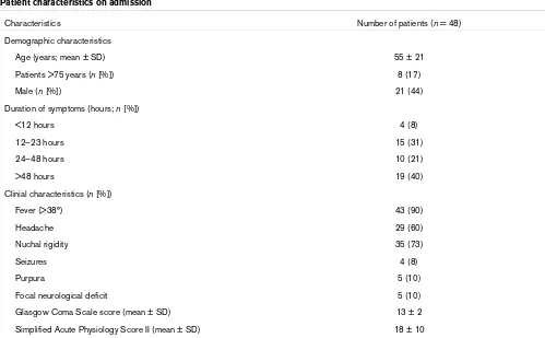
The clinical characteristics of the 48 patients are summarized in Table 1. The mean interval between admission and lumbar puncture was 90 ± 40 min and the mean time elapsing from admission to injection of the first dose of antibiotic was 120 ± 70 min.
start of treatment showing sterilization in all cases. Antibiotic treatment was simplified eight times out of 21 on the basis of the results of microbiological analysis of CSF samples.
All of the other bacteria identified were sensitive to amoxicillin, with the initial antibiotic therapy being appropriate in all cases. The treatment was simplified between 24 and 72 hours after
the start of treatment in six patients out of 24 on the basis of the results of microbiological analysis of the CSF.
[image:3.612.56.555.105.414.2]For the three patients with a CSF culture not showing any evi-dence of bacteria, antibiotic treatment with amoxicillin and ceftriaxone was started on admission and continued for 15– 20 days.
Table 1
Patient characteristics on admission
Characteristics Number of patients (n = 48)
Demographic characteristics
Age (years; mean ± SD) 55 ± 21
Patients >75 years (n [%]) 8 (17)
Male (n [%]) 21 (44)
Duration of symptoms (hours; n [%])
<12 hours 4 (8)
12–23 hours 15 (31)
24–48 hours 10 (21)
>48 hours 19 (40)
Clinial characteristics (n [%])
Fever (>38°) 43 (90)
Headache 29 (60)
Nuchal rigidity 35 (73)
Seizures 4 (8)
Purpura 5 (10)
Focal neurological deficit 5 (10)
Glasgow Coma Scale score (mean ± SD) 13 ± 2
Simplified Acute Physiology Score II (mean ± SD) 18 ± 10
SD, standard deviation.
Table 2
Bacteriology of CSF on admission
Organism CSF Gram stain (n = 48) Culture (n = 48)
Streptococcus pneumoniae (n [%]) 12 (25) 21 (44)
Neisseria meningitidis (n [%]) 6 (12) 9 (19)
Other streptococci (n [%]) 0 5 (10)
Listeria monocytogenes (n [%]) 0 6 (13)
Haemophilus influenzae (n [%]) 0 2 (4)
Escherichia coli (n [%]) 0 1 (2)
Staphylococcus aureus (n [%]) 0 1 (2)
Total (n [%]) 18 (38) 45 (94)
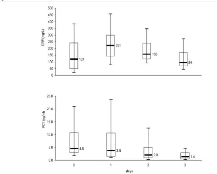
[image:3.612.54.557.482.624.2]The changes in serum and CSF cytochemical parameters are shown in Tables 3 and 4 and in Fig. 1. With regard to the CSF, only the lactate concentration differed significantly between D0 and D2. Sterilization of the CSF was noted in the 34 patients who underwent a second lumbar puncture. In the 14 patients who did not undergo a repeat lumbar puncture, the duration of antibiotic treatment was 12–16 days, resulting in cure in all cases, and the duration of hospital stay was between 13 and 18 days. With respect to serum parameters, the decrease in PCT level was the only significant difference observed between D0 and D2.
Among the 48 patients, five patients (9%) died between 12 and 28 days after their admission to hospital. Only one of these patients was younger than 75 years. All of these patients underwent a second lumbar puncture during treatment, with analysis of the resulting sample showing sterilization of the CSF in all cases, which was confirmed by a third lumbar punc-ture in three of the five patients. A serum PCT concentration below 0.5 ng/ml was observed in all patients between 6 and 9 days after admission. The cause of death was multiple organ failure (n = 1), cerebral thrombophlebitis (n = 1) and cerebral oedema (n = 3). Four patients had neurological sequelae at 30 days.
Discussion
In the present study a significant and early decrease in serum PCT concentration was associated with cure of meningitis. In contrast, analysis of CSF showed a significant decrease only
in lactate concentration between 48 and 72 hours after the first lumbar puncture.
The value of repeat lumbar puncture at 48 hours remains debatable, and second-line antibiotic treatment is based essentially on the MIC of various antibiotics for the bacteria identified or on the clinical course [2,6,12,13,18]. Apart from the microbiological data, the CSF parameters traditionally described during ABM appear to be little modified by appropriate antibiotic therapy within 48 hours. Blazer and coworkers [16], studied the effect of antibiotic treatment on the CSF parameters of 68 children presenting with ABM. None of the cytochemical parameters studied (proteins, glu-cose, total and polymorphonuclear leucocytes) exhibited a sig-nificant decrease between the first lumbar puncture and a second lumbar puncture performed 44–68 hours after the start of antibiotic therapy, whereas two bacteria were still detectable in the repeat CSF samples drawn. Similar findings were reported by Bland and coworkers [19] concerning the changes in these cytochemical parameters after 24–72 hours of treatment for ABM in 15 children.
[image:4.612.55.556.115.245.2]Different results were obtained in an animal study [20]. In five sheep, treatment for an experimentally induced meningitis due to Escherichia coli resulted in a rapid decrease in polymorpho-nuclear leucocyte count in the CSF, which was associated with an increase in glucose concentration and a decrease in protein concentration. However, in that study antibiotic treat-ment was administered intrathecally. In the present study there
Table 3
Cytochemical parameters of CSF and CSF/serum ratio on admission and after 2–3 days of treatment
Parameter Day 0 (admission; n = 48) Day 2 (2–3 days of treatment; n = 34)
Leucocyte count (cells/mm3) 757 (366–2730) 580 (309–2025)
Polymorphonuclear leucocyte count (cells/mm3) 605 (258–2482) 417 (254–1762)
Protein level (g/l) 4.2 (2–6.2) 3.9 (1.8–5)
Glucose CSF level (mmol/l) 2.4 (0.8–3.6) 2.5 (1.2–3)
CSF/serum glucose ratio 0.31 (0.1–0.48) 0.35 (0.17–0.5)
Lactate CSF level (mmol/l) 8.74 (5.5–13) 5* (3–9)
CSF/serum lactate ratio 3.22 (2.4–4.7) 2.73 (1.5–3.3)
Values are expressed as median (interquartile range). CSF, cerebrospinal fluid. *P < 0.001.
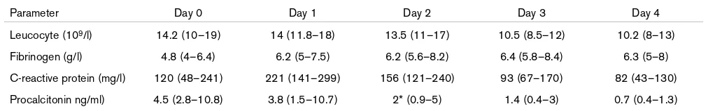
Table 4
Change in serum biological parameters from admission to day 4 of treatment
Parameter Day 0 Day 1 Day 2 Day 3 Day 4
Leucocyte (109/l) 14.2 (10–19) 14 (11.8–18) 13.5 (11–17) 10.5 (8.5–12) 10.2 (8–13)
Fibrinogen (g/l) 4.8 (4–6.4) 6.2 (5–7.5) 6.2 (5.6–8.2) 6.4 (5.8–8.4) 6.3 (5–8)
C-reactive protein (mg/l) 120 (48–241) 221 (141–299) 156 (121–240) 93 (67–170) 82 (43–130)
Procalcitonin ng/ml) 4.5 (2.8–10.8) 3.8 (1.5–10.7) 2* (0.9–5) 1.4 (0.4–3) 0.7 (0.4–1.3)
[image:4.612.53.569.297.377.2]was no significant decrease in the polymorphonuclear leuco-cyte count or protein concentration in the CSF after 48–72 hours of appropriate antibiotic treatment. The glucose concen-tration measured in the CSF remained stable, but there was a significant decrease in lactate concentration.
Although numerous articles have demonstrated the value of assaying lactate during the course of ABM [19,21-27], few data exist concerning the changes in this parameter during the treatment of this disease. In 21 patients with ABM, Gontroni and coworkers [23] showed a rapid decrease in lactate con-centration in the CSF during the first 24 hours of treatment. Gould [22], Bland [19] and Genton [21] and their groups obtained similar results concerning the change in lactate con-centration in CSF after 24–72 hours of treatment in 6, 15 and 25 patients with ABM, respectively. In the study reported by Bland and coworkers [19], the mean lactate concentration in the CSF was 75.1 ± 6.6 mg/100 ml at the time of the first lum-bar puncture and 49.5 ± 5.7 mg/100 ml after 24–72 hours of treatment.
With regard to the changes in serum parameters, the present study revealed a rapid decrease in PCT concentration within the first 24 hours of treatment, which was accompanied by an increase in CRP, with the level of CRP diminishing only after 2–3 days.
[image:5.612.60.500.92.444.2]In 1993, Assicot and coworkers [17] demonstrated that serum PCT concentration was a marker of infectious states of bacte-rial origin in children, exhibiting a rapid decrease following anti-biotic treatment. Although several studies have demonstrated the value of serum PCT concentration in the differential diag-nosis of ABM and viral or aseptic meningitis [28-31], few data are available concerning the change in serum PCT during treatment for ABM. Schwartz and colleagues [31] reported a reduction in median serum PCT concentration from 1.75 mg/ ml at baseline to 1.05 mg/ml after 48 hours of treatment in 11 patients with ABM. In three of these patients, the PCT concen-tration remained unchanged, or increased, in conjunction with an unfavourable clinical course. In the study reported by Gen-drel and coworkers [28], conducted in eight children receiving
Figure 1
Evolution of CRP and PCT levels over 72 hours
treatment for ABM, the serum PCT concentration diminished within 24 hours of treatment in all but two cases.
Although appropriate antibiotic therapy appears to be corre-lated with a rapid decrease in PCT levels, the absence of patients receiving an inappropriate treatment in our series did not allow us to determine the change in PCT levels under these circumstances. What are the arguments in support of a relationship between decrease in PCT levels and appropriate antibiotic treatment? Smith and coworkers [32] investigated the value of PCT in 43 patients presenting with melioidiosis of various grades of severity. Among the 16 patients with a severe infection, 13 exhibited a decrease in PCT levels from the first day of treatment. In the three other patients an increase in PCT levels was observed in relation to infectious complications (pulmonary abscess, septic arthritis, splenic abscess). In two patients the Pseudomonas pseudomallei
infection detected was resistant to the initial antibiotic therapy.
Although the change in serum levels of CRP has been shown to be of value for tracking the course of a bacterial infection during treatment [33,34], the characteristics of this protein are such that its concentration reaches a maximum only after 24– 48 hours [35]; this is in contrast to PCT, which attains a peak serum concentration more rapidly. After injection of endotoxin, the peak serum concentration of PCT is reached within approximately 8 hours [36].
Certain limitations of the present study should be mentioned. This was a descriptive study of the variation in serum PCT con-centrations over time in patients who had received appropriate antibiotic treatment from the moment they were admitted to hospital. We currently have no data on changes in serum PCT levels occurring in patients who did not receive suitable treat-ment. Two patients with bacterial meningitis were not included in the study on the grounds that they presented with a serum procalcitonin level below 0.5 ng/ml on admission. At present there is no clear explanation for this finding. Several studies have reported low levels of serum PCT during ABM [30,31,37]. For the most part, this occurred in patients pre-senting with bacterial meningitis caused by intracellular bacte-ria or nosocomial infections [31,37].
Conclusion
The change in serum PCT level during treatment for commu-nity-acquired ABM appears to be a valuable parameter for evaluating the efficacy of antibiotic therapy. This hypothesis needs confirmation, particularly in patients presenting with bacterial meningitis that is not microbiologically documented.
Competing interests
The author(s) declare that they have no competing interests.
Authors' contributions
AV conceived of the study, and participated in its design and coordination and drafted the manuscript. PG participated in the inclusion and treatment of patients and drafted the manu-script. SG performed the statistical analysis. BT participated in the inclusion and treatment of patients. FR participated in the inclusion and treatment of patients. OM participated in the inclusion and treatment of patients. AC carried out the the microbiology. CL carried out the immunoassays. FZ partici-pated in the design of the study and drafted the manuscript. JCB helped to draft the manuscript. All authors read and approved the final manuscript.
References
1. Durand ML, Caldewood SB, Weber DJ, Miller SI, Southwick FS, Caviness VS, Swartz MN: Acute bacterial meningitis in adults. A review of 493 episodes. N Engl J Med 1993, 328:21-28. 2. Quagliarello VJ, Scheld WM: Treatment of bacterial meningitis.
N Eng J Med 1997, 336:708-716.
3. De Gans J, van de Beek D: Dexamethasone in adults with bac-terial meningitis. N Engl J Med 2002, 347:1549-1556. 4. Lindvall P, Ahlm C, Ericsson M, Gothefors L, Naredi S, Koskinen
LO: Reducing intracranial pressure may increase survival among patients with bacterial meningitis. Clin Infect Dis 2004, 38:384-390.
5. Rivers E, Nguyen B, Havstad S, Ressler J, Muzzin A, Knoblich B, Peterson E, Tomlanovich M: Early goal-directed therapy in the treatment of severe sepsis and septic shock. N Engl J Med 2001, 345:1368-1377.
6. Tunkel AR, Scheld WM: Acute bacterial meningitis. Lancet 1995, 346:1675-1680.
7. Aronin SI, Peduzzi P, Quagliarello VJ: Community-acquired bac-terial meningitis: risk stratification for adverse clinical out-come and effect of antibiotic timing. Ann Intern Med 1998, 129:862-869.
8. Meyer CN, Samuelsson IS, Galle M, Bangsborg JM: Adult bacte-rial meningitis: aetiology, penicillin susceptibility, risk factors, prognostic factors and guidelines for empirical antibiotic treatment. Clin Microbiol Infect 2004, 10:709-717.
9. Kipli T, Anttila M, Kallio MJ, Peltola H: Lenght of prediagnostic history related to the course and sequelae of childhood bacte-rial meningits. Pediatr Infect Dis J 1993, 12:184-188.
10. Kallio MJ, Kilpi T, Anttila M, Peltola H: The effect of a recent pre-vious visit to a physician on outcome after childhood bacterial meningitis. JAMA 1994, 272:787-791.
11. Lebel MH, McCracken GH Jr: Delayed cerebrospinal fluid steri-lization and adverse outcome of bacterial meningitis in infants and children. Pediatrics 1989, 83:161-167.
Key messages
• After appropriate antibiotic treatment, serum PCT level decrease within the first 24 hours.
• After appropriate antibiotic teatment, serum CRP level decrease between days 2 and 3.
• The value of repeat lumbar puncture at 48 hours remains debatable.
• We have no data on changes in serum PCT levels in patients who do not receive an appropriate antibiotic.
12. Anonymous: Community-acquired purulent meningitis. Short text of the 9th consensus conference on anti-infectious ther-apy [in French]. Presse Med 1998, 27:1145-1150.
13. Begg N, Cartwright KAV, Cohen J, Kaczmarski EB, Innes JA, Leen CL, Nathwani D, Singer M, Southgate L, Todd WT: Consensus statement on diagnosis, investigation, treatment and preven-tion of acute bacterial meningitis in immunocompetent adults. J Infect 1999, 39:1-15.
14. Van de Beek D, de Gans J, Spanjaard L, Vermeulen M, Dankert J: Antibiotic guidelines and antibiotic use in adult bactérial men-ingitis in The Nertherlands. J Antimicrob Chemother 2002, 49:661-666.
15. Moller K, Skinhoj P: Guidelines for managing acute bacterial meningitis. BMJ 2003, 320:1290-1292.
16. Blazer S, Berant M, Alon U: Bacterial meningitis. Effect of anti-biotic treatment on cerebrospinal fluid. Am J Clin Pathol 1983, 80:386-387.
17. Assicot M, Gendrel D, Carsin H, Raymond J, Guilbaud J, Bohuon C: High serum procalcitonin concentrations in patients with sepsis and infection. Lancet 1993, 341:515-518.
18. Heyderman RS, Lambert HP, O'Sullivan I, Stuart JM, Taylor BL, Wall RA: Early management of suspected bacterial meningitis and meningococcal septicaemia in adults. J Infect 2003, 46:75-77.
19. Bland RD, Lister RC, Ries JP: Cerebrospinal fluid lactic acid level and pH in meningitis. Am J Dis Child 1974, 128:151-156. 20. Nazifi S, Rezakhani A, Badran M: Evaluation of hematological, serum biochemical and cerebrospinal fluid parameters in experimental bacterial meningitis in the calf. J Vet Med A 1997, 44:55-63.
21. Genton B, Berger JP: Cerebrospinal fluid lactate in 78 cases of adult meningitis. Intensive Care Med 1990, 16:196-200. 22. Gould IM, Irwin WJ, Wadhwani RR: The use of cerebrospinal
fluid lactate determination in the diagnosis of meningitis. Scand J Infect Dis 1980, 12:185-188.
23. Gontroni G, Rodriguez WJ, Deane CA, Hicks JM, Ross S: Cere-brospinal fluid lactate determination: a new parameter for the diagnosis of acute and partially treated meningitis. Chemother-apy 1976, 1:175-182.
24. Berg B, Gärdsell P, Skansberg P: Cerebrospinal fluid lactate in the diagnosis of meningitis. Diagnostic value compared to standard biochemical methods. Scand J Infect Dis 1982, 14:111-115.
25. Curtis GDW, Slack MPE, Tompkins DS: Cerebrospinal fluid lac-tate and the diagnosis of meningitis. J Infect 1981, 3:159-165. 26. Spranger M, Schwab S, Krempien S, Maiwald M, Bruno K, Hacke W: Excess glutamate levels in the cerebrospinal fluid predict clinical outcome of bacterial meningitis. Arch Neurol 1996, 53:992-996.
27. Knight JA, Dudek SM, Haymond RE: Early (chemical) diagnosis of bacterial meningitis: cerebrospinal fluid glucose, lactate, and lactate dehydrogenase compared. Clin Chem 1981, 27:1431-1434.
28. Gendrel D, Raymond J, Assicot M, Moulin F, Iniguez JL, Lebon P, Bohuon C: Measurement of procalcitonin levels in children with bacterial or viral meningitis. Clin Inf Dis 1997, 24:1240-1242.
29. Viallon A, Zeni F, Lambert C, Pozzetto B, Tardy B, Venet C, Ber-trand JC: High sensitivity and specificity of serum procalcitonin levels in adults with bacterial meningitis. Clin Infect Dis 1999, 28:1313-1316.
30. Jereb M, Muzlovic I, Hojker S, Strle F: Predictive value of serum and cerebrospinal fluid procalcitonin levels for the diagnosis of bacterial meningitis. Infection 2001, 29:209-212.
31. Schwarz S, Bertram M, Schwab S, Andrassy K, Hache W: Serum procalcitonin levels in bacterial and abacterial meningitis. Crit Care Med 2000, 28:1828-1832.
32. Smith MD, Suputtamongkol Y, Chaowagul W, Assicot M, Bohuon C, Petitjean S, White NJ: Elevated serum procalcitonin levels in patients with melioidosis. Clin Infect Dis 1995, 20:641-645. 33. Povoa P: C-reactive protein: a valuable marker of sepsis.
Inten-sive Care Med 2002, 28:235-243.
34. Cox ML, Rudd AG, Gallimore R, Hodkinson HM, Pepys MB: Real-time measurement of serum C-reactive protein in the man-agement of infection in the elderly. Age Ageing 1986, 15:257-266.
35. Mary P, Veinberg F, Couderc R: Acute meningitis, acute phase proteins and procalcitonin. Ann Biol Clin 2003, 61:127-137. 36. Dandona P, Nix D, Wilson MF, Aljada A, Love J, Assicot M, Bohuon
C: Procalcitonin increase after endotoxin injection in normal subjects. J Clin Endocrinol Metab 1994, 79:1605-1608. 37. Hoffmann O, Reuter U, Masuhr F, Holtkamp M, Kassim N, Weber