Neuroscience Institute Dissertations Neuroscience Institute
Spring 4-26-2012
Peripheral and central mechanisms through which high energy
Peripheral and central mechanisms through which high energy
diets impair hippocampal-dependent memory in male rats
diets impair hippocampal-dependent memory in male rats
Amy Patricia Ross
Follow this and additional works at: https://scholarworks.gsu.edu/neurosci_diss
Recommended Citation Recommended Citation
Ross, Amy Patricia, "Peripheral and central mechanisms through which high energy diets impair hippocampal-dependent memory in male rats." Dissertation, Georgia State University, 2012. https://scholarworks.gsu.edu/neurosci_diss/5
This Dissertation is brought to you for free and open access by the Neuroscience Institute at ScholarWorks @ Georgia State University. It has been accepted for inclusion in Neuroscience Institute Dissertations by an authorized administrator of ScholarWorks @ Georgia State University. For more information, please contact
HIGH ENERGY DIETS IMPAIR HIPPOCAMPAL-DEPENDENT MEMORY
IN MALE RATS
by
AMY PATRICIA ROSS
Under the Direction of Dr. Marise Parent
ABSTRACT
Over the past five decades, per capita caloric intake has increased by
approxi-mately 28% in the United States. A hallmark of the current standard American diet is an
excess of energy sources from saturated fat and refined carbohydrates. High energy
diets such as the “Western” diet cause numerous pathologies, including non-alcoholic
fatty liver disease (NAFLD), high blood pressure, dyslipidemia, and peripheral insulin
resistance. High energy diets also negatively impact the hippocampus, a brain area
im-portant for learning and memory. It is not surprising, then, that high energy diets impair
hippocampal-dependent memory. The experiments in this dissertation investigate
en-induced NAFLD impairs hippocampal-dependent memory, but these cognitive deficits
were not due to decreases in insulin-like growth factor-1 (IGF-1) or hippocampal insulin
signaling. Next, we found that a high energy diet increased the ability of epinephrine to
increase blood glucose concentrations, indicating a novel way in which high energy
di-ets impair liver function. The final set of experiments found that high energy didi-ets do not
necessarily impair memory but instead may prevent the memory-enhancing effects of
acute stress. Taken together, these results indicate that high energy diets interact with
acute stress to negatively impact hippocampal-dependent memory, and that
hippocam-pal insulin resistance and IGF-1are not likely involved.
PERIPHERAL AND CENTRAL MECHANISMS THROUGH WHICH
HIGH ENERGY DIETS IMPAIR HIPPOCAMPAL-DEPENDENT MEMORY
IN MALE RATS
by
AMY PATRICIA ROSS
A Dissertation Submitted in Partial Fulfillment of the Requirements for the Degree of
Doctor of Philosophy
in the College of Arts and Sciences
Georgia State University
Copyright by Amy Patricia Ross
HIGH ENERGY DIETS IMPAIR HIPPOCAMPAL-DEPENDENT MEMORY
IN MALE RATS
by
AMY PATRICIA ROSS
Committee Chair: Dr. Marise Parent
Committee: Dr. Timothy Bartness
Dr. Charles Derby
Dr. Kim Huhman
Electronic Version Approved:
Office of Graduate Studies
College of Arts and Sciences
Georgia State University
There are many people I would like to thank because I could not have
accom-plished everything on my own. First, I would like to thank my dissertation committee.
Marise Parent, I appreciate your guidance and patience over the past 7 years. I am a
better scientist because of you. Thank you for being a great mentor and friend. Tim
Bartness, when designing experiments or presenting findings, I often find myself
ask-ing, “What would Tim do?” Thank you for being the tiny voice of strong inference in my
head. Kim Huhman, thank you for always being willing to meet with me to guide me in
the right direction. Chuck Derby, I appreciate your thoughtful comments and
encour-agement to think outside the box. I would also like to thank our collaborator, John
Miel-ke, for sharing his knowledge and always having the most diplomatic responses to
re-viewer comments.
I would also like to thank past and present Parent lab members. Krista Wild, you
kept me sane during the most difficult year of graduate school. I miss having you near
me. Jenna Darling, Yoko Ogawa, and Emily Bruggeman, thank you for providing
opinions, support, assistance, and humor any time I needed. I could not ask for better
labmates. During the past 7 years I have had the pleasure of working with many
under-graduate and master’s students. I could not have been nearly as productive without
their assistance. I thank Nathan Waldron, Mariana Silva, Dorothy Bota, Ade
Ka-sumu, Kevin Fernander, Megan Krench, Aja Muldrow, Walid Radwan, Bethany
Yee, Lalita Balakrishnan, Amanda Arnold, Jenine Ampudia, Eseosaserea
Igbi-nigie, and Saima Masud and wish them the best in their own scientific careers.
I would like to thank everyone in the Neuroscience Institute for creating a
chal-lenging and supportive environment. This includes all the faculty who served as
teach-ers and mentors as well as the staff who made accomplishing daily lab chores, taking
care of the rats, and keeping track of college deadlines much easier.
I am so lucky to have such an amazing group of friends. Amanda McAvoy, Greg
Marcinko, Chad Mandichak, Josh Faish, Laura Been, Mahin Shahbazi, Tizeta
Tadesse, Leslie Dunham, Cameron Miller, Luis Martinez, Susan Paige, Laura
Turner, Nick Turner, Carrie Lippy, Jenna Darling, Justin Darling, Marc Badura,
and Amanda Arnold thank you for always being there to brighten my day. Seth
Auf-derheide, your patience and support has been amazing over the past few years. I don’t
know what I would have done without you.
Finally, I could not have made it this far without my family. Thank you to my
par-ents, Ken and Karen Ross, for always believing in me and encouraging me to be the
best person I can be. Thank you to my sister, Kelly Mays, for having more confidence
in me than I ever had for myself. I will always strive to be as intelligent and as beautiful
TABLE OF CONTENTS
ACKNOWLEDGEMENTS ... i
LIST OF TABLES ... ix
LIST OF FIGURES... x
LIST OF ABBREVIATIONS ... xii
CHAPTER 1: GENERAL INTRODUCTION ... 1
1.1 Specific Aims ... 7
1.2 References ... 10
CHAPTER 2: NON-ALCOHOLIC FATTY LIVER DISEASE IMPAIRS HIPPOCAMPAL-DEPENDENT MEMORY IN MALE RATS ... 21
2.1 Abstract ... 21
2.2 Introduction... 22
2.3 Materials and Methods ... 25
2.4 Results... 31
2.5 Discussion... 32
2.6 Acknowledgements ... 37
2.7 References ... 37
2.8 Chapter 2 Table ... 47
2.9 Chapter 2 Figures ... 48
CHAPTER 3: A HIGH ENERGY DIET, BUT NOT STEATOSIS, POTENTIATES THE ABILITY OF EPINEPHRINE TO INCREASE BLOOD GLUCOSE CONCENTRATIONS... 55
3.2 Introduction... 56
3.3 Methods ... 58
3.4 Results... 61
3.5 Discussion... 64
3.6 Acknowlegements ... 67
3.7 References ... 67
3.8 Chapter 3 Figures ... 73
CHAPTER 4: THE INTERACTION BETWEEN THE EFFECTS OF A HIGH ENERGY DIET AND ACUTE STRESS ON HIPPOCAMPAL-DEPENDENT MEMORY IN MALE RATS ... 80
4.1 Abstract ... 80
4.2 Introduction... 81
4.3 Experiment 1 Methods... 86
4.4 Experiment 1 Results ... 91
4.5 Experiment 2 Methods... 93
4.6 Experiment 2 Results ... 94
4.7 Experiment 3 Methods... 94
4.8 Experiment 3 Results ... 95
4.9 Experiment 4 Methods... 95
4.10 Experiment 4 Results ... 95
4.11 Experiment 5 Methods... 96
4.12 Experiment 5 Results ... 96
4.14 Experiment 6 Results ... 98
4.15 Experiment 7 Methods... 98
4.16 Experiment 7 Results ... 99
4.17 Discussion... 100
4.18 Acknowledgements ... 105
4.19 References ... 105
4.20 Chapter 4 Figures ... 113
CHAPTER 5: GENERAL DISCUSSION ... 127
5.1 References ... 135
APPENDICES ... 158
LIST OF TABLES
LIST OF FIGURES
Figure 2.1: Body mass and caloric consumption 48
Figure 2.2: Confirmation of non-alcoholic fatty liver disease 49
Figure 2.3: Plasma triglycerides and hormone concentrations 50
Figure 2.4: Spatial water maze training 51
Figure 2.5: Spatial water maze testing 52
Figure 2.6: Insulin-stimulated phosphorylation of IR-β and PKB/AKT 53
Figure 2.7: Hippocampal growth factor measurements 54
Figure 3.1: Percent change in body mass 73
Figure 3.2: Confirmation of non-alcoholic fatty liver disease 74
Figure 3.3: Baseline blood glucose concentrations 75
Figure 3.4: Epinephrine-stimulated blood glucose concentrations 76
Figure 3.5: Percent change in body mass and lipid accumulation reversal 77
Figure 3.6: Baseline blood glucose concentrations 78
Figure 3.7: Epinephrine-stimulated blood glucose concentrations 79
Figure 4.1: Percent change in body mass and caloric consumption 113
Figure 4.2: Spatial object recognition training in rats with scores above the
median
114
Figure 4.3: Spatial object recognition testing in rats with scores above the
median
115
Figure 4.4: Spatial object recognition testing in rats with scores below the
median
116
Figure 4.5: Spatial object recognition testing in all rats 117
Figure 4.6: Spatial object recognition testing in rats injected with
epineph-rine
Figure 4.7: Spatial object recognition training in rats injected with sotalol 119
Figure 4.8: Spatial object recognition testing in rats injected with sotalol 120
Figure 4.9: Spatial object recognition training in rats injected with
metyrapone
121
Figure 4.10: Spatial object recognition testing in rats injected with
metyrapone
122
Figure 4.11: Spatial object recognition training in rats fed a high energy diet
then standard chow
123
Figure 4.12: Spatial object recognition testing in rats fed a high energy diet
then standard chow
124
Figure 4.13: Plasma stress hormone concentrations 125
Figure 4.14: Plasma stress hormone concentrations and open field
differ-ence scores
LIST OF ABBREVIATIONS
aCSF, artificial cerebral spinal fluid
BBB, blood brain barrier
BDNF, brain-derived neurotrophic factor
ELISA, enzyme-linked immunosorbent assay
HED, high energy diet
HPLC, high performance liquid chromatography
IGF-1, insulin-like growth factor-1
IR, insulin resistance
IR-β, insulin receptor beta-subunit
kcal, kilocalorie
LTD, long term depression
LTP, long term potentiation
NAFLD, non-alcoholic fatty liver disease
NMB, non-fat milk blocker
PKB/AKT, protein kinase B
RIA, radioimmunoassay
SC, standard chow
SOR, spatial object recognition
TBS-T, tris-buffered saline with Tween 20
CHAPTER 1: GENERAL INTRODUCTION
Over the past five decades, per capita caloric intake has increased by
approxi-mately 28% in the United States (USDA 2011). A hallmark of the current standard
American diet is an excess of energy sources from saturated fat and refined
carbohy-drates (Grotto and Zied 2010; Hu, Rimm et al. 1999; Iqbal, Anand et al. 2008). High
en-ergy diets such as the “Western” diet cause numerous pathologies, including
non-alcoholic fatty liver disease (NAFLD; Ackerman, Oron-Herman et al. 2005; Fu, Sun et al.
2009), high blood pressure (Hwang, Ho et al. 1987; Elliott, Keim et al. 2002; Catena,
Giacchetti et al. 2003; Delbosc, Paizanis et al. 2005; Panchal, Poudyal et al. 2010;
Poudyal, Campbell et al. 2010), dyslipidemia (Sleder, Chen et al. 1980; Kelley, Allan et
al. 2004; Couchepin, Le et al. 2008), and peripheral insulin resistance (Zavaroni,
Sand-er et al. 1980; Tobey, Mondon et al. 1982; BezSand-erra, Ueno et al. 2000; Panchal, Poudyal
et al. 2010). Many of these pathologies are components of the metabolic syndrome,
which now affects both children and adults (Kohen-Avramoglu, Theriault et al. 2003).
Although most research has focused on the peripheral consequences of high energy
diets, there is a growing body of literature demonstrating high energy diets impact the
brain (Greenwood and Winocur 1990; Molteni, Barnard et al. 2002; Stranahan, Norman
et al. 2008; Ross, Bartness et al. 2009). The goal of the following studies was to
deter-mine if certain peripheral and central effects of high energy diets are involved in the
memory-impairing effects of these diets.
High energy diets negatively impact the hippocampus.
Previous research has identified a number of ways in which high energy diets
and memory (Ergorul and Eichenbaum 2004). High energy diets decrease brain derived
neurotrophic factor (BDNF; Molteni, Barnard et al. 2002), synaptic communication
(Stranahan, Norman et al. 2008), and neurogenesis (Lindqvist, Mohapel et al. 2006) in
the hippocampus. In addition, high energy diets decrease long-term potentiation (LTP;
Stranahan, Norman et al. 2008) and long-term depression (LTD; Mielke, Taghibiglou et
al. 2005), cellular mechanisms of plasticity that are thought to underlie learning and
memory processes (Malenka and Bear 2004; Massey and Bashir 2007).
It is not surprising, then, that high energy diets impair performance in memory
tasks that depend on the hippocampus. For example, feeding rats a high fructose diet
(Ross, Bartness et al. 2009) or a high fat, high sugar diet (Molteni, Barnard et al. 2002;
Stranahan, Norman et al. 2008; Darling et. al unpublished) causes memory impairments
in the water maze task. In addition, fat-fed rats are impaired in the water maze task
(Pa-than, Gaikwad et al. 2008), radial arm maze, and a variable-interval delayed alternation
task (Greenwood and Winocur 1990).
High energy diets may impair memory via central insulin resistance.
It has been well documented that high energy diets cause peripheral insulin
re-sistance (Zavaroni, Sander et al. 1980; Tobey, Mondon et al. 1982; Bezerra, Ueno et al.
2000; Panchal, Poudyal et al. 2010). Of interest, impaired insulin signaling in the
pe-riphery is related to cognitive deficits in rats fed high energy diets (Chen, Xie et al.
2009). High energy diets also cause central insulin resistance (Mielke, Taghibiglou et al.
2005). Mielke and colleagues (2005) found that feeding hamsters a diet of 60% fructose
for 6 weeks decreases phosphorylation of the insulin receptor-β subunit and protein
important for learning and memory (Moosavi, Naghdi et al. 2007; Moosavi, Naghdi et al.
2007; McNay, Ong et al. 2010). In addition, diet-induced insulin resistance reduces the
ability of insulin to stimulate LTD (Mielke, Taghibiglou et al. 2005), and high energy diets
prevent exogenous insulin from enhancing memory (McNay, Ong et al. 2010). These
findings raise the possibility that high energy diets may impair memory by causing
hip-pocampal insulin resistance.
High energy diets may impair hippocampal-dependent memory via non-alcoholic fatty
liver disease-induced decreases in insulin-like growth factor-1.
Chronic consumption of high energy diets causes NAFLD (Ackerman,
Oron-Herman et al. 2005; Ahmed, Redgrave et al. 2009; Panchal, Poudyal et al. 2010). The
liver sends excess energy from high energy diets through the bloodstream to adipose
tissue (Flint 1998) or stores them as lipids (Stein and Shapiro 1960). NAFLD is an
ac-cumulation of lipids in the liver, ranging from simple steatosis to fibrosis (McCullough
2004). Hepatic lipid accumulation causes insulin resistance (Samuel, Liu et al. 2004),
oxidative stress (Raso, Esposito et al. 2009; Ruiz-Ramirez, Chavez-Salgado et al.
2011) and impairs liver functioning (Ai, Zhu et al. 2011; Ha, Kim et al. 2011; Pasarin,
Abraldes et al. 2011).
It is possible that the NAFLD-induced disturbances in liver functioning could
sult in impaired cognition. NAFLD could impair hippocampal-dependent memory by
re-ducing hepatic insulin-like growth factor-1 (IGF-1) production and/or by decreasing the
amount of IGF-1 transported across the blood brain barrier (BBB). The liver is the main
source of IGF-1 (Yakar, Liu et al. 1999), a hormone capable of crossing the BBB
O'Kusky, Ye et al. 2000; Lichtenwalner, Forbes et al. 2001). In addition, IGF-1 is
impor-tant for learning and memory. For example, spatial memory deficits observed in rodent
models of type 2 diabetes are prevented by exogenous administration of IGF-1 (Lupien,
Bluhm et al. 2003), and blocking IGF-1 receptors in the brain impairs performance of
hippocampal-dependent memory tasks (Lupien, Bluhm et al. 2003). Increases in
en-dogenous IGF-1 cause exercise-induced memory enhancement in rats (Ding, Vaynman
et al. 2006; Cassilhas, Lee et al. 2012). In addition, decreases in IGF-1 concentrations
are correlated with cognitive function in aging individuals (Tan and Baxter 1986;
Ale-man, de Vries et al. 2000; Bellar, Glickman et al. 2011). Of interest, patients with
NAFLD exhibit decreases in circulating IGF-1 (Arturi, Succurro et al. 2011) and
experi-ence cognitive impairments (Newton 2010).
High energy diets may impair memory through non-alcoholic fatty liver disease-induced
decreases in the ability of epinephrine to release hepatic glucose.
Diet-induced NAFLD may interfere with the capacity of the liver to mobilize
glu-cose during the fight or flight response. During this response, stress stimulates the
ad-renal gland to release epinephrine. Epinephrine, in turn, stimulates the liver to release
glucose, which then circulates throughout the body (Sacca, Vigorito et al. 1983). Given
that hepatic lipid accumulation impedes the ability of insulin to decrease hepatic glucose
secretion (Yadav, Jain et al. 2009), these excess lipids may interfere with the ability of
epinephrine to increase hepatic glucose release.
Emotionally arousing events enhance memory, at least in part, via the release of
endogenous epinephrine (Gold and van Buskirk 1978; King and Williams 2009).
receiv-ing a footshock in an inhibitory avoidance task (McCarty and Gold 1981), and bereceiv-ing
placed in a novel environment (de Boer, Koopmans et al. 1990) cause the release of
endogenous epinephrine. Given that epinephrine cannot cross the BBB (Weil-Malherbe,
Axelrod et al. 1959), it is thought that epinephrine enhances memory, in part, through
the hepatic release of glucose (Sacca, Vigorito et al. 1983; Hall and Gold 1986). This
idea is supported by the fact that epinephrine and glucose both impact memory in a
similar dose-dependent manner. To be specific, optimal memory performance occurs
with mid-range doses of either hormone, but too little or too much glucose or
epineph-rine has no or negative effects on memory (Gold and Van Buskirk 1975; Gold, van
Buskirk et al. 1977; Gold 1986; Gold, Vogt et al. 1986). In addition, eliminating
endoge-nous epinephrine release through adrenalectomy reduces stress-induced increases in
blood glucose concentrations and impairs performance in an inhibitory avoidance task
(Hall and Gold 1990). Injections of exogenous glucose restore memory performance in
adrenalectomized rats (Hall and Gold 1990). Emotionally arousing events also enhance
memory through the release of endogenous corticosterone (Sandi, Loscertales et al.
1997). Stressful events stimulate the adrenal gland to secrete corticosterone, which,
un-like epinephrine, crosses the BBB (McEwen, Weiss et al. 1968; McEwen, Weiss et al.
1969). Once corticosterone is in the brain, limbic structures, including the hippocampus,
absorb and retain the hormone (McEwen, Weiss et al. 1968; McEwen, Weiss et al.
1969). Corticosterone injections enhance memory in object recognition (Okuda,
Rooz-endaal et al. 2004) and the spatial water maze (Sandi, Loscertales et al. 1997). In
addi-tion, eliminating endogenous corticosterone through adrenalectomy impairs water maze
It is possible that high energy diets only impair memory in tasks that stimulate the
release of stress hormones. High energy diets increase epinephrine (Kaufman, Li et al.
1993) and corticosterone (Tannenbaum, Brindley et al. 1997; Legendre and Harris
2006; Cano, Jimenez-Ortega et al. 2008) secretion and decrease adrenal gland weight
(Boukouvalas, Antoniou et al. 2008). In addition, behavioral data from our lab and
oth-ers suggest that rats fed a high fructose or high fat/high sugar diet have impaired
mem-ory during tasks that are stressful, such as the water maze task (Molteni, Barnard et al.
2002; Stranahan, Norman et al. 2008; Ross, Bartness et al. 2009), but not during less
stressful tasks such as object recognition (Darling et al., unpublished) or spontaneous
alternation (Ross 2008). The existing data do not directly address the possibility that
high energy diets only impair memory in stressful tasks because there are many
ex-perimental differences in these studies in addition to stress, such as different handling
and training protocols, testing apparatus, retention intervals, motivation, performance
requirements, and light cycle.
Are the high energy diet-induced memory impairments reversible?
To our knowledge, there are no studies that have investigated whether removing
the high energy dietary components reverses the diet-induced memory impairments.
One study has found that giving rats an antidiabetic drug reverses the cognitive deficits
induced by a high energy diet (Pathan, Gaikwad et al. 2008). Beyond this finding, most
research in this area has been dedicated to determining whether the peripheral effects
of high energy diets can be attenuated with interventions such as exercise (Brandt, De
Bock et al. 2010; da Luz, Frederico et al. 2011) or dietary supplements (Shetty, Mengi
al. 2011). Such interventions reverse diet-induced non-alcoholic fatty liver disease
(Shetty, Mengi et al. 2010; Marsman, Heger et al. 2011; Panchal, Poudyal et al. 2011)
and peripheral insulin resistance (Brandt, De Bock et al. 2010; da Luz, Frederico et al.
2011; Tan, Kamal et al. 2011) in high energy-fed rats. Given that the high energy
diet-induced metabolic and cognitive consequences are not permanent, it is possible that
the diet-induced memory impairments could be overturned by simply removing the high
energy dietary components.
1.1 Specific Aims
The evidence reviewed above suggests that the high energy diet-induced
mem-ory impairments are caused by metabolic disturbances in the periphery and in the brain.
Therefore, the experiments in this dissertation investigate possible diet-induced
conse-quences that may contribute to the impairing effects of the diets on
hippocampal-dependent memory. These experiments are outlined in the following aims:
Specific Aim 1: Are the memory deficits induced by high energy diets caused, at least in
part, by hippocampal insulin resistance?
High energy diets produce hippocampal insulin resistance in hamsters (Mielke,
Taghibiglou et al. 2005); however, it is not known if these diets impair central insulin
signaling in rats. Given that hippocampal insulin signaling is important for learning and
memory (Moosavi, Naghdi et al. 2007; Moosavi, Naghdi et al. 2007; McNay, Ong et al.
2010), and high energy diets impair hippocampal-dependent memory (Greenwood and
Winocur 1990; Molteni, Barnard et al. 2002; Pathan, Gaikwad et al. 2008; Stranahan,
high energy diet decreases phosphorylation of the insulin receptor-β subunit and protein
kinase B/AKT in rats, and if so, whether this insulin resistance is related to the
diet-induced memory impairments.
Specific Aim 2: Are the memory deficits produced by high energy diets caused, at least
in part, by NAFLD-induced decreases in IGF-1?
High energy diets are used to create models of non-alcoholic fatty liver disease
(NAFLD; Ackerman, Oron-Herman et al. 2005; Fu, Sun et al. 2009), a metabolic
distur-bance that results in impaired liver function (Samuel, Liu et al. 2004; Ai, Zhu et al. 2011;
Ha, Kim et al. 2011; Pasarin, Abraldes et al. 2011). Of importance, the liver is the main
source of insulin-like growth factor-1 (IGF-1; Yakar, Liu et al. 1999), a hormone
impor-tant for learning and memory (Tan and Baxter 1986; Aleman, de Vries et al. 2000;
Lu-pien, Bluhm et al. 2003). In addition, NAFLD increases circulating triglycerides (TGs;
Ackerman, Oron-Herman et al. 2005), which prevent IGF-1 from crossing the BBB
(Die-trich, Muller et al. 2007). It is possible that high energy diets impair memory through
NAFLD-induced decreases in hepatic IGF-1 production and/or the amount of IGF-1
transported to the brain. Therefore, Experiment 1 tested whether high energy
diet-induced NAFLD decreases hepatic IGF-1. A second experiment tested whether NAFLD
decreases hippocampal IGF-1.
Specific Aim 3: Do high energy diets impair epinephrine-stimulated glucose release?
Sympathetic nervous system activation mobilizes resources for the fight or flight
response, in part, through the release of epinephrine, which increases hepatic glucose
release into the bloodstream (Sacca, Vigorito et al. 1983). In addition, the ability of
epinephrine-stimulated glucose release (Hall and Gold 1986). High energy diets produce memory
impairments (Greenwood and Winocur 1990; Molteni, Barnard et al. 2002; Pathan,
Gaikwad et al. 2008; Stranahan, Norman et al. 2008; Ross, Bartness et al. 2009) and
NAFLD (Ackerman, Oron-Herman et al. 2005; Svegliati-Baroni, Candelaresi et al. 2006;
Ahmed, Redgrave et al. 2009), and NAFLD impairs liver functioning (Ai, Zhu et al. 2011;
Ha, Kim et al. 2011; Pasarin, Abraldes et al. 2011). Therefore, this experiment tested
whether a high energy diet impairs the ability of epinephrine to increase blood glucose
concentrations. Given that previous studies demonstrate that the effects of high energy
diets, such as NAFLD, are not permanent, (Shetty, Mengi et al. 2010; Marsman, Heger
et al. 2011; Panchal, Poudyal et al. 2011), a second experiment tested whether
remov-ing excess fat and sugar from the diet reverses hepatic lipid accumulation and restores
the ability of epinephrine to increase blood glucose concentrations.
Specific Aim 4: Do high energy diets impair the stress hormone modulation of memory?
High energy diets increase the secretion of epinephrine (Kaufman, Li et al. 1993)
and corticosterone (Tannenbaum, Brindley et al. 1997; Legendre and Harris 2006;
Ca-no, Jimenez-Ortega et al. 2008), which are two hormones important for the modulation
of memory (Gold and Van Buskirk 1975; Gold and van Buskirk 1978; Sandi, Loscertales
et al. 1997; King and Williams 2009). Given that rats fed high energy diets are impaired
on stressful memory tasks (Molteni, Barnard et al. 2002; Stranahan, Norman et al.
2008; Ross, Bartness et al. 2009), but not on relatively unstressful memory tasks (Ross
2008; Darling et al., unpublished), it is possible that high energy diets impair the stress
hormone modulation of memory. Experiment 1 tested whether a high energy diet
in a low stress version of the same task. Experiment 2 tested whether the diet-induced
memory impairments could be replicated by injecting the rats with epinephrine in a
moderate stress version of the SOR task, and Experiments 3 and 4 tested whether the
diet-induced memory impairments could be prevented by blocking the effects of
epi-nephrine and corticosterone in the high stress version of the SOR task.
Specific Aim 5: Are the memory-impairing effects of high energy diets reversible?
Thus far, to our knowledge, no one has tested whether eliminating access to fat
and sugar reverses the cognitive effects of high energy diets. Therefore, this experiment
tested whether removing excess fat and sugar from the diet restores the ability of stress
to enhance hippocampal-dependent memory.
In summary, the following experiments use behavioral, biochemical, and
histo-logical techniques to test whether the memory-impairing effects of high energy diets are
caused by (1) hippocampal insulin resistance and (2) NAFLD-induced decreases in
hip-pocampal and/or hepatic IGF-1, (3) NAFLD-induced decreases in
epinephrine-stimulated glucose release, (4) the result of altered adrenal hormone modulation of
memory and (5) whether the diet-induced memory impairments are reversible. These
findings add to the growing body of literature demonstrating that high energy diets are
harmful to the brain as well as the body.
1.2 References
Aberg, M. A., N. D. Aberg, et al. (2000). "Peripheral infusion of IGF-I selectively induces
Ackerman, Z., M. Oron-Herman, et al. (2005). "Fructose-induced fatty liver disease:
he-patic effects of blood pressure and plasma triglyceride reduction." Hypertension
45(5): 1012-8.
Ahmed, U., T. G. Redgrave, et al. (2009). "Effect of dietary fat to produce non-alcoholic
fatty liver in the rat." J Gastroenterol Hepatol 24(8): 1463-71.
Ai, Z. L., C. H. Zhu, et al. (2011). "The role of hepatic liver X receptor alpha- and sterol
regulatory element binding protein-1c-mediated lipid disorder in the pathogenesis
of non-alcoholic steatohepatitis in rats." J Int Med Res 39(4): 1219-29.
Aleman, A., W. R. de Vries, et al. (2000). "Age-sensitive cognitive function, growth
hor-mone and insulin-like growth factor 1 plasma levels in healthy older men."
Neu-ropsychobiology 41(2): 73-8.
Arturi, F., E. Succurro, et al. (2011). "Nonalcoholic fatty liver disease is associated with
low circulating levels of insulin-like growth factor-I." J Clin Endocrinol Metab
96(10): E1640-4.
Bellar, D., E. L. Glickman, et al. (2011). "Serum insulin like growth factor-1 is associated
with working memory, executive function and selective attention in a sample of
healthy, fit older adults." Neuroscience 178: 133-7.
Bezerra, R. M., M. Ueno, et al. (2000). "A high fructose diet affects the early steps of
insulin action in muscle and liver of rats." J Nutr 130(6): 1531-5.
Boukouvalas, G., K. Antoniou, et al. (2008). "Post weaning high fat feeding affects rats'
behavior and hypothalamic pituitary adrenal axis at the onset of puberty in a
Brandt, N., K. De Bock, et al. (2010). "Cafeteria diet-induced insulin resistance is not
associated with decreased insulin signaling or AMPK activity and is alleviated by
physical training in rats." Am J Physiol Endocrinol Metab 299(2): E215-24.
Cano, P., V. Jimenez-Ortega, et al. (2008). "Effect of a high-fat diet on 24-h pattern of
circulating levels of prolactin, luteinizing hormone, testosterone, corticosterone,
thyroid-stimulating hormone and glucose, and pineal melatonin content, in rats."
Endocrine 33(2): 118-25.
Cassilhas, R. C., K. S. Lee, et al. (2012). "Spatial memory is improved by aerobic and
resistance exercise through divergent molecular mechanisms." Neuroscience
202: 309-17.
Catena, C., G. Giacchetti, et al. (2003). "Cellular mechanisms of insulin resistance in
rats with fructose-induced hypertension." Am J Hypertens 16(11 Pt 1): 973-8.
Chen, S., H. Xie, et al. (2009). "Relationship between insulin sensitivity index and
cogni-tive function in diet-induced insulin resistant rats." Pharmazie 64(6): 410-4.
Couchepin, C., K. A. Le, et al. (2008). "Markedly blunted metabolic effects of fructose in
healthy young female subjects compared with male subjects." Diabetes Care
31(6): 1254-6.
da Luz, G., M. J. Frederico, et al. (2011). "Endurance exercise training ameliorates
insu-lin resistance and reticulum stress in adipose and hepatic tissue in obese rats."
Eur J Appl Physiol 111(9): 2015-23.
de Boer, S. F., S. J. Koopmans, et al. (1990). "Plasma catecholamine, corticosterone
and glucose responses to repeated stress in rats: Effect of interstressor interval
Delbosc, S., E. Paizanis, et al. (2005). "Involvement of oxidative stress and NADPH
oxi-dase activation in the development of cardiovascular complications in a model of
insulin resistance, the fructose-fed rat." Atherosclerosis 179(1): 43-9.
Dietrich, M. O., A. Muller, et al. (2007). "Western style diet impairs entrance of
blood-borne insulin-like growth factor-1 into the brain." Neuromolecular Med 9(4):
324-30.
Ding, Q., S. Vaynman, et al. (2006). "Insulin-like growth factor I interfaces with
brain-derived neurotrophic factor-mediated synaptic plasticity to modulate aspects of
exercise-induced cognitive function." Neuroscience 140(3): 823-33.
Economic Research Service (ERS), US Department of Agriculture (USDA). (July 2011).
"Food Availability (Per Capita) Data System." Retrieved March 31, 2012, from
http://www.ers.usda.gov/Data/FoodConsumption.
Elliott, S. S., N. L. Keim, et al. (2002). "Fructose, weight gain, and the insulin resistance
syndrome." Am J Clin Nutr 76(5): 911-22.
Ergorul, C. and H. Eichenbaum (2004). "The hippocampus and memory for "what,"
"where," and "when"." Learn Mem 11(4): 397-405.
Flint, D. J. (1998). "Effects of antibodies to adipocytes on body weight, food intake, and
adipose tissue cellularity in obese rats." Biochem Biophys Res Commun 252(1):
263-8.
Fu, J. H., H. S. Sun, et al. (2009). "The effects of a fat- and sugar-enriched diet and
chronic stress on nonalcoholic fatty liver disease in male Wistar rats." Dig Dis Sci
Gold, P. E. (1986). "Glucose modulation of memory storage processing." Behav Neural
Biol 45(3): 342-9.
Gold, P. E. and R. van Buskirk (1978). "Effects of alpha- and beta-adrenergic receptor
antagonists on trial epinephrine modulation of memory: relationship to
post-training brain norepinephrine concentrations." Behav Biol 24(2): 168-84.
Gold, P. E., R. van Buskirk, et al. (1977). "Effects of posttraining epinephrine injections
on retention of avoidance training in mice." Behav Biol 20(2): 197-204.
Gold, P. E. and R. B. Van Buskirk (1975). "Facilitation of time-dependent memory
proc-esses with posttrial epinephrine injections." Behav Biol 13(2): 145-53.
Gold, P. E., J. Vogt, et al. (1986). "Glucose effects on memory: behavioral and
pharma-cological characteristics." Behav Neural Biol 46(2): 145-55.
Greenwood, C. E. and G. Winocur (1990). "Learning and memory impairment in rats fed
a high saturated fat diet." Behav Neural Biol 53(1): 74-87.
Grotto, D. and E. Zied (2010). "The standard American diet and its relationship to the
health status of Americans." Nutrition in Clinical Practice 25(6): 603-612.
Ha, S. K., J. Kim, et al. (2011). "Role of AMP-activated protein kinase and adiponectin
during development of hepatic steatosis in high-fat diet-induced obesity in rats." J
Comp Pathol 145(1): 88-94.
Hall, J. L. and P. E. Gold (1986). "The effects of training, epinephrine, and glucose
in-jections on plasma glucose levels in rats." Behav Neural Biol 46(2): 156-67.
Hall, J. L. and P. E. Gold (1990). "Adrenalectomy-induced memory deficits: role of
Hu, F. B., E. Rimm, et al. (1999). "Reproducibility and validity of dietary patterns
as-sessed with a food-frequency questionnaire." Am J Clin Nutr 69(2): 243-9.
Hwang, I. S., H. Ho, et al. (1987). "Fructose-induced insulin resistance and hypertension
in rats." Hypertension 10(5): 512-6.
Iqbal, R., S. Anand, et al. (2008). "Dietary patterns and the risk of acute myocardial
in-farction in 52 countries: results of the INTERHEART study." Circulation 118(19):
1929-37.
Kaufman, L. N., H. Y. Li, et al. (1993). "Adrenal medulla as a mediator of diet-induced
hypertension." Am J Physiol 265(1 Pt 2): R1-6.
Kelley, G. L., G. Allan, et al. (2004). "High dietary fructose induces a hepatic stress
re-sponse resulting in cholesterol and lipid dysregulation." Endocrinology 145(2):
548-55.
King, S. O., 2nd and C. L. Williams (2009). "Novelty-induced arousal enhances memory
for cued classical fear conditioning: interactions between peripheral adrenergic
and brainstem glutamatergic systems." Learn Mem 16(10): 625-34.
Kohen-Avramoglu, R., A. Theriault, et al. (2003). "Emergence of the metabolic
syn-drome in childhood: an epidemiological overview and mechanistic link to
dyslipi-demia." Clin Biochem 36(6): 413-20.
Legendre, A. and R. B. Harris (2006). "Exaggerated response to mild stress in rats fed
high-fat diet." Am J Physiol Regul Integr Comp Physiol 291(5): R1288-1294.
Lichtenwalner, R. J., M. E. Forbes, et al. (2001). "Intracerebroventricular infusion of
in-sulin-like growth factor-I ameliorates the age-related decline in hippocampal
Lindqvist, A., P. Mohapel, et al. (2006). "High-fat diet impairs hippocampal neurogenesis
in male rats." Eur J Neurol 13(12): 1385-8.
Lupien, S. B., E. J. Bluhm, et al. (2003). "Systemic insulin-like growth factor-I
admini-stration prevents cognitive impairment in diabetic rats, and brain IGF regulates
learning/memory in normal adult rats." J Neurosci Res 74(4): 512-23.
Mabry, T. R., P. E. Gold, et al. (1995). "Age-related changes in plasma catecholamine
responses to acute swim stress." Neurobiol Learn Mem 63(3): 260-8.
Malenka, R. C. and M. F. Bear (2004). "LTP and LTD: an embarrassment of riches."
Neuron 44(1): 5-21.
Marsman, H. A., M. Heger, et al. (2011). "Reversal of hepatic steatosis by omega-3 fatty
acids measured non-invasively by (1) H-magnetic resonance spectroscopy in a
rat model." J Gastroenterol Hepatol 26(2): 356-63.
Massey, P. V. and Z. I. Bashir (2007). "Long-term depression: multiple forms and
impli-cations for brain function." Trends Neurosci 30(4): 176-84.
McCarty, R. and P. E. Gold (1981). "Plasma catecholamines: effects of footshock level
and hormonal modulators of memory storage." Horm Behav 15(2): 168-82.
McCullough, A. J. (2004). "The clinical features, diagnosis and natural history of
nonal-coholic fatty liver disease." Clin Liver Dis 8(3): 521-33, viii.
McEwen, B. S., J. M. Weiss, et al. (1968). "Selective retention of corticosterone by
lim-bic structures in rat brain." Nature 220(5170): 911-2.
McEwen, B. S., J. M. Weiss, et al. (1969). "Uptake of corticosterone by rat brain and its
McNay, E. C., C. T. Ong, et al. (2010). "Hippocampal memory processes are modulated
by insulin and high-fat-induced insulin resistance." Neurobiol Learn Mem 93(4):
546-53.
Mielke, J. G., C. Taghibiglou, et al. (2005). "A biochemical and functional
characteriza-tion of diet-induced brain insulin resistance." J Neurochem 93(6): 1568-78.
Molteni, R., R. J. Barnard, et al. (2002). "A high-fat, refined sugar diet reduces
hippo-campal brain-derived neurotrophic factor, neuronal plasticity, and learning."
Neu-roscience 112(4): 803-14.
Moosavi, M., N. Naghdi, et al. (2007). "Intra CA1 insulin microinjection improves
mem-ory consolidation and retrieval." Peptides 28(5): 1029-34.
Moosavi, M., N. Naghdi, et al. (2007). "Insulin protects against stress-induced
impair-ments in water maze performance." Behav Brain Res 176(2): 230-6.
Newton, J. L. (2010). "Systemic symptoms in non-alcoholic fatty liver disease." Dig Dis
28(1): 214-9.
O'Kusky, J. R., P. Ye, et al. (2000). "Insulin-like growth factor-I promotes neurogenesis
and synaptogenesis in the hippocampal dentate gyrus during postnatal
develop-ment." J Neurosci 20(22): 8435-42.
Oitzl, M. S. and E. R. de Kloet (1992). "Selective corticosteroid antagonists modulate
specific aspects of spatial orientation learning." Behav Neurosci 106(1): 62-71.
Okuda, S., B. Roozendaal, et al. (2004). "Glucocorticoid effects on object recognition
memory require training-associated emotional arousal." Proc Natl Acad Sci U S A
Panchal, S., H. Poudyal, et al. (2010). "High Carbohydrate-High Fat Diet-Induced
Meta-bolic Syndrome and Cardiovascular Remodeling in Rats." J Cardiovasc
Pharma-col.
Panchal, S. K., H. Poudyal, et al. (2011). "Rutin attenuates metabolic changes,
nonal-coholic steatohepatitis, and cardiovascular remodeling in high-carbohydrate,
high-fat diet-fed rats." J Nutr 141(6): 1062-9.
Pasarin, M., J. G. Abraldes, et al. (2011). "Insulin resistance and liver microcirculation in
a rat model of early NAFLD." J Hepatol 55(5): 1095-102.
Pathan, A. R., A. B. Gaikwad, et al. (2008). "Rosiglitazone attenuates the cognitive
defi-cits induced by high fat diet feeding in rats." Eur J Pharmacol 589(1-3): 176-9.
Poudyal, H., F. Campbell, et al. (2010). "Olive leaf extract attenuates cardiac, hepatic,
and metabolic changes in high carbohydrate-, high fat-fed rats." J Nutr 140(5):
946-53.
Raso, G. M., E. Esposito, et al. (2009). "Comparative therapeutic effects of metformin
and vitamin E in a model of non-alcoholic steatohepatitis in the young rat." Eur J
Pharmacol 604(1-3): 125-31.
Reinhardt, R. R. and C. A. Bondy (1994). "Insulin-like growth factors cross the
blood-brain barrier." Endocrinology 135(5): 1753-61.
Ross, A. P. (2008). The effects of a high fructose diet on physiology and cognition in
male Sprague-Dawley rats. Master of Arts Thesis. Atlanta, Georgia State
Univer-sity. Master of Arts.
Ross, A. P., T. J. Bartness, et al. (2009). "A high fructose diet impairs spatial memory in
Ruiz-Ramirez, A., M. Chavez-Salgado, et al. (2011). "High-sucrose diet increases ROS
generation, FFA accumulation, UCP2 level, and proton leak in liver
mitochon-dria." Am J Physiol Endocrinol Metab 301(6): E1198-207.
Sacca, L., C. Vigorito, et al. (1983). "Role of gluconeogenesis in epinephrine-stimulated
hepatic glucose production in humans." Am J Physiol 245(3): E294-302.
Samuel, V. T., Z. X. Liu, et al. (2004). "Mechanism of hepatic insulin resistance in
non-alcoholic fatty liver disease." J Biol Chem 279(31): 32345-53.
Sandi, C., M. Loscertales, et al. (1997). "Experience-dependent facilitating effect of
cor-ticosterone on spatial memory formation in the water maze." Eur J Neurosci 9(4):
637-42.
Shetty, S. N., S. Mengi, et al. (2010). "A study of standardized extracts of Picrorhiza
kur-roa Royle ex Benth in experimental nonalcoholic fatty liver disease." J Ayurveda
Integr Med 1(3): 203-10.
Sleder, J., Y. D. Chen, et al. (1980). "Hyperinsulinemia in fructose-induced
hypertriglyc-eridemia in the rat." Metabolism 29(4): 303-5.
Stein, Y. and B. Shapiro (1960). "Uptake and metabolism of triglycerides by the rat
liv-er." J Lipid Res 1: 326-31.
Stranahan, A. M., E. D. Norman, et al. (2008). "Diet-induced insulin resistance impairs
hippocampal synaptic plasticity and cognition in middle-aged rats." Hippocampus
18(11): 1085-8.
Svegliati-Baroni, G., C. Candelaresi, et al. (2006). "A model of insulin resistance and
re-ceptor-alpha and n-3 polyunsaturated fatty acid treatment on liver injury." Am J
Pathol 169(3): 846-60.
Tan, K. and R. C. Baxter (1986). "Serum insulin-like growth factor I levels in adult
dia-betic patients: the effect of age." J Clin Endocrinol Metab 63(3): 651-5.
Tan, Y., M. A. Kamal, et al. (2011). "Chinese herbal extracts (SK0506) as a potential
candidate for the therapy of the metabolic syndrome." Clin Sci (Lond) 120(7):
297-305.
Tannenbaum, B. M., D. N. Brindley, et al. (1997). "High-fat feeding alters both basal and
stress-induced hypothalamic-pituitary-adrenal activity in the rat." Am J Physiol
273(6 Pt 1): E1168-77.
Tobey, T. A., C. E. Mondon, et al. (1982). "Mechanism of insulin resistance in
fructose-fed rats." Metabolism 31(6): 608-12.
Weil-Malherbe, H., J. Axelrod, et al. (1959). "Blood-brain barrier for adrenaline." Science
129(3357): 1226-7.
Yadav, H., S. Jain, et al. (2009). "Epigenomic derangement of hepatic glucose
metabo-lism by feeding of high fructose diet and its prevention by Rosiglitazone in rats."
Dig Liver Dis 41(7): 500-8.
Yakar, S., J. L. Liu, et al. (1999). "Normal growth and development in the absence of
hepatic insulin-like growth factor I." Proc Natl Acad Sci U S A 96(13): 7324-9.
Zavaroni, I., S. Sander, et al. (1980). "Effect of fructose feeding on insulin secretion and
CHAPTER 2: NON-ALCOHOLIC FATTY LIVER DISEASE IMPAIRS
HIPPOCAMPAL-DEPENDENT MEMORY IN MALE RATS
A.P. Ross a, E.C. Bruggeman a, A.W. Kasumu a, J.G. Mielke b, and M.B. Parent a
a Neuroscience Institute, Georgia State University, P.O. Box 5030, Atlanta,
Georgia, 30302-5030, United States
b School of Public Health and Health Systems, University of Waterloo, 200
University Avenue West, Waterloo, Ontario, N2L, 3G1, Canada
Originally published in Physiology & Behavior, 106, 133-141 (2012).
2.1 Abstract
Non-alcoholic fatty liver disease (NAFLD) is a disorder observed in children and
adults characterized by an accumulation of liver fat (> 5% wet weight) in the absence of
excessive alcohol intake. NAFLD affects 10 to 30% of the American population and is
the most common cause of liver disease in the United States. NAFLD leads to serious
disturbances in cardiovascular and hormonal function; however, possible effects on
brain function have been overlooked. The aims of the present study were to test
wheth-er diet-induced NAFLD impairs hippocampal-dependent memory and to detwheth-ermine
whether any observed deficits are associated with changes in hippocampal insulin
sig-naling or concentrations of brain-derived neurotrophic factor (BDNF) and insulin-like
growth factor-1 (IGF-1). Post-weanling male Sprague-Dawley rats were fed a high
spa-tial water maze. NAFLD was confirmed with postmortem measures of liver mass and
liver lipid concentrations. NAFLD did not affect acquisition of the spatial water maze, but
did impair retention tested 48 h later. Specifically, both groups demonstrated similar
de-creases in latency to swim to the escape platform over training trials, but on the memory
test NAFLD rats took longer to reach the platform and made fewer visits to the platform
location than control diet rats. There were no differences between the groups in terms of
insulin-stimulated phosphorylation of insulin receptor β subunit (IR-β) and protein kinase
B (PKB/AKT) in hippocampal slices or hippocampal BDNF or IGF-1 concentrations.
Thus, these data indicate that NAFLD impairs hippocampal-dependent memory function
and that the deficit does not appear attributable to alterations in hippocampal insulin
signaling or hippocampal BDNF or IGF-1 concentrations.
2.2 Introduction
Non-alcoholic fatty liver disease (NAFLD) is observed in children and adults and
is characterized by an accumulation of liver fat (> 5% wet weight) in the absence of
ex-cessive alcohol intake (Neuschwander-Tetri and Caldwell 2003; Patton, Sirlin et al.
2006). The prevalence of pediatric and adult NAFLD is alarmingly high, and its
inci-dence is increasing worldwide. For example, in the United States, over 30% of adults
and 10% of children suffer from NAFLD (Bloomgarden 2005; Schwimmer, Deutsch et al.
2006). In fact, NAFLD is the most common cause of liver pathology in adults and
chil-dren and causes serious disturbances in cardiovascular and hormonal function
(Pacifi-co, Cantisani et al. 2008; Mager, Patterson et al. 2010; Lee, Shim et al. 2011; Lucero,
Over-nutrition is considered the main cause of NAFLD (Ackerman, Oron-Herman
et al. 2005; Ahmed, Redgrave et al. 2009; Safwat, Pisano et al. 2009; Larter, Chitturi et
al. 2010). In the US, children and adults over consume high energy foods, particularly
those that are high in fat and sugar (Hu, Rimm et al. 1999; Iqbal, Anand et al. 2008;
Mager, Patterson et al. 2010). These macronutrients are metabolized mainly by the liver
and converted to energy, and the surplus is stored by the liver as lipids. Given that
NAFLD is primarily a disorder of over-nutrition, feeding rats high-energy diets is a
com-monly accepted animal model of NAFLD (Ackerman, Oron-Herman et al. 2005;
Sve-gliati-Baroni, Candelaresi et al. 2006; Ahmed, Redgrave et al. 2009). For example,
Sprague-Dawley rats fed a 60% fructose diet or a diet of at least 57% fat develop
symp-toms of NAFLD, including hepatomegaly (Ross, Bartness et al. 2009),
hypertriglyc-eridemia (Ackerman, Oron-Herman et al. 2005; Ross, Bartness et al. 2009), and
in-creased hepatic lipids (Ackerman, Oron-Herman et al. 2005; Svegliati-Baroni,
Cande-laresi et al. 2006; Ahmed, Redgrave et al. 2009).
Virtually all research on NAFLD thus far has focused on the peripheral effects of
the disease, whereas the effects of NAFLD on central function have been largely
over-looked. For a number of reasons, we hypothesized that NAFLD impairs
hippocampal-dependent memory. For instance, NAFLD is characterized by disruptions in lipid
me-tabolism, which result in increased circulating triglycerides (TGs; Kelley, Allan et al.
2004). Elevated TGs, in turn, prevent substances important for learning and memory,
such as leptin (Farr, Banks et al. 2006; Oomura, Hori et al. 2006) and insulin-like growth
factor-1 (IGF-1; Lupien, Bluhm et al. 2003; Ding, Vaynman et al. 2006), from crossing
addition, direct administration of TGs into the brain impairs hippocampal-dependent
memory (Farr, Yamada et al. 2008). Diet-induced NAFLD could also impact cognition by
impairing hepatic glucose control, or by producing central insulin resistance. Our data
indicate that NAFLD potentiates epinephrine-induced hepatic glucose release (Ross,
Darling et al. 2010), which is significant because acute and chronic hyperglycemia are
associated with memory deficits (Gold, Vogt et al. 1986; Meneilly, Cheung et al. 1993;
Rodriguez, Horne et al. 1994; Vanhanen, Koivisto et al. 1997; Awad, Gagnon et al.
2002). In addition, elevations in glucose result in simultaneous increases in circulating
insulin, and hippocampal infusions of insulin enhance memory (Moosavi, Naghdi et al.
2007; Moosavi, Naghdi et al. 2007; McNay, Ong et al. 2010). Given that insulin
signal-ing in the hippocampus is important for learnsignal-ing and memory (Moosavi, Naghdi et al.
2007; Moosavi, Naghdi et al. 2007; McNay, Ong et al. 2010), NAFLD-induced
interrup-tions in hippocampal insulin signaling could result in learning and memory deficits.
No-tably, rats fed a high fructose diet and patients with NAFLD often experience peripheral
insulin resistance (Zavaroni, Sander et al. 1980; Tobey, Mondon et al. 1982; Bezerra,
Ueno et al. 2000; Manco, Marcellini et al. 2008; Manco, Giordano et al. 2009), but it is
not known if they also suffer from central insulin resistance.
Diet-induced NAFLD could also impair cognition through changes in the levels of
growth hormones found in the hippocampus. Over-nutrition is often associated with
de-creases in hippocampal brain-derived neurotrophic factor (BDNF; Molteni, Barnard et al.
2002; Kanoski, Meisel et al. 2007; Stranahan, Norman et al. 2008), which is significant
because BDNF promotes hippocampal cell growth and is involved in learning and
addi-tion, the liver is responsible for releasing IGF-1 (Yakar, Liu et al. 1999), a hormone also
important for the growth and repair of hippocampal neurons and learning and memory
(Aleman, de Vries et al. 2000; Lupien, Bluhm et al. 2003). As a result, NAFLD-induced
decreases in the availability of IGF-1 and/or BDNF could negatively impact
hippocam-pal-dependent learning and memory.
Given the extensive plasticity that takes place in the brains of developing animals
(Eayrs and Goodhead 1959; Aghajanian and Bloom 1967; McIlwain and Bachelard
1971), and the high prevalence of NAFLD in childhood and adolescence (Schwimmer,
Deutsch et al. 2006), we hypothesized that over-nutrition-induced NAFLD during
devel-opment has long term consequences on brain function. As a result, we induced NAFLD
in post-weanling rats using a diet high in fructose and then tested
hippocampal-dependent learning and memory once they reached adulthood. NAFLD was confirmed
with postmortem measures of liver mass and liver lipid concentrations. We also
deter-mined whether NAFLD impaired the ability of insulin to stimulate phosphorylation of the
insulin receptor β-subunit (IR-β; the first protein in the signaling cascade; Kahn, White et
al. 1993) and protein kinase B (PKB/AKT; a downstream molecule involved in learning
and memory; Dou, Chen et al. 2005). Finally, we determined whether NAFLD
de-creased hippocampal BDNF or IGF-1 concentrations.
2.3 Materials and Methods
Animals and housing
Twelve timed-pregnant dams (Sprague-Dawley rats, Charles River, Wilmington,
MA) arrived 7-10 days before delivering litters. The animal care facility was controlled
All procedures were approved by Georgia State University’s Institutional Animal Care
and Use Committee and were in accordance with PHS guidelines.
Diets
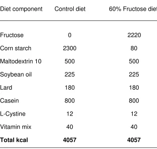
On postnatal day 21, male pups were weaned and placed singly in suspended,
wire-bottom cages (Allentown, Allentown, NJ) with plastic inserts that covered half the
cage floor. The rats were weighed, matched on absolute body mass, and then assigned
to the control or diet-induced NAFLD (NAFLD) groups. To control for possible litter
ef-fects, both the control and NAFLD groups were comprised of rats from each litter. The
NAFLD group was provided ad libitum with pellets composed of 60% fructose
(Re-search Diets, New Brunswick, NJ). The 60% fructose concentration was chosen
be-cause this amount is an established model of NAFLD (Ackerman, Oron-Herman et al.
2005; Mohamed, Nallasamy et al. 2009; Chen, Fang et al. 2010), produces
hippocam-pal insulin resistance in hamsters (Mielke, Taghibiglou et al. 2005), and causes memory
deficits in rats fed fructose starting in adulthood (Ross, Bartness et al. 2009). The
con-trol group was fed ad libitum with isocaloric pellets that contained no fructose (60%
vegetable starch; Research Diets, New Brunswick, NJ), but an equal percentage of
car-bohydrates (70%), proteins (20%), and lipids (10%; see Table 1). Rat body mass and
food intake were recorded daily for 1 week out of every 3 weeks until behavioral tests
were performed. To measure food intake, pellets in each hopper and dried spillage from
under each cage were weighed and then subtracted from the amount placed in the
hopper the previous day. Average daily caloric consumption was calculated by
Spatial water maze
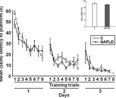
After 12 weeks on the diet, the spatial water maze task was used to assess
learning and memory. The task depends on the integrity of the hippocampus for
suc-cessful performance (Morris, Garrud et al. 1982; Clark, Broadbent et al. 2005), and rats
fed fructose during adulthood have impaired retention performance in the spatial water
maze (Ross, Bartness et al. 2009). For water maze acquisition, the rats were trained to
locate a clear, Plexiglas platform (26 cm in height and 11.5 cm in diameter) that was
submerged 1 cm below the surface of the water (20± 3ºC) in a circular pool (0.46 m in
depth and 1.35 m in diameter). Curtains were hung on two sides of the pool, and one
visual pattern was pasted on each curtain.For purposes of analysis, the water maze
was divided into four virtual quadrants, with one quadrant containing the platform.
Ac-quisition training began by placing the rat on the platform for 30 s and then placing it in
the water in each non-platform containing quadrant in a random order and allowing it a
maximum of 60 s to reach the platform. Latency to reach the platform on each training
trial was used as the measure of acquisition. If the rat did not reach the platform within
60 s, it was guided gently by hand to the platform. The rat was allowed to remain on the
platform for 15 s at the end of each trial and was then placed in an empty cage with a
heat lamp for a 30 s inter-trial interval. All rats were given 8 training trials on the first
day. On the second day, they were trained until they swam to the platform in less than 8
s for 3 consecutive trials for a maximum of 8 trials. Rats that did not reach the criterion
on the second day were given a third day of training with the same criterion for a
maxi-mum of 8 trials. This criterion was determined on the basis of pilot studies in which
group performance plateaued at 8 seconds. This training protocol was adopted in order
to ensure that learning occurred and to avoid overtraining, which could mask any
mem-ory impairments. Eight NAFLD and 9 control rats did not meet the criterion by the end of
the third day and thus were excluded from all of the memory and biochemical
meas-ures, resulting in 19 NAFLD and 21 control rats.
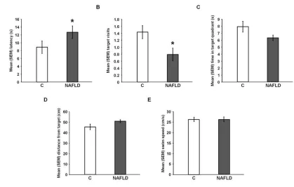
Retention of the training was tested 48 h after the last training day. Each rat was
placed in the pool facing the wall in a randomly determined non-platform quadrant and
allowed to swim for 20 s. The platform was not present, and memory measures during
the probe test included: 1) latency to cross the target (previous platform location), 2)
time spent in the target quadrant, 3) number of target approaches and 4) average
prox-imity to the target. Swim speed was also measured. All trials were recorded and
ana-lyzed using TopScan software (CleverSys, Reston, VA).
Tissue collection and assays
Forty-eight hours after water maze testing, rats were fasted for 4 h, anesthetized
with isoflurane (5% in 95% O2) and then euthanized by decapitation. Brains were
re-moved, and in one subset of rats (NAFLD: n = 6; control: n = 6), the hippocampus was
extracted and frozen on dry ice for measures of IGF-1 and BDNF. In the other group
(NAFLD: n = 7; control: n = 7), hippocampal tissue was collected and sliced for
meas-ures of hippocampal insulin signaling (see below). After decapitation, trunk blood from
all rats was collected in heparinized tubes and centrifuged to obtain plasma for
meas-ures of IGF-1, TGs, and glucose. Livers from all rats were removed, weighed, and flash
frozen for measures of lipids and IGF-1. All tissue was stored at -80ºC until assays were
Folch method (Folch, Lees et al. 1957), and a subset of liver samples were sectioned
and stained with oil red-O and hematoxylin for assessment of lipid droplets (Ackerman,
Oron-Herman et al. 2005).
Following the manufacturer’s recommended protocols, enzyme-linked
immu-nosorbent assays (ELISAs) were used to measure IGF-1 (R&D Systems, Minneapolis,
MN) and BDNF (Promega, Madison, WI). In addition, a protein assay (BioRad,
Hercu-les, CA) was used to measure the total protein concentration present in the samples.
Plasma samples were assayed for TGs (Sigma, St. Louis, MO) using a colorimetric
as-say and spectrophotometry. Glucose was measured using an Accu-Chek glucose meter
(Roche, Indianapolis, IN).
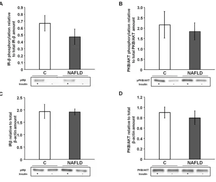
Hippocampal insulin signaling
Preparation of homogenates
The solutions used for the preparation of hippocampal slices and homogenates
were as described by Mielke and colleagues (Mielke, Taghibiglou et al. 2005). Each
brain was dissected in chilled artificial cerebrospinal fluid (aCSF). The hippocampus
was then sectioned into 12 to 16 400 µm transverse slices, which were suspended on
mesh in oxygenated aCSF (5% CO2/95%O2; 33 – 34ºC). After a 60 min post-slicing
re-covery period, half the slices were placed in aCSF plus insulin (100 nM) for 10 min, and
the other half remained in aCSF. Slices from both conditions were then manually
ho-mogenized over ice in separate tubes containing 5 mL of lysis buffer. The homogenates
were centrifuged at 1000 x g for 10 min at 4ºC, and a detergent-compatible protein
as-say (BioRad, Hercules, CA) was used to measure the protein concentration present in
Electrophoresis and immunoblotting
To measure the effect of NAFLD on insulin-stimulated phosphorylation of IR-β
and PKB/AKT, the protein samples were separated electrophoretically on 10%
SDS-polyacrylamide gels and transferred to polyvinylidene fluoride (PVDF) membranes. All
membranes were blocked in 5% non-fat milk blocker (NMB)/tris-buffered saline with
Tween-20 (TBS-T) and probed with primary antibodies against phospho-IR-β (mouse
monoclonal directed against Tyr1150 and Tyr1151; 1:500; Santa Cruz Biotechnology,
Santa Cruz, CA) and phospho-PKB/AKT (rabbit monoclonal directed against Ser473;
1:6,000; Cell Signaling Technology, Danvers, MA) followed by the appropriate
horserad-ish peroxidase (HRP) linked secondary antibody (goat anti-mouse or goat anti-rabbit;
1:20,000). All antibody solutions were prepared in 5% NMB/TBS-T. The membranes
were stripped and re-probed for total IR-β (rabbit polyclonal; 1:5,000; Santa Cruz
Bio-technology, Santa Cruz, CA), PKB/AKT (rabbit monoclonal; 1:6,000; Cell Signaling
Technology, Danvers, MA), and β-actin (rabbit polyclonal; 1:50,000; Novus Biologicals,
Littleton, CO). Membranes were then incubated in chemiluminescent HRP substrate
(Millipore, Billerica, MA) to activate the secondary antibody. Densitometry was
per-formed using the FluorChem 8800 Imaging System (Alpha Innotech, Santa Clara, CA).
Data analysis
The data were stored and analyzed using Microsoft Excel, Version 5.0 and
Sta-tistical Package for the Social Sciences (SPSS), Version 18.0. A Student’s t-test was
performed to determine whether there were differences between the means of the
con-trol and NAFLD rats for acquisition, time spent in the target quadrant, average proximity
and BDNF concentrations, and IR-β and PKB/AKT levels. Trials to criterion, latency to
cross the target, the number of target approaches, and swim speed were not normally
distributed (Kolmogorov- Smirnov test). As a result, a Mann-Whitney U-test was used to
analyze these measures. A repeated measures analysis of variance was performed to
determine whether there were differences in body mass across time between the
con-trol and NAFLD rats. Differences among groups were considered statistically significant
if p < 0.05.
2.4 Results
Chronic high fructose consumption increased liver mass and lipid content
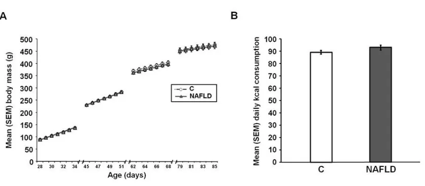
Body mass across the experiment [F (1, 38) = 0.05, p = 0.83; Figure 2.1A] and
average kcal consumption [t(38) = -1.63, p = 0.11; Figure 2.1B] did not differ between
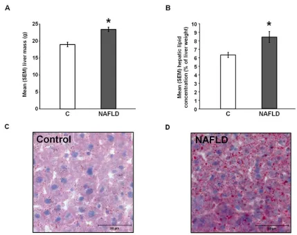
groups. Liver mass [t(38) = -4.65, p = 0.00; Figure 2.2A] and hepatic lipid concentrations
[t(37) = -3.49, p = 0.00; Figure 2.2B] were significantly higher in the NAFLD rats than in
control rats. In addition, visual inspection of oil red-O staining suggests that lipid
drop-lets were larger and more numerous in the NAFLD rats [Figures 2.2C and 2.2D].
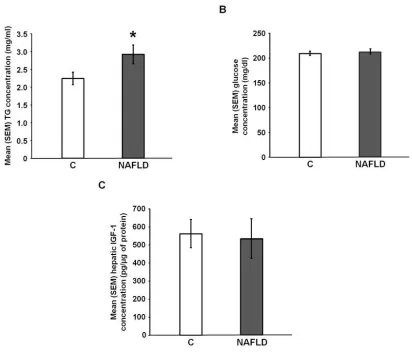
NAFLD significantly increased circulating TGs [t(38) = -2.07, p = 0.05; Figure 2.3A], but
did not significantly affect plasma glucose concentrations [t(32) = -0.45, p = 0.66; Figure
2.3B] or hepatic IGF-1 concentrations [t(10) = 0.20, p = 0.84; Figure 2.3C].
NAFLD impaired retention performance in a spatial water maze
NAFLD did not impair acquisition of the spatial water maze task. Decreases in
the latency to reach the platform across trials were comparable across groups [all p >
0.05; Figure 2.4]; moreover, the number of trials to criterion did not differ between
NAFLD impaired performance on the memory test. NAFLD rats displayed
signifi-cantly longer latencies to reach the target on the retention test [U(38) = 279.00, p =
0.03; Figure 2.5A] and made significantly fewer target approaches [U(38) = 121.00, p =
0.03; Figure 2.5B] than did control rats. In addition, NAFLD rats tended to spend less
time in the target quadrant [t(38) = 1.80, p = 0.07; Figure 2.5C] and tended to swim
far-ther away from the target [t(38) = -1.78, p = 0.07; Figure 2.5D] than did control rats.
Swim speed did not differ significantly between the two groups on the retention test
[U(38) = 202.00, p = 0.95; Figure 2.5E].
NAFLD altered neither hippocampal insulin signaling nor growth hormone levels
NAFLD did not significantly affect IR-β phosphorylation [t(9) = 1.09, p = 0.30;
Figure 2.6A] or PKB/AKT phosphorylation [t(9) = 0.41, p = 0.69; Figure 2.6B]. In
addi-tion, the total amount of IR-β [t(9) = 0.59, p = 0.57; Figure 2.6C] and PKB/AKT protein
[t(9) = 1.55, p = 0.69; Figure 2.6D] was not significantly different between control and
NAFLD rats. NAFLD also did not significantly affect hippocampal BDNF [t(10) = 1.16, p
= 0.27; Figure 2.7A] or IGF-1 concentrations [t(10) = -1.19, p = 0.26; Figure 2.7B].
2.5 Discussion
In this study, we replicated the finding that rats fed a high fructose diet displayed
higher plasma TG concentrations, larger livers, and more hepatic lipids than animals fed
a control diet. Of importance, the present results indicate that NAFLD impairs
hippo-campal-dependent memory. Although rats fed the high-fructose diet required the same
number of trials to learn the location of the submerged platform in the spatial water
maze task, they did not remember this location as well as control rats 48 h later. To be