0095-1137/07/$08.00
⫹
0
doi:10.1128/JCM.01155-07
Copyright © 2007, American Society for Microbiology. All Rights Reserved.
Evaluation of a Novel PCR-Based Assay for Detection and
Identification of
Chlamydia trachomatis
Serovars in
Cervical Specimens
䌤
Koen Quint,
1Carolina Porras,
2Mahboobeh Safaeian,
3* Paula Gonza
´lez,
2Allan Hildesheim,
3Wim Quint,
1Leen-Jan van Doorn,
1Sandra Silva,
2Willem Melchers,
5Mark Schiffman,
3Ana Cecilia Rodrı´guez,
2,3Sholom Wacholder,
3Enrique Freer,
4Bernal Cortes,
2and Rolando Herrero
2for the
Costa Rican Vaccine Trial Group
DDL Diagnostic Laboratory, Voorburg, The Netherlands
1; Proyecto Epidemiolo
´gico Guanacaste, Fundacio
´n INCIENSA, San Jose´,
Costa Rica
2; Division of Cancer Epidemiology and Genetics, National Cancer Institute, Bethesda, Maryland
3; Centro de
Investigacio
´n Estructuras Microsco
´picas, Universidad de Costa Rica, San Jose´, Costa Rica
4; and Department of
Medical Microbiology, Medical Centre, Radboud University Nijmegen, Nijmegen, The Netherlands
5Received 8 June 2007/Returned for modification 20 August 2007/Accepted 13 October 2007
The aims of this study were to compare a novel PCR-based
Chlamydia trachomatis
detection and genotyping
(Ct-DT) assay with the FDA-approved, commercially available
C
.
trachomatis
detection Hybrid Capture 2
(HC2) assay and to investigate the
C
.
trachomatis
serovar distribution among young women in a rural Costa
Rican study population. A total of 5,828 sexually active women participating in a community-based trial in
Costa Rica were tested for
C
.
trachomatis
by HC2. A sample of 1,229 specimens consisting of 100% HC2
C
.
trachomatis
-positive specimens (
n
ⴝ
827) and a random sample of 8% HC2
C
.
trachomatis
-negative specimens
(
n
ⴝ
402) were tested with the Ct-DT assay. Agreement between the two assays was determined by the
unweighted kappa statistic. Discrepant specimens were tested with a second commercially available test
(COBAS TaqMan). The Ct-DT-positive specimens were further analyzed with the Ct-DT genotyping step to
investigate the distribution of 14 different
C
.
trachomatis
serovars (A, B/Ba, C, D/Da, E, F, G/Ga, H, I/Ia, J, K,
L1, L2/L2a, and L3). After accounting for the sampling fraction selected for Ct-DT testing, crude agreement
with the HC2 assay was 98% and the kappa was 0.92 (95% confidence interval [CI], 0.89 to 0.97). The 33
discordant samples that were further analyzed with the COBAS TaqMan test showed better agreement with the
Ct-DT assay (31/33,
P
< 0.001). Among the 806 Ct-DT-positive samples, serovar E was the most common
serovar (31%), followed by serovars F and D (both 21%) and serovar I (15%). In conclusion, the novel Ct-DT
assay permits reliable detection and identification of
C
.
trachomatis
serovars.
Chlamydia trachomatis
is the most prevalent sexually
trans-mitted bacterial pathogen. Annually, an estimated 90 million
new cases occur worldwide (32). Up to 50 and 75% of the
urogenital
C
.
trachomatis
infections in men and women,
re-spectively, remain asymptomatic. Infections can persist for
months and, if untreated, can lead to severe reproductive
prob-lems such as pelvic inflammatory disease in women (10, 23, 31)
and epididymitis in men (2). Furthermore,
C
.
trachomatis
infec-tion facilitates the transmission of human immunodeficiency virus
(6), and some studies have also suggested a potential role for
C
.
trachomatis
as a cofactor in cervical cancer among human
papil-lomavirus (HPV)-positive women (18, 26, 27).
Strains of
C
.
trachomatis
have traditionally been classified
into serovars based on reactivity with specific monoclonal
an-tibodies after culture of the strain (14, 21, 30). In contrast, the
current routine laboratory diagnosis of
C
.
trachomatis
infection
often involves molecular methods such as PCR or nucleic acid
hybridization assays. In general, these assays aim at detection
of the cryptic plasmid (COBAS TaqMan and COBAS
Ampli-cor; Roche Diagnostics) or rRNA (Pace 2 System; Gen-Probe
Corporation). The Hybrid Capture 2 (HC2)
C
.
trachomatis
detection method is a non-PCR nucleic acid hybridization
as-say that is based on detection of the
omp1
gene, as well as the
cryptic plasmid. While these commercial assays are highly
ef-fective for detection of
C
.
trachomatis
infection, they do not
permit identification of the different serovars.
Nonetheless, the different serovars of
C
.
trachomatis
display
diverse clinical manifestations that merit consideration. When
identification of specific
C
.
trachomatis
serovars is desired, they
can be identified on the basis of nucleotide sequence
differ-ences in the
ompA
gene, which encodes the major outer
mem-brane protein. Phylogenetic analysis of
ompA
divides strains
into three major serogroups and many serovars, including
se-rogroup B (comprising serovars B/Ba, D/Da, E, L1, and L2/
L2a), serogroup C (comprising serovars A, C, H, I/Ia, J, K, and
L3), and intermediate serogroup I (comprising serovars F
and G/Ga) (3, 30, 33).
Serovars A and B, certain serovar Ba strains, and serovar C
are commonly associated with the ocular disease trachoma
(17). While serovars B and C are rarely found in the urogenital
tract (19), serovars Ba (specifically, the genovariant A7), D/Da,
* Corresponding author. Mailing address: Division of Cancer
Epi-demiology and Genetics, Hormonal and Reproductive EpiEpi-demiology
Branch, National Cancer Institute, 6120 Executive Boulevard, Suite
550, Rockville, MD 20852. Phone: (301) 594-2934. Fax: (301)
402-0916. E-mail: [email protected].
䌤
Published ahead of print on 24 October 2007.
3986
on May 16, 2020 by guest
http://jcm.asm.org/
E, H, I/Ia, J, K, F, and G/Ga are common in the urogenital
tract and can sometimes be detected in the respiratory tracts of
infants because of transmission during delivery (4). Serovars
D/Da and E are the most commonly recovered types. Serovars
L1, L2/L2a, and L3 are associated with a specific and distinct
condition, lymphogranuloma venereum (16).
A large epidemiologic investigation was initiated to
investi-gate
C
.
trachomatis
serovar distribution among women in
re-lationship to several characteristics such as cervical
inflamma-tion, parity, and cervical neoplasia in a province of Costa Rica
that has a traditionally high risk of cervical cancer (9). We
considered the novel PCR-based
C
.
trachomatis
Detection and
genoTyping (Ct-DT) assay as a promising candidate approach
that combines sensitive detection of
C
.
trachomatis
infection by
a multiplex broad-spectrum PCR with microtiter plate
hybrid-ization (DNA enzyme immunoassay [DEIA]) with
C
.
tracho-matis
typing based on reverse hybridization assay (RHA) (25).
Before we applied the assay to the large epidemiologic
anal-yses, the new Ct-DT assay was validated by comparing the
assay to an FDA-approved, commercially available
C
.
tracho-matis
detection assay (HC2; Digene, Gaithersburg, MD). This
report describes the evaluation of the Ct-DT assay and also
reports our findings on
C
.
trachomatis
serovar distribution in
the Costa Rican study population.
MATERIALS AND METHODS
Study population and specimen collection.Cervical specimens were collected from women participating in the enrolment visit of a community-based double-blind randomized clinical trial investigating the efficacy of an HPV type 16/18 vaccine to prevent cervical intraepithelial neoplasia grade 2 or 3 or cancer. Women were identified through a door-to-door population census conducted in the Province of Guanacaste and adjacent Puntarenas in Costa Rica. Eligible participants were women 18 to 25 years old living in Guanacaste and adjacent Puntarenas who were in good general health, had no history of chronic condi-tions that required treatment, were willing to use a birth control method for a period covering the 6-month vaccination phase, and lived in the study area with no plans of imminent departure from the study area. Recruitment began in June 2004 and ended in December 2005. A total of 7,466 women, approximately 30% of the census, fulfilled the inclusion criteria and were enrolled to receive the candidate vaccine against HPV type 16/18 or hepatitis A vaccine. All study protocols were reviewed and approved by the National Cancer Institute (NCI) and Costa Rican Institutional Review Boards.
At enrolment, women provided written informed consent, and prior to ran-domization, a questionnaire that inquired about demographics, sexual activity, contraceptive use, reproductive history, cigarette use, and family history of can-cers was administered.
At enrolment, a pelvic exam was performed on all consenting, sexually expe-rienced women, during which exfoliated cervical cells were collected with a Cervex brush (Rovers Medical Devices BV, Oss, The Netherlands). The cells were placed in 20 ml of liquid cytology medium (PreservCyt; Cytyc Corporation, Marlborough, MA) and kept at room temperature. At the cytology laboratory, two 0.5-ml aliquots were drawn and stored in liquid nitrogen. After aliquoting, liquid-based cytology samples (ThinPrep; Cytyc Corporation) were prepared. The remaining PreservCyt sample was used to detectC.trachomatis,Neisseria gonorrhoeae, and HPV by the HC2 assay (Digene Corporation). Women who tested positive forC.trachomatisand/orN.gonorrhoeaewere offered counseling and treatment with a single 1-g dose of azithromycin for them and their partners free of charge as part of the trial protocol. Treatment efficacy was confirmed at the next study visit with additionalC.trachomatisandN. gonorrhoeaetests.
C.trachomatisdetection and genotyping.As mentioned, the HC2C. tracho-matisassay was performed on all available specimens. One of the two 0.5-ml aliquots was used to testC.trachomatisby the Ct-DT system (Labo Biomedical Products, Rijswijk, The Netherlands). The Ct-DT assay was performed on all HC2C.trachomatis-positive specimens and 8% of the randomly selected HC2C.
trachomatis-negative samples. To further investigate discrepant HC2 and Ct-DT results, a second commercially availableC.trachomatisassay (COBAS TaqMan; Roche Diagnostics) was chosen for adjudication. Neither the HC2 nor the
COBAS TaqMan assay provides information onC.trachomatisserovars. COBAS TaqMan was used to test all (n⫽33) samples with discrepant assay results, Ct-DT borderline positive samples (n⫽7), 10% of the randomly selected samples that were negative by both HC2C.trachomatisand Ct-DT (n⫽39), and 10% of the randomly selected samples that were positive by both the HC2C.
trachomatisand Ct-DT assays (n⫽79).
C.trachomatistesting by HC2.Commercially available assays were performed according to the manufacturers’ instructions.
HC2 (Digene Corporation, Gaithersburg, MD) is an FDA-approved nucleic acid hybridization assay with signal amplification that combines antibody capture of target DNA with RNA probe hybrids and chemiluminescence for signal detection. The number of relative light units measured compared to a positive standard (RLU/PC) is used to discriminate against positive and negative sam-ples.
C.trachomatistesting by HC2 was done in a tiered approach. All samples were first subjected to a combined HC2C.trachomatis-N.gonorrhoeaeDNA test according to the manufacturer’s instructions. Briefly, theC.trachomatis-N. gon-orrhoeaetest contains a probe cocktail mixture that is complementary to a total of approximately 39,300 bp (4%) of theC.trachomatisgenomic DNA, 7,500 bp (100%) of theC.trachomatiscryptic plasmid, 9,700 bp (0.5%) of theN. gonor-rhoeaegenomic DNA, and 4,200 bp (100%) of theN.gonorrhoeaecryptic plas-mid. This is a qualitative test, and a positive result indicates the presence ofC.
trachomatisand/orN.gonorrhoeaeDNA in the specimen. A negative test indi-cates the absence of bothC.trachomatisandN.gonorrhoeaeor the presence of DNA at levels below the detection limit of the assay.
Subsequently, all samples positive by the combinedC.trachomatis-N. gonor-rhoeaeDNA test were further tested by the HC2C.trachomatis-specific assay, which uses the same hybrid capture technology to confirm the presence ofC.
trachomatisin each sample. As in all HC2 assays, target DNA is hybridized with a cRNA probe cocktail and the RNA-DNA hybrids are captured onto an anti-body-coated microplate well. Immobilized hybrids are incubated with alkaline phosphatase-conjugated antibodies and detected with a chemiluminescent sub-strate. Specimens with RLU/PC cutoff value ratios ofⱖ1 are considered positive forC.trachomatisDNA. The two HC2 assays were performed in the laboratory at the University of Costa Rica in San Jose on residual PreservCyt samples.
C.trachomatisdetection and genotyping by Ct-DT.The new Ct-DT assay is a commercially available assay and was performed according to the manufacturer’s (Laboratory Biomedical Products BV, Rijswijk, The Netherlands) instructions. The Ct-DT assay comprises an amplification step, followed by a detection step by a DEIA. All samples positive at the amplification and detection steps were subsequently genotyped by RHA by using the same PCR product generated for the detection assay.
DNA isolation method.Total DNA was isolated from 200-l PreservCyt ali-quots with the MagNA Pure LC instrument (Roche Diagnostics, Almere, The Netherlands) and the Total DNA isolation kit (Roche Diagnostics). DNA was eluted in 100l of water. Each DNA extraction run contained positive and negative controls to monitor the DNA isolation procedure.
Ct-DT amplification step.The first part of the Ct-DT assay comprises a PCR amplification step which uses aC.trachomatismultiplex broad-spectrum PCR primer mixture with multiple forward and reverse primers targeting theomp1
VD2 region and the cryptic plasmid. TheC.trachomatisPCR primer set is designed to amplify all of the known serovars available in GenBank. Briefly, this multiplex primer set amplifies a fragment of 241 bp from the cryptic plasmid and a fragment of 160/157 bp from variable region 2 of theomp1gene (25).
Briefly the PCR mixture consist of 10l of isolated DNA, 2.5 mM MgCL2, 1⫻
GeneAmp PCR buffer II, 1.5 U AmpliTaq Gold DNA polymerase (Applied Biosystems, Foster City, CA), 0.2 mM deoxynucleoside triphosphates (Invitro-gen, Carlsbad, CA), and 15 pmol of each primer (Eurogentec S.A., Seraing, Belgium) in a total volume of 50l. The standard PCR program is a 9-min preheating step at 94°C, followed by 40 cycles of amplification (30 s at 94°C, 45 s at 55°C, and 45 s at 72°C) and a final 5-min elongation step at 72°C.
Ct-DT detection step.The specific detection ofC.trachomatisamplicons is performed by a DEIA. PCR products were hybridized to a mixture of conserved probes for the cryptic plasmid, as well as theomp1gene (25), to permit detection of all of the genotypes available in GenBank. Reverse primers contain a biotin label at the 5⬘end, enabling capture of the reverse strand onto streptavidin-coated microtiter plates. Captured amplicons are denatured by alkaline treat-ment, and the captured strand is detected by a defined cocktail of digoxigenin-labeled probes. After washing, the hybrids are detected by an enzymatic reaction, resulting in a colored product. The DEIA provides an optical density (OD) value at 450 nm. Each DEIA run contained separate titrated positive, borderline-positive, and negative controls and a PCR-positive control containing isolated DNA from a cell culture of serovar E. Samples yielding OD values equal to or
on May 16, 2020 by guest
http://jcm.asm.org/
higher than the borderline are considered positive. The borderline positive samples areC.trachomatis-positive samples that contained the lowest amount of
C.trachomatisamplicon detectable with the Ct-DT assay. The OD value of the borderline range depends on the titrated borderline internal control and differs for every single run.
Ct-DT genotyping step.TheC.trachomatis-genotyping step from the Ct-DT assay is based on the RHA technology. Biotin-labeled PCR amplicons from Ct-DT-positive samples at the detection step were subsequently used to differ-entiate between theC.trachomatisserovars by RHA on a nitrocellulose strip, which contains probes for the cryptic plasmid, for the three differentC. tracho-matisserogroups (B, C, and I), and for the 19 serovars (A, B/Ba, C, D/Da, E, F, G/Ga, H, I/Ia, J, K, L1, L2/L2a, and L3). One extra probe is added to detect a genovariant of serovar J that otherwise remains undetected. Each RHA run contains a negative and a positive control (serovar E). The Ct-DT genotyping step was performed according to the manufacturer’s instructions.
C.trachomatisdetection by the COBAS TaqMan assay.The COBAS TaqMan
C.trachomatistest (Roche Molecular Systems, Branchburg, NJ) was performed according to the manufacturer’s instructions in the laboratory for Medical Mi-crobiology, Radboud University Nijmegen, Nijmegen, The Netherlands, on re-sidual isolated DNA samples. This test was run on the Roche Diagnostics COBAS TaqMan 48 Analyzer, an instrument that offers automated real-time PCR amplification and detection in a closed system. Systematic internal controls and built-in cross-contamination prevention mechanisms further enhance the reliability of the results obtained.
Statistical analysis.The primary outcome wasC.trachomatisprevalence de-termined by the HC2 assay compared with the Ct-DT amplification and detec-tion step (PCR and DEIA).
Agreement between the two assays was determined by unweighted kappa () statistics and 95% confidence intervals (CI), which test percent agreement be-yond that expected by chance alone. Generally, the kappa values are interpreted as follows:ⱕ0.20, poor agreement; 0.21 to 0.40, fair agreement; 0.41 to 0.60, moderate agreement; 0.61 to 0.80, good agreement;⬎0.80, excellent agreement. In order to maximize information on serovars efficiently, all HC2C. tracho-matis-positive samples were tested by the novel Ct-DT assay while we chose an 8% random sample of HC2C.trachomatis-negative samples to test by Ct-DT. The percentage of HC2C.trachomatis-negative samples was decided by labora-tory resources. In testing a random sample of HC2 C.trachomatis-negative samples, we presumed that most of the HC2-negative specimens would be neg-ative by Ct-DT as well and that we could validly estimate the characteristics of the remaining 92% of the HC2C.trachomatis-negative samples that were not tested with Ct-DT by reference to the randomly selected 8% subset. Accordingly, to report population level data, we extrapolated the observed data to the entire set of HC2C.trachomatis-negative samples.
In the extrapolated data, the nonparametric test for matched data (McNemar’s
2test) was used to determine whether the proportion of samples classified as
positive by the HC2 assay and negative by the Ct-DT assay was equal to the proportion of samples classified as negative by the HC2 assay and positive by the Ct-DT assay. We report the results based on observed data that were tested by both assays, and additionally, we report results based on the extrapolated data adjusting for the sampling fraction.
RESULTS
Pelvic examinations in which exfoliated cervical cells were
obtained were performed on 5,871 sexually experienced
women enrolled in the trial. HC2 testing was performed for
5,828 specimens; 42 samples were excluded because of an
insufficient specimen volume. The median age of the women
was 21 years (interquartile range, 19 to 23 years), and the
median age at first sexual intercourse was 17 years
(interquar-tile range, 15 to 18 years). Forty-four percent (
n
⫽
2,564) of
the women were single, 52% (
n
⫽
3,059) were married, and
3.4% (
n
⫽
199) reported being separated, divorced, or
wid-owed. Only 16% of the women reported ever having smoked,
and 42% of the women reported only one lifetime sexual
partner.
C
.
trachomatis
detection.
C
.
trachomatis
prevalence in the
cohort, as measured by the HC2 assay, was 14% (827/5,828;
95% CI, 13.3 to 15.1). All 827 HC2
C
.
trachomatis
-positive
samples and an additional 402 randomly selected, HC2
C
.
trachomatis
-negative samples were further analyzed by the
Ct-DT system. Data for the 1,229 paired observations are
shown in Table 1. Of the 1,229 observations, the Ct-DT assay
detected 806 as
C
.
trachomatis
positive and 423 as
C
.
tracho-matis
negative.
In the paired data without extrapolation (
n
⫽
1,229), the
crude agreement between the two assays for
C
.
trachomatis
detection was 97% and the kappa was 0.94 (95% CI, 0.92 to
0.96). It would not be proper to calculate the population-wide
percentage of
C
.
trachomatis
-positive specimens, or the relative
positivity of the two assays with McNemar’s test, on the basis of
the data in Table 1 because only a small percentage of the HC2
C
.
trachomatis
-negative samples were tested. Hence, we
ex-trapolated the observed HC2
C
.
trachomatis
-negative samples
to the entire study population. By maintaining the fraction of
samples positive by Ct-DT, we could confidently estimate that
if all of the specimens had been tested, the crude agreement
between the assays would be 98% and the kappa would
de-crease slightly to 0.92 (95% CI, 0.89 to 0.97). The 27 (0.5%)
samples that were HC2
C
.
trachomatis
positive but negative by
the Ct-DT assay were not changed when we reconstituted the
whole Costa Rican study population, but these 27 would be
properly compared to an extrapolated estimate of 75 (1.5%)
samples that would be
C
.
trachomatis
positive by Ct-DT assay
but HC2
C
.
trachomatis
negative (McNemar’s
P
⫽
0.03).
We also examined the agreement between the HC2 and
Ct-DT assays, stratified by age, lifetime number of sexual
part-ners, and marital status. Kappa values remained stable across
age, number of sexual partners, and marital status (data not
shown).
Discrepant analysis by the Roche COBAS TaqMan
C
.
[image:3.585.302.541.80.212.2]
tra-chomatis
test.
One hundred fifty-eight samples were further
tested by the commercially available
C
.
trachomatis
assay
(COBAS TaqMan) to understand discrepant assay findings,
which included 33 observed discordant samples, 7
border-line positive by Ct-DT, 79 randomly selected HC2 and
Ct-DT assay-positive samples, and 39 randomly selected
HC2 and Ct-DT-negative samples. Table 2 shows the results
of this analysis. All 79 samples positive by both the HC2 and
Ct-DT assays were also positive by COBAS TaqMan, while
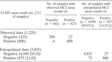
TABLE 1. Agreement between HC2 and Ct-DT assays
aCt-DT assay result (no.关%兴 of samples)
No. of samples with observed HC2 assay
results of:
No. of samples with extrapolated HC2
assay results of:
Negative (n⫽402)
Positive (n⫽827)
Negative (n⫽4,998
关85.8%兴)
Positive (n⫽827
关14.2%兴)
Observed data (1,229)
Negative (423)
396
27
Positive (806)
6
800
Extrapolated data (5,825)
Negative (4,950
关
85.0
兴
)
4,923
27
Positive (875
关
15.0
兴
)
75
800
aThe agreement between observed assay results was 97% (unweighted
kappa⫽0.94关CI, 0.92 to 0.96兴). The agreement between extrapolated assay results was 98% (unweighted kappa⫽0.92关CI, 0.89 to 0.97兴). McNemar’s
P⫽0.03.
on May 16, 2020 by guest
http://jcm.asm.org/
only 1 of the 39 samples negative by both the HC2 and Ct-DT
assays was positive by COBAS TaqMan. Among the 27 HC2
C
.
trachomatis
-positive, Ct-DT-negative samples, 25 were
nega-tive by COBAS TaqMan. All five HC2
C
.
trachomatis
-negative,
Ct-DT-positive samples were positive by COBAS (Table 2).
There were eight Ct-DT borderline-positive samples; six were
confirmed as positive by COBAS TaqMan and seven were
confirmed as positive by the HC2 assay. Thus, among the 33
specimens where the HC2 and Ct-DT assays disagreed, COBAS
TaqMan was significantly more likely to agree with the Ct-DT
assay (31/33,
P
⬍
0.001) than with the HC2 assay.
C
.
trachomatis
serogroups and serovars.
Results of serovar
distribution among the 806 women positive by Ct-DT are
pre-sented in Table 3. There were 12 specimens that were positive
for the
C
.
trachomatis
plasmid but negative by the genotyping
assay for the 14 serovars that were tested. Among the 806
infections that were Ct-DT plasmid positive, serovar E was the
most common (31%), followed by serovars F and D (both
21%) and serovar I (15%). Sequence analysis of the 10 serovar
B/Ba samples revealed that all belonged to the Ba serotype. As
expected, serovars A, C, L1, L2/L2a, and L3 were not identified
in these samples from the genital tract. The HC2 probe
iden-tified 97 to 100% of all of the
C
.
trachomatis
serovars as
positive. There were 14 samples in which multiple
C
.
tracho-matis
serovars were identified, without any evident pattern
(Table 3).
DISCUSSION
We compared the performance of the novel Ct-DT assay
with that of the FDA-approved commercially available HC2
C
.
trachomatis
assay and found excellent agreement between the
two assays for detection of
C
.
trachomatis
.
A particular strength of this interassay comparison is that it
was performed on a large, community-based sample of women.
The study population was large and representative of young
women participating in a clinical trial evaluating an HPV type
16 and 18 prophylactic vaccine. The women were initially
screened for
C
.
trachomatis
infection with the HC2
C
.
tracho-matis
-
N
.
gonorrhoeae
assay. When a sample was positive, the
presence of
C
.
trachomatis
specifically was determined by an
additional HC2 test for
C
.
trachomatis
only. Subsequently, all
HC2
C
.
trachomatis
-positive samples, as well as 8% of the
randomly selected HC2
C
.
trachomatis
-negative samples, were
analyzed with the novel Ct-DT assay. Both assays were
per-formed on samples derived from one specimen from the same
participant, collected in the same medium, thus reducing the
chance that differences could be attributed to procedural
vari-ations. We presented observed and extrapolated agreement
adjusting for the sampling fraction.
In 94% (31/33) of the discordant HC2 and Ct-DT assay results,
the findings of the Ct-DT assay were corroborated by the Roche
COBAS TaqMan assay, which is based on amplification of the
cryptic plasmid and has a sensitivity of approximately 20 copies
per PCR. In particular, all five Ct-DT-positive, HC2-negative
results were also positive by the Roche COBAS TaqMan assay,
suggesting that they were true positive results missed by the
HC2 assay.
The majority of the discordant results were HC2
C
.
tracho-matis
positive and Ct-DT negative. When we used the HC2
[image:4.585.300.541.89.426.2]RLU value as a proxy for the burden of infection, we observed
that 25 of these 27 specimens yielded lower bacterial DNA
quantities (values in the lowest RLU quartile between 1 and
27) than those positive by both assays (data not shown).
HC2-positive, Ct-DT-negative findings may be explained either by
sampling error related to a low
C
.
trachomatis
load, by false
negativity of the Ct-DT assay, or by false positivity of the HC2
assay. False positivity of the HC2 assay could theoretically be
explained by a lack of specificity and cross-hybridization of the
very long RNA probes used in the HC2 assay with DNA from
TABLE 2. COBAS TaqMan
C.
trachomatis
assay findings for
adjudication of discrepant HC2 and Ct-DT assay results
HC2 result Ct DT result
No. of samples
testeda
No. of samples with Roche COBAS
result of:
Negative Positive
Negative
Negative
39
38
1
Positive
Positive
79
0
79
Negative
Positive
5
0
5
Negative
Borderline positive
1
1
0
Positive
Borderline positive
7
1
6
Positive
Negative
27
25
2
aThe total number of samples was 158.
TABLE 3. Ct-DT genotyping (serogroup and serovar) assay and
HC2 test results for 806
C.
trachomatis-positive samples
Test group and serogroup(s)
No. of samples
tested
Serovar(s) No. (%) of samples
% HC2 positive
Single infection
B
417
E
245 (30.4)
100.0
D/Da
162 (20.1)
100.0
B/Ba
10 (1.2)
100.0
C
191
I/Ia
121 (15.0)
100.0
J
30 (3.7)
96.7
H
28 (3.5)
100.0
K
12 (1.5)
100.0
I
172
F
165 (20.5)
98.8
G/Ga
7 (0.8)
100.0
Multiple infections
B
1
D/Da
⫹
E
1 (0.1)
100.0
B
⫹
C
6
D/Da
⫹
H
2 (0.2)
100.0
D/Da
⫹
I/Ia
1 (0.1)
100.0
D/Da
⫹
J
2 (0.2)
100.0
E
⫹
I/Ia
1 (0.1)
100.0
B
⫹
I
3
D/Da
⫹
F
1 (0.1)
100.0
E
⫹
F
2 (0.2)
100.0
I
1
F
⫹
G
1 (0.1)
100.0
I
⫹
C
3
F
⫹
I/Ia
1 (0.1)
100.0
F
⫹
J
1 (0.1)
100.0
F
⫹
K
1 (0.1)
100.0
Plasmid only
12 (1.5)
75.0
on May 16, 2020 by guest
http://jcm.asm.org/
[image:4.585.43.284.89.196.2]other microorganisms present in the specimen. In the HC2
C
.
trachomatis
test, 4% of the genome (containing stretches of
conserved and repeat sequences) is covered by a range of RNA
probes which might lead to false positive reactivity.
In addition to detecting the
C
.
trachomatis
plasmid, the
Ct-DT assay has been designed to determine the
C
.
trachoma-tis
serogroup and serovar. Although the prevalence of
C
.
tra-chomatis
serovars described in the literature shows
consider-able variation, our results are similar to those of other studies
in Australia (32), Alabama, (8), Sweden (13), Taiwan (11),
China (7), and Korea (15). As in those populations, serovar E
also was the most prevalent type in the present study cohort. In
contrast, in Uganda (25), Thailand (1), and Colombia (20),
serovar E was far less prevalent. This variation may be due to
the use of different methods of
C
.
trachomatis
serovar
identi-fication (1) or to the use of a small isolated study population in
Uganda (20, 25). However, there is laboratory evidence
sug-gesting that serovar E can outcompete other strains for
nutri-ents and growth factors, which would suggest a reason for a
rapid expansion of serovar E if, in fact, an expansion can be
inferred from cross-sectional data (12). To address such
hy-potheses, we are currently testing 1,000 age-stratified
speci-mens collected more than 10 years ago in the same region of
Costa Rica from women more than 18 years old.
In the present study, 10 of the 806 Ct-DT-positive samples
tested contained serovar B/Ba. The Ct-DT genotyping test
cannot discriminate between these two strains. Serotype B
strains have only once been found in urogenital samples (B/
Alpha-95) (19). The occurrence of serovar B in urogenital
samples, although observed, is a very rare event. However, the
Ba serotypes have regularly been found at both urogenital and
ocular sites (19). Further sequence analysis of the amplicons
from the 10 samples in this study revealed the presence of
serovar Ba in all cases.
Among the 806 Ct-DT-positive samples that were further
analyzed with the Ct-DT genotyping assay, 12 showed only
positivity for the cryptic plasmid, with no serogroups or
sero-vars identified. The absence of amplification might be due to
either sequence variation in the primer target region of the
omp1
gene or inhibition of
omp1
PCR amplification. An
alter-native explanation for the absence of
omp1
amplification might
be related to the occurrence of sampling variation. This may
play an important role when samples contain very low numbers
of target molecules or when heterogeneous clinical materials,
such as cervical swabs or biopsy specimens, are used. Every
C
.
trachomatis
bacterium contains 10 to 20 copies of the cryptic
plasmid but only one single
omp1
gene. If the isolated DNA
contains only a very low concentration of bacterial DNA, it is
possible that only cryptic-plasmid DNA but no genomic DNA
would be included in the PCR mixture and that only the
plas-mid PCR yields positive results. Similar findings of a positive
plasmid-directed PCR and a negative
omp1
PCR have been
reported for genital and conjunctival samples (22, 28).
Both the Ct-DT and HC2
C
.
trachomatis
assays detect the
cryptic plasmid, as well as genomic sequences of
C
.
trachoma-tis
, in a combined assay. In our study, analysis with the Ct-DT
genotyping assay revealed positivity for the cryptic plasmid in
all
C
.
trachomatis
-positive samples; however, we could not
con-firm the existence of plasmid-negative strains as previously
reported (5, 24, 29).
In conclusion, we observed excellent agreement between the
HC2 and Ct-DT systems for detection of
C
.
trachomatis
. In
addition, the Ct-DT assay permits easy and rapid identification
of the serotype in the positive samples, with the same
am-plimers as used for detection of
C
.
trachomatis
positivity. The
present study shows that the Ct-DT assay is a robust method
which can be used for studying the natural history of
C
.
tra-chomatis
and
C
.
trachomatis
serovars. We will now use this
assay to perform large-scale epidemiologic investigations
re-garding
C
.
trachomatis
serovars and inflammation, parity, and
risk of cervical cancer among HPV-infected women.
ACKNOWLEDGMENTS
The Costa Rican Vaccine Trial is a long-standing collaboration
between investigators in Costa Rica and NCI. The trial is sponsored
and funded by NCI and conducted in agreement with the Ministry of
Health of Costa Rica. Vaccine was provided for our trial by GSK
Biologicals under a Clinical Trials Agreement with NCI. GSK also
provided support for aspects of the trial associated with regulatory
submission needs of the company under FDA BB-IND 7920.
NCI and Costa Rica investigators make final editorial decisions on
this presentation and subsequent publications.
Additional names and affiliations of investigators in the Costa Rican
Vaccine Trial group are as follows: Proyecto Epidemiolo
´gico
Guana-caste, Fundacio
´n INCIENSA, San Jose
´, Costa Rica, Mario Alfaro
(cytologist), Manuel Barrantes (field supervisor), M. Concepcion
Bratti (coinvestigator), Fernando Ca
´rdenas (general field supervisor),
Bernal Corte
´s (specimen and repository manager), Albert Espinoza
(head, coding and data entry), Yenory Estrada (pharmacist), Paula
Gonzalez (coinvestigator), Diego Guille
´n (pathologist), Rolando
Her-rero (co-principal investigator), Silvia E. Jimenez (trial coordinator),
Jorge Morales (colposcopist), Lidia Ana Morera (head study nurse),
Elmer Pe
´rez (field supervisor), Carolina Porras (coinvestigator), Ana
Cecilia Rodriguez (coinvestigator), and Maricela Villegas (clinic
M.D.); University of Costa Rica, San Jose
´, Costa Rica, Enrique Freer
(director, HPV Diagnostics Laboratory), Jose Bonilla (head, HPV
Immunology Laboratory), Sandra Silva (head technician, HPV
Diag-nostics Laboratory), Ivannia Atmella (immunology technician), and
Margarita Ramı´rez (immunology technician); U.S. NCI, Bethesda,
MD, Pamala Gahr (trial coordinator), Allan Hildesheim (co-principal
investigator and NCI co-project officer), Douglas R. Lowy (HPV
vi-rologist), Mark Schiffman (medical monitor and NCI co-project
offi-cer), John T. Schiller (HPV virologist), Mark Sherman (quality control
pathologist), Diane Solomon (medical monitor and quality control
pathologist), Sholom Wacholder (statistician); Science Applications
International Corporation, NCI—Frederick, Frederick, MD, Ligia
Pinto (head, HPV Immunology Laboratory) and Alfonso
Garcia-Pi-neres (scientist, HPV Immunology Laboratory); Women’s and Infants’
Hospital, Providence, RI, Claire Eklund (quality control cytology) and
Martha Hutchinson (quality control cytology); Delft Diagnostics
Lab-oratory, Delft, The Netherlands, Wim Quint (HPV DNA testing) and
Leen-Jan van Doorn (HPV DNA testing).
REFERENCES
1.Bandea, C. I., K. Kubota, T. M. Brown, P. H. Kilmarx, V. Bhullar, S. Yanpaisarn, P. Chaisilwattana, W. Siriwasin, and C. M. Black.2001. Typing of Chlamydia trachomatis strains from urine samples by amplification and sequencing the major outer membrane protein gene (omp1). Sex. Transm. Infect.77:419–422.
2.Berger, R. E., E. R. Alexander, G. D. Monda, J. Ansell, G. McCormick, and K. K. Holmes.1978. Chlamydia trachomatis as a cause of acute “idiopathic” epididymitis. N. Engl. J. Med.298:301–304.
3.Caldwell, H. D., and J. Schachter.1982. Antigenic analysis of the major outer membrane protein ofChlamydiaspp. Infect. Immun.35:1024–1031. 4.Darville, T.2005. Chlamydia trachomatis infections in neonates and young
children. Semin. Pediatr. Infect. Dis.16:235–244.
5.Farencena, A., M. Comanducci, M. Donati, G. Ratti, and R. Cevenini.1997. Characterization of a new isolate ofChlamydia trachomatiswhich lacks the common plasmid and has properties of biovar trachoma. Infect. Immun.
65:2965–2969.
6.Fleming, D. T., and J. N. Wasserheit.1999. From epidemiological synergy to
on May 16, 2020 by guest
http://jcm.asm.org/
public health policy and practice: the contribution of other sexually trans-mitted diseases to sexual transmission of HIV infection. Sex. Transm. Infect.
75:3–17.
7.Gao, X., X. S. Chen, Y. P. Yin, M. Y. Zhong, M. Q. Shi, W. H. Wei, Q. Chen, R. W. Peeling, and D. Mabey.2007. Distribution study ofChlamydia tracho-matisserovars among high-risk women in China performed using PCR-restriction fragment length polymorphism genotyping. J. Clin. Microbiol.
45:1185–1189.
8.Geisler, W. M., R. J. Suchland, and W. E. Stamm.2006. Association of Chlamydia trachomatis serovar Ia infection with black race in a sexually transmitted diseases clinic patient population in Birmingham, Alabama. Sex. Transm. Dis.33:621–624.
9.Herrero, R., M. H. Schiffman, C. Bratti, A. Hildesheim, I. Balmaceda, M. E. Sherman, M. Greenberg, F. Cardenas, V. Gomez, K. Helgesen, J. Morales, M. Hutchinson, L. Mango, M. Alfaro, N. W. Potischman, S. Wacholder, C. Swanson, and L. A. Brinton.1997. Design and methods of a population-based natural history study of cervical neoplasia in a rural province of Costa Rica: the Guanacaste Project. Rev. Panam. Salud Publica1:362–375. 10.Hillis, S. D., L. M. Owens, P. A. Marchbanks, L. F. Amsterdam, and W. R.
Mac Kenzie.1997. Recurrent chlamydial infections increase the risks of hospitalization for ectopic pregnancy and pelvic inflammatory disease. Am. J. Obstet. Gynecol.176:103–107.
11.Hsu, M. C., P. Y. Tsai, K. T. Chen, L. H. Li, C. C. Chiang, J. J. Tsai, L. Y. Ke, H. Y. Chen, and S. Y. Li.2006. Genotyping of Chlamydia trachomatis from clinical specimens in Taiwan. J. Med. Microbiol.55:301–308. 12.Jones, R. B., J. A. Williams, and B. van der Pol.1998. Competitive growth of
serovars E and F combined in mixed tissue culture infections, p. 523–526.In
R. S. Stephens, G. I. Byrne, G. Christiansen, I. N. Clarke, J. T. Grayston, R. G. Rank, G. L. Ridgway, P. Saikku, J. Schachter, and W. E. Stamm (ed.), Chlamydial infections. Proceedings of the Ninth International Symposium on Human Chlamydial Infection. International Chlamydia Symposium, Napa, CA.
13.Jurstrand, M., L. Falk, H. Fredlund, M. Lindberg, P. Olcen, S. Andersson, K. Persson, J. Albert, and A. Backman.2001. Characterization ofChlamydia trachomatis omp1genotypes among sexually transmitted disease patients in Sweden. J. Clin. Microbiol.39:3915–3919.
14.Kuo, C. C., S. P. Wang, K. K. Holmes, and J. T. Grayston.1983. Immuno-types ofChlamydia trachomatisisolates in Seattle, Washington. Infect. Im-mun.41:865–868.
15.Lee, G., J. Park, B. Kim, S. A. Kim, C. K. Yoo, and W. K. Seong.2006. OmpA genotyping of Chlamydia trachomatis from Korean female sex workers. J. Infect.52:451–454.
16.Mabey, D., and R. W. Peeling.2002. Lymphogranuloma venereum. Sex. Transm. Infect.78:90–92.
17.Mabey, D., and A. Solomon.2003. The effect of antibiotic treatment on active trachoma and ocular Chlamydia trachomatis infection. Expert Rev. Anti-infect. Ther.1:209–216.
18.Madeleine, M. M., T. Anttila, S. M. Schwartz, P. Saikku, M. Leinonen, J. J. Carter, M. Wurscher, L. G. Johnson, D. A. Galloway, and J. R. Daling.2007. Risk of cervical cancer associated with Chlamydia trachomatis antibodies by histology, HPV type and HPV cofactors. Int. J. Cancer120:650–655. 19.Millman, K., C. M. Black, R. E. Johnson, W. E. Stamm, R. B. Jones, E. W.
Hook, D. H. Martin, G. Bolan, S. Tavare, and D. Dean.2004.
Population-based genetic and evolutionary analysis ofChlamydia trachomatisurogenital strain variation in the United States. J. Bacteriol.186:2457–2465. 20.Molano, M., C. J. Meijer, S. A. Morre, R. Pol, and A. J. van den Brule.2004.
Combination of PCR targeting the VD2 of omp1and reverse line blot analysis for typing of urogenitalChlamydia trachomatisserovars in cervical scrape specimens. J. Clin. Microbiol.42:2935–2939.
21.Ossewaarde, J. M., M. Rieffe, A. de Vries, R. P. Derksen-Nawrocki, H. J. Hooft, G. J. van Doornum, and A. M. van Loon.1994. Comparison of two panels of monoclonal antibodies for determination ofChlamydia trachomatis
serovars. J. Clin. Microbiol.32:2968–2974.
22.Pedersen, L. N., H. O. Kjaer, J. K. Moller, T. F. Orntoft, and L. Ostergaard.
2000. High-resolution genotyping ofChlamydia trachomatisfrom recurrent urogenital infections. J. Clin. Microbiol.38:3068–3071.
23.Peipert, J. F.2003. Clinical practice. Genital chlamydial infections. N. Engl. J. Med.349:2424–2430.
24.Peterson, E. M., B. A. Markoff, J. Schachter, and L. M. de la Maza.1990. The 7.5-kb plasmid present in Chlamydia trachomatis is not essential for the growth of this microorganism. Plasmid23:144–148.
25.Quint, K. D., L. J. van Doorn, B. Kleter, M. de Koning, H. A. van den Munckhof, S. A. Morre, B. ter Harmsel, E. Weiderpass, G. Harbers, W. J. Melchers, and W. G. V. Quint.2007. A highly sensitive, multiplex broad-spectrum PCR-DNA-enzyme immunoassay and reverse hybridization assay for rapid detection and identification of Chlamydia trachomatis serovars. J. Mol. Diagn.9:631–638.
26.Silins, I., W. Ryd, A. Strand, G. Wadell, S. Tornberg, B. G. Hansson, X. Wang, L. Arnheim, V. Dahl, D. Bremell, K. Persson, J. Dillner, and E. Rylander.2005. Chlamydia trachomatis infection and persistence of human papillomavirus. Int. J. Cancer116:110–115.
27.Smith, J. S., C. Bosetti, N. Munoz, R. Herrero, F. X. Bosch, J. Eluf-Neto, C. J. Meijer, A. J. van den Brule, S. Franceschi, and R. W. Peeling.2004. Chlamydia trachomatis and invasive cervical cancer: a pooled analysis of the IARC multicentric case-control study. Int. J. Cancer111:431–439. 28.Stevens, M. P., S. N. Tabrizi, R. Muller, V. Krause, and S. M. Garland.2004.
Characterization ofChlamydia trachomatis omp1genotypes detected in eye swab samples from remote Australian communities. J. Clin. Microbiol.42:
2501–2507.
29.Stothard, D. R., J. A. Williams, B. Van Der Pol, and R. B. Jones.1998. Identification of aChlamydia trachomatisserovar E urogenital isolate which lacks the cryptic plasmid. Infect. Immun.66:6010–6013.
30.Wang, S. P., C. C. Kuo, R. C. Barnes, R. S. Stephens, and J. T. Grayston.
1985. Immunotyping of Chlamydia trachomatis with monoclonal antibodies. J. Infect. Dis.152:791–800.
31.Westrom, L., R. Joesoef, G. Reynolds, A. Hagdu, and S. E. Thompson.1992. Pelvic inflammatory disease and fertility. A cohort study of 1,844 women with laparoscopically verified disease and 657 control women with normal lapa-roscopic results. Sex. Transm. Dis.19:185–192.
32.Xiong, L., F. Kong, H. Zhou, and G. L. Gilbert.2006. Use of PCR and reverse line blot hybridization assay for rapid simultaneous detection and serovar identification ofChlamydia trachomatis. J. Clin. Microbiol.44:1413– 1418.
33.Yuan, Y., Y. X. Zhang, N. G. Watkins, and H. D. Caldwell.1989. Nucleotide and deduced amino acid sequences for the four variable domains of the major outer membrane proteins of the 15Chlamydia trachomatisserovars. Infect. Immun.57:1040–1049.

![2 [(4 Chlorobenzyl)carbonylmethyl]benzoic acid](data:image/gif;base64,R0lGODlhAQABAIAAAP///wAAACH5BAEAAAAALAAAAAABAAEAAAICRAEAOw==)