Copyright © 2002, American Society for Microbiology. All Rights Reserved.
Identification of the
Brucella melitensis
Vaccine Strain Rev.1 in
Animals and Humans in Israel by PCR Analysis of the
Pst
I Site Polymorphism of Its
omp2
Gene
Svetlana Bardenstein,
1Michal Mandelboim,
1† Thomas A. Ficht,
2Miriam Baum,
1and Menachem Banai
1*
Department of Bacteriology, Kimron Veterinary Institute, Bet Dagan 50250, Israel,1and
Department of Veterinary Pathobiology, College of Veterinary Medicine, Texas A&M University, College Station, Texas 77843-44672
Received 24 September 2001/Returned for modification 16 December 2001/Accepted 27 January 2002
Adverse effects of strain persistence and secretion in milk have been encountered with theBrucella melitensis
vaccine strain Rev.1. Field isolates obtained from vaccinated animals and from a human resembled the vaccine strain Rev.1 by conventional bacteriological tests. The lack of a specific molecular marker that could specif-ically characterize the commercial vaccine strain prevented confirmation of the homology of the Rev.1-like field isolates to the vaccine strain. The composition of theomp2locus from two gene copies with differences in their
PstI restriction endonuclease sites was used to establish an epidemiologic fingerprint for theomp2gene in the Rev.1 vaccine strain. Primers designed to amplify DNA sequences that overlap thePstI site revealed a single 282-bp DNA band common to all Brucellaspp. Agarose gel electrophoresis of the PstI digests of the PCR products from strains 16M and the vaccine strain Rev.1 revealed a distinctive profile that included three bands: one band for the intact 282-bp fragment amplified fromomp2aand two bands resulting from the digestion of the amplifiedomp2bgene fragment, 238- and 44-bp DNA fragments, respectively. Amplified fragments of 37 Rev.1-like isolates, including 2 human isolates, also exhibited this pattern. In contrast, DNA digests of all other Israeli field isolates, including atypical B. melitensisbiotype 1 and representatives of the biotype 2 and 3 isolates, produced two bands of 238 and 44 bp, respectively, corresponding with the digestion of bothomp2a
andomp2bgenes. This method facilitates identification of the Rev.1 vaccine strain in both animals and humans in Israel.
Brucella melitensiscauses a worldwide zoonosis. It is one of the major causes of abortions in sheep and goats, and the organism is secreted in the milk of infected animals. People contract the disease by direct contact with contaminated fetal membranes or, more commonly, as a result of the consumption of contaminated unpasteurized milk and cheese products. The organisms are small, gram-negative coccobacilli that grow in the host as nonobligatory intracellular pathogens of the reticu-loendothelial system. Derivatives of tetracycline are often used to treat human infection, while a slaughter policy is recom-mended for livestock in order to eradicate the disease (11).
In the late 1950s, Elberg developed a live attenuated vac-cine, strain Rev.1 (12, 13). It was shown that although the vaccine prevented abortion, it did not provide protection against infection. Bosseray demonstrated that different lots of Rev.1 vaccines showed variable immunogenicity in mice ac-cording to their level of virulence (5, 6). This study emphasized the instability of the biological properties of the vaccine strain, stressing the need for stringent control of vaccine production (7).
The problems in the laboratory were reflected by similar
results in the field. In South Africa, selection of a few smooth colonies as seed stock led to production of a virulent vaccine strain which infected sheep and caused human disease (22, 27, 28).
Throughout the last decade Israel maintained a conservative vaccination policy in which only young female livestock be-tween the ages of 2 to 6 months were vaccinated, using a full dose by the subcutaneous route. Nevertheless, retrospective data demonstrated that the Rev.1 vaccine led to the adverse effects of strain persistence in the vaccinated animals and was occasionally spread horizontally (4, 30). Moreover, in two cases it was shown that the vaccine strain caused human infection, demonstrating the zoonotic hazards of its virulence. The fact that vaccination did not always protect the animals in the field and the several cases of secretion of the field strain in milk had proven the inefficacy of the whole vaccination program.
International agencies, in their assistance to developing countries, suggested that national control programs should de-pend on a whole-flock vaccination scheme as a cost-effective method until the prevalence of the disease was reduced. Only then should test and slaughter be implemented to eradicate the disease (10). There was opposition to this proposal (4) due to the adverse effects encountered in the field and the public concerns of possible risks to the human population following secretion of the vaccine strain in milk. The absence of a specific molecular marker that could be associated with the identity of the commercial vaccine strain prevented those opposing the
* Corresponding author. Mailing address: Department of Bacteriol-ogy, Kimron Veterinary Institute, P.O. Box 12, Bet Dagan 50250, Israel. Phone: 972-3-9681698. Fax: 972-3-9681753. E-mail: mbana_vs @netvision.net.il.
† Present address: Faculty of Life Science, Bar-Ilan University, Ra-mat Gan 52900, Israel.
1475
on May 15, 2020 by guest
http://jcm.asm.org/
vaccination program from linking Rev.1-like field isolates to the vaccine strain. By the same token, those in favor of the program could ignore the risks posed by the Rev.1 vaccine, using the same rationale.
Data presented below provide evidence supporting the
ex-istence of aPstI site polymorphism in theBrucella omp2 gene.
ThePstI digestion pattern of PCR-amplified fragments from
the Israeli isolates was different from that of the prototype
strain 16M. Curiously, thePstI digestion profile of theomp2
amplified fragments from the vaccine strain Rev.1 resembled that of strain 16M, allowing the differentiation of Rev.1
iso-lates fromB. melitensisfield strains in Israel. This achievement
could specifically address the potential misdiagnosis of the
atypicalB. melitensisbiovar 1 strains as Rev.1 isolates due to
similarities in their phenotypic susceptibility to penicillin.
MATERIALS AND METHODS
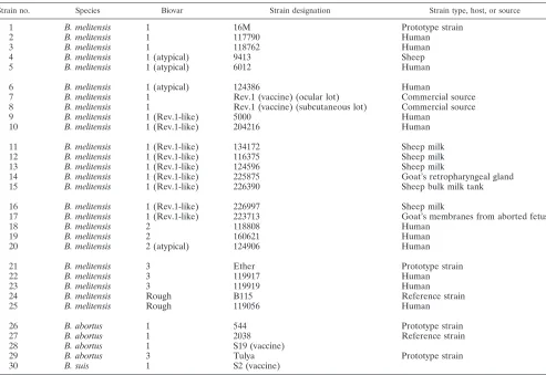
Bacterial strains.The bacterial strains used in this study (Table 1) were from the collection maintained in the Israeli reference laboratory.Brucellafield strains were isolated by conventional methods (1) from milk samples, aborted fetuses, and placentas. Human isolates were obtained from local medical laboratories. Strain 16M was obtained from J.-M. Verger, Institut National de la Recherche Agronomique, Nouzilly, France. Strain biotyping was performed by standard methods (1).Escherichia colistrain NCTC 9001 was used as a control (for non-BrucellaDNA).Rhizobium melilotistrains 158M and 161M were obtained from D. Kishinevsky, andAgrobacterium radiobacterstrains At5/r and At96216 were obtained from D. Zutra, The Volcani Center, Beit Dagan, Israel. Bacterial
strains other thanBrucellawere obtained from the Department of Bacteriology, The Kimron Veterinary Institute, Beit Dagan, Israel, or from the Department of Clinical Microbiology, the Hebrew University-Hadassah Medical School, Jerusa-lem, Israel.
Bacterial DNA.To prepare chromosomal DNA, bacterial cells were harvested in saline and incubated for 20 min at 4°C with lysozyme (4 mg/ml). Sodium dodecyl sulfate (0.5% [wt/vol]) and proteinase K (200 mg/ml) were then added, and incubation was continued at 37°C for 1 h. The cell lysate was extracted once with phenol-isoamyl alcohol (1:1:49) and once with chloroform-isoamyl alcohol (1:24). The purified DNA was alcohol precipitated, resuspended in TE (50 mM Tris-HCl, 1 mM EDTA [pH 8.0]), and stored at 4°C.
PCR and oligonucleotide primers.TheBrucella omp2 gene was used as target DNA. The forward 5⬘primer (p1 [TGGAGGTCAGAAATGAAC]) and reverse 3⬘primer (p2 [GAGTGCGAAACGAGCGC]) of anomp2gene segment were obtained from National Biosciences, Inc., Plymouth, Minn.
PCR amplification was performed by the method of Mullis and Faloona (26). A typical reaction mixture contained 50 mM KCl, 1.5 mM MgCl2, 0.1% (wt/vol)
[image:2.587.46.539.82.421.2]Triton X-100, 0.2 mg of bovine serum albumin (fraction IV; Sigma) per ml, and 10 mM Tris-HCl (pH 8.5). Each reaction mixture was supplemented with 100 mM each of the four deoxyribonucleotides, 100 ng of sample DNA, and each oligonucleotide primer. For slide PCR, sample DNA was replaced with brucellae that were laid on a glass slide, air dried, and fixed by being heated. A sample of the dried cells was then collected with a needle, the needle was dipped in 10l of double-distilled water, and 2l from this solution was put in the PCR mixture. Otherwise, sample DNA (2l from a bacterial cell suspension in double-distilled water boiled at 100°C for 20 min) was used. Reactions were initiated by adding 0.5 U ofTaqpolymerase (Appligene, Illkirch, France). The reaction mixture was covered with 15l of mineral oil (Sigma) to prevent evaporation. Following hot start treatment at 95°C for 3 min, PCR was performed with an Eppendorf Thermocycler (Eppendorf, Hamburg, Germany) as follows: 35 cycles of PCR, with 1 cycle consisting of 20 s at 95°C for DNA denaturation, 1 min at 50°C for TABLE 1. Brucellastrains used in this study
Strain no. Species Biovar Strain designation Strain type, host, or source
1 B. melitensis 1 16M Prototype strain
2 B. melitensis 1 117790 Human
3 B. melitensis 1 118762 Human
4 B. melitensis 1 (atypical) 9413 Sheep
5 B. melitensis 1 (atypical) 6012 Human
6 B. melitensis 1 (atypical) 124386 Human
7 B. melitensis 1 Rev.1 (vaccine) (ocular lot) Commercial source
8 B. melitensis 1 Rev.1 (vaccine) (subcutaneous lot) Commercial source
9 B. melitensis 1 (Rev.1-like) 5000 Human
10 B. melitensis 1 (Rev.1-like) 204216 Human
11 B. melitensis 1 (Rev.1-like) 134172 Sheep milk
12 B. melitensis 1 (Rev.1-like) 116375 Sheep milk
13 B. melitensis 1 (Rev.1-like) 124596 Sheep milk
14 B. melitensis 1 (Rev.1-like) 225875 Goat’s retropharyngeal gland
15 B. melitensis 1 (Rev.1-like) 226390 Sheep bulk milk tank
16 B. melitensis 1 (Rev.1-like) 226997 Sheep milk
17 B. melitensis 1 (Rev.1-like) 223713 Goat’s membranes from aborted fetus
18 B. melitensis 2 118808 Human
19 B. melitensis 2 160621 Human
20 B. melitensis 2 (atypical) 124906 Human
21 B. melitensis 3 Ether Prototype strain
22 B. melitensis 3 119917 Human
23 B. melitensis 3 119919 Human
24 B. melitensis Rough B115 Reference strain
25 B. melitensis Rough 119056 Human
26 B. abortus 1 544 Prototype strain
27 B. abortus 1 2038 Reference strain
28 B. abortus 1 S19 (vaccine)
29 B. abortus 3 Tulya Prototype strain
30 B. suis 1 S2 (vaccine)
on May 15, 2020 by guest
http://jcm.asm.org/
DNA annealing, and 1 min at 72°C for polymerase-mediated primer extension. The last cycle included incubation of the sample at 72°C for 7 min. Ten micro-liters of the amplified product was analyzed by electrophoresis in 1.5% agarose gels in TEA buffer (20 mM Tris-acetate, 1 mM EDTA [pH 8.0]).
DNA digestion.Restriction enzymes were used according to the manufactur-er’s instructions (Boehringer GmbH, Mannheim, Germany). The digested DNA was separated by electrophoresis on either 1.5% agarose gels (wt/vol in Tris-acetate buffer) or 10% polyacrylamide gels (wt/vol in Tris-borate buffer). DNA fragments were visualized by staining with ethidium bromide (1.5g/ml).
RESULTS
Validation of the method with prototype strains.The PCR
was first performed to test specificity by comparing Brucella
species DNAs to the DNAs from several other bacteria,
in-cluding the taxonomically closely related Agrobacterium and
Rhizobiumstrains (29). A single band with the expected size of
282 bp (19) was obtained only whenBrucellaDNA was used as
a template. All other bacterial strains and a water sample failed to produce an amplified fragment (data not shown).
The PCR test was studied with Brucella prototype strains
from two of the three important species, namely,B. melitensis
andB. abortus(field and vaccine strains). In addition,B. suis
vaccine strain S2 was included as a representative of this spe-cies. As shown in Fig. 1, the DNAs from all the strains
pro-duced a 282-bp band. As shown in Fig. 2,PstI digestions of the
amplified fragments from the strains gave different bands on
agarose gels.B. abortus(lanes 1, 3, 4, and 5),B. suisS2 (lane
8), andB. melitensisbiovar 3 (lane 12) digests revealed a single
band, a 238-bp band. Other possible smaller fragments are not
shown on the gel. In comparison,PstI digestion ofB. melitensis
strain 16M (prototype forB. melitensisbiovar 1 virulent strain
[lane 9]) and strain Rev.1 (a vaccine strain from two different producers administered by a subcutaneous and an ocular route [lanes 10 and 11, respectively]) amplified DNAs, included two visible bands: a large band, which was uncut DNA (lanes 6 and
8), and another band, a 238-bpB. abortusfragment. Other
pos-sible smaller bands are not shown on the gel. ThePstI digestion
pattern of the amplified fragment obtained fromB. melitensis
strain B115 (a stable rough form obtained from an infected
goat in Malta in the early 1970s) (lane 2) was similar to those for strains 16M and Rev.1 (lanes 9, 10, and 11, respectively).
The digestion profiles of the same DNAs were analyzed by polyacrylamide gel electrophoresis, as shown in Fig. 3. The purpose of this analysis was to identify possible smaller frag-ments that were not shown by agarose gel electrophoresis. As can be seen in Fig. 3, besides the 282- and 238-bp DNA bands, all samples produced an additional identical smaller fragment which was calculated to be 44 bp. It was calculated that the two smaller bands together were the same size as the uncut DNA,
confirming the expectedPstI restriction pattern forB. abortus
biovar 1 (19).
PstI digestion profile of theomp2 gene amplified fragment from atypical B. melitensis biotype 1 strains. Atypical B. melitensis biotype 1 isolates from a human source (isolate 6012) and sheep source (isolate 9413) were described
[image:3.587.48.280.75.208.2]previ-FIG. 1. Agarose gel electrophoresis of PCR-amplifiedomp2gene fragments fromBrucellaprototype strains. The figure shows a single band, a 282-bp DNA fragment. Lanes: M, molecular size ladder (in base pairs); 1,B. abortusstrain 2308; 2,B. melitensisstrain B115; 3,B. abortusstrain Tulya; 4,B. abortusstrain 544; 5,B. abortusvaccine S19; 6,B. melitensisstrain 16M; 7,B. suisvaccine S2; 8,B. melitensisvaccine strain Rev.1 (subcutaneous lot); 9,B. melitensisvaccine strain Rev.1 (ocular lot); 10,B. melitensisstrain Ether.
FIG. 2. Agarose gel electrophoresis ofPstI digests of amplified
omp2 gene fragments from Brucella prototype strains. The figure shows the uncut 282-bp DNA and the larger,PstI-digested DNA frag-ments. The smaller, 44-bp DNA fragment is not shown. Lanes: 1,B. abortusstrain 2308; 2, B. melitensisstrain B115; 3,B. abortusstrain Tulya; 4,B. abortusstrain 544; 5,B. abortusvaccine S19; 6,B. melitensis
strain 16M (uncut); M, molecular size ladder (in base pairs); 7,B. abortusstrain 544 (uncut); 8,B. suisS2; 9,B. melitensisstrain 16M; 10,
B. melitensisvaccine strain Rev.1 (subcutaneous lot); 11,B. melitensis
[image:3.587.304.537.76.203.2]vaccine strain Rev.1 (ocular lot); 12,B. melitensisstrain Ether.
FIG. 3. Polyacrylamide gel electrophoresis ofPstI digests of ampli-fiedomp2gene fragments fromBrucellaprototype strains. The figure shows the three DNA fragments, the uncut DNA and the twoPst I-digested DNA fragments, with sizes of 282, 238, and 44 bp, respec-tively. Lanes: M, molecular size ladder (in base pairs); 1,B. melitensis
strain 16M (uncut); 2,B. melitensisstrain 16M; 3,B. melitensisvaccine strain Rev.1 (subcutaneous lot); 4,B. abortusvaccine S19; 5,B. abortus
strain 544; 6,B. abortusstrain 2308; 7,B. melitensisstrain Ether.
on May 15, 2020 by guest
http://jcm.asm.org/
[image:3.587.322.516.521.652.2]ously (2). We compared thePstI digestion profiles of theomp2
gene amplified fragment obtained from these strains and those
obtained from prototype strains. AllBrucellastrains produced
identical amplified 282-bp fragments (data not shown). Figure 4 depicts a polyacrylamide gel analysis of the digestion profile
of these DNAs. As can be seen,PstI digestion of the two B.
melitensisatypical strains (lanes 2 and 3) produced a uniform
pattern identical to that obtained forB. abortusstrains S19 and
544 (lanes 6 and 7) and to that ofB. melitensisbiotype 3 (lane
8). The PstI digestions of the amplified fragments from the
commercial vaccine strain Rev.1 and the prototype strain 16M yielded a different pattern (lanes 4 and 5, respectively). The gel also shows the smaller, 44-bp band common to all digests.
We then analyzed the digestion profile of theomp2DNA
fragment of selected human isolates as shown in Fig. 5. The samples included biovar 1 strains (lanes 1, 2, 3, 4, and 14), a rough isolate (lane 7), atypical biotype 1 (lane 15) and atypical biotype 2 (lane 16) strains, and two Rev.1 human isolates (lanes 10 and 12). In addition, we included the prototype strain 16M (lane 11) and two uncut DNAs from strain Rev.1 (lane 8) and strain 16M (lane 9). Besides strain Rev.1 (lanes 5 and 6),
Rev.1-like isolates (lanes 10 and 12), andB. melitensisstrain
16M (lane 11), all other isolates had similar digestion profiles
(as shown in Fig. 2 for B. abortus strains) and B. melitensis
biotype 3 strain Ether.
PstI digestion profile of theomp2 gene amplified fragment fromB. melitensisRev.1-like isolates.A total of 23B. melitensis
biotype 1 field isolates, 5B. melitensisatypical biovar 1 field
isolates (2), 13B. melitensisbiotype 2 field isolates including 3
atypical strains, and 13 B. melitensis biotype 3 field isolates
were studied. All exhibited the pattern shown forB. abortus
strains (data not shown). In contrast, 37 field isolates with Rev.1-like bacteriological characteristics produced an identical
PstI digestion pattern (as did the commercial vaccine, strain
Rev.1). A representative selection is shown in Fig. 6, lanes 1, 2, 3, 8, 9, 10, and 11, compared to lanes 4 and 7, respectively.
DISCUSSION
The PCR technique has increasingly been used as a
supple-mentary method inBrucella diagnosis (8, 14, 15, 21, 23, 25).
Recently, a molecular biotyping approach has been proposed on the basis of restriction endonuclease polymorphism in the genes encoding the major 25- and 36-kDa outer membrane
proteins ofBrucella(9, 17, 20). Theomp2gene exists as a locus
of two nearly homologous repeated copies that differ slightly
amongBrucellaspp. and biotypes (18). We used this
[image:4.587.64.260.74.209.2]informa-tion to design specific primers that amplify a 282-bp fragment
FIG. 4. Polyacrylamide gel electrophoresis ofPstI digests of ampli-fiedomp2gene fragments from atypicalB.melitensisbiotype 1 isolates from humans and animals compared to prototype strains. The figure shows the three DNA fragments, the uncut DNA and the twoPst I-digested DNA fragments, with sizes of 282, 238, and 44 bp, respec-tively. Lanes: M, molecular size ladder (in base pairs); 1,B. melitensis
strain 6012 (human isolate) (uncut); 2,B. melitensisstrain 6012 (human isolate); 3,B. melitensisstrain 9413 (sheep isolate); 4,B. melitensis
vaccine strain Rev.1 (subcutaneous lot); 5,B. melitensisstrain 16M; 6,
B. abortusvaccine S19; 7,B. abortusstrain 544; 8,B. melitensisstrain Ether.
FIG. 5. Agarose gel electrophoresis ofPstI digests of amplified
omp2gene fragments fromBrucellafield strains. The figure shows the uncut 282-bp DNA and the larger,PstI-digested DNA fragment. The smaller, 44-bp DNA fragment is not shown. Lanes: 1,B. melitensis
biotype 1 strain 117790; 2,B. melitensisbiotype 1 strain 118762; 3,B. melitensis biotype 2 strain 118808; 4,B. melitensis biotype 2 strain 160621; 5,B. melitensisvaccine strain Rev.1 (ocular lot); 6,B. melitensis
vaccine strain Rev.1 (subcutaneous lot); 7,B. melitensisrough strain 119056; 8,B. melitensisvaccine strain Rev.1 (subcutaneous lot) (un-cut); M, molecular size ladder (in base pairs); 9,B. melitensisstrain 16M (uncut); 10,B. melitensisbiotype 1 strain 5000 (human Rev.1-like isolate); 11,B. melitensisstrain 16M; 12,B. melitensisbiotype 1 strain 204215 (sheep Rev.1-like isolate); 13,B. melitensisbiotype 3 strain 119917; 14, B. melitensis biotype 3 strain 119919; 15, B. melitensis
[image:4.587.306.533.74.186.2]biotype 1 atypical strain 124386; 16,B. melitensisbiotype 2 atypical strain 124906.
FIG. 6. Agarose gel electrophoresis ofPstI digests of amplified
omp2gene fragments fromBrucellafield strains that resemble Rev.1 according to biochemical biotyping. The figure shows the uncut 282-bp DNA and the larger,PstI-digested DNA fragment. The smaller, 44-bp DNA fragment is not shown. Lanes: 1,B. melitensisstrain 134172; 2,B. melitensisstrain 116375; 3,B. melitensisstrain 124596; 4,B. melitensis
vaccine strain Rev.1 (subcutaneous lot); 5,B. melitensisvaccine strain Rev.1 (subcutaneous lot) (uncut); M, molecular size ladder (in base pairs); 6,B. melitensisvaccine strain (ocular lot) (uncut); 7,B. meliten-sisvaccine strain Rev.1 (ocular lot); 8,B. melitensisstrain 225877; 9,B. melitensisstrain 226390; 10,B. melitensisstrain 226997; 11,B. melitensis
strain 223713.
on May 15, 2020 by guest
http://jcm.asm.org/
[image:4.587.307.536.503.617.2](Fig. 1), flanking upstream sequences of the 5⬘terminus of the
two genes and expanding downstream of the PstI andKpnI
sites (17). We assumed that the sensitivity of the test would be doubled by selecting duplicated DNA sequences of the two
genes. Moreover, we assumed that because of the existingPstI
site polymorphism between B. melitensis and B. abortus, the
test would distinguish between the two species. According to
the working hypothesis, DNA fragments obtained from B.
melitensis strain 16M should produce three bands, an intact
282-bp fragment from the amplifiedomp2agene that lacks the
PstI site and two smaller fragments of 238 and 44 bp, the
products obtained from digestion of theomp2bamplified
frag-ment (17). In contrast,B. abortusDNA should produce only
the two smaller fragments from both genes, a 238-bp fragment and a 44-bp fragment, respectively (Fig. 2 and 3).
We used this method to study the stability of theomp2gene
among local B. melitensis isolates derived from small
rumi-nants, cattle, and humans and representing the current
Bru-cellapopulation in Israel. Our data confirmed the expected
paradigm forB. melitensisstrains B115, 16M, and the vaccine
strain Rev.1 (Fig. 2, lanes 2, 9, 10, and 11), as well as forB.
abortus(Fig. 2, lanes 3, 4, and 5) andB. suisstrain 2 (Fig. 2,
lane 8). The IsraeliB. melitensis field isolates from the three
biotypes, including the atypical biotype 1 strains, unexpectedly
exhibited thePstI digestion profile which occurs inB. abortus,
i.e., two bands of 238 and 44 bp, respectively (Fig. 4, lane 3, and Fig. 5, lanes 1, 2, 3, 4, 7, 13, 14, 15, and 16; also data not shown).
In a comprehensive study, Meyer has shown that unlikeB.
abortus, B. melitensislacked plasticity in the features charac-terized by the conventional biotyping methods (24). Our data
indicated that in contrast to these findings,B. melitensis has
undergone genetic diversions in a pattern similar to that
pre-viously shown to occur in otherBrucella spp. From the data,
one could infer that the prevailing Israeli biotype 1 strains have
acquired a newPstI site in theomp2agene (compared to the
sequences established for strain 16M). On the other hand, strains belonging to biotypes 2 and 3 acquired this change earlier, since all isolates demonstrated the same pattern, which
was similar to theB. melitensisbiovar 3 prototype strain Ether
(Fig. 2, lane 12).
Results obtained by Cloeckaert et al. (9) confirmed these
data, showing that B. melitensis isolates were split between
those with a singlePstI site located in the omp2bgene and
those with twoPstI sites, one inomp2aand one inomp2b.
Interestingly, from the list presented by Cloeckaert et al., it can be seen that even Israeli isolates from the 19702 all had
twoPstI sites, one inomp2aand the second inomp2b, similar
to the results presented above for isolates from the later
pe-riod. This suggests that the IsraeliB. melitensisbiotype 1 strains
emerged separately from 16M, a strain that originated in the United States.
It is interesting that recent field isolates in Mexico produced
a digestion profile of the omp2 amplified gene fragments
similar to that of strain 16M and strain Rev.1 (T. A. Ficht, personal communication). Our data and those obtained by Cloeckaert et al. have further shown that the described phe-nomenon applied not only to strain 16M and Rev.1 but also to the rough strain B115 and H38 as well (Fig. 2, lane 2) (9). We
could propose, therefore, that at least two separateB.
meliten-sisbiotype 1 lines have evolved, one represented by strain 16M
and the other represented by the Israeli isolates. A similar conclusion was drawn by Cloeckaert et al. regarding the
ab-sence of aBglII restriction site in theomp2bgenes of the Israeli
isolates (9).
This study included 37 isolates that according to conven-tional bacteriological methods were characterized as vaccine
strain Rev.1. ThePstI digestion pattern of theomp2amplified
gene fragments resembled that of strain 16M, the prototype
strain for virulentB. melitensisbiovar 1, and that of the vaccine
strain Rev.1 (Fig. 2, lanes 9, 10, and 11). In contrast, thePstI
digestion profile of theomp2agene amplified fragments from
all other Israeli isolates, representing the three biotypes, de-picted a reproducible and conserved pattern that was different from that shown for strains 16M and Rev.1 (Fig. 5, lanes 1, 2, 3, 4, 7, 13, 14, 15, and 16). This suggests that a genetic link might be established between the prototype strain 16M and the vaccine strain Rev.1. A few other geographically remote iso-lates may have shared the same ancestral strain.
Human infection with the vaccine strain Rev.1 in South Africa has been reported, following horizontal infection among sheep. Clonal selection of virulent colonies during the prepa-ration of a working seed stock probably led to production of a vaccine lot with undesirable characteristics (22, 27, 28). It is interesting that, in Israel, we also identified a human case of infection with the vaccine strain Rev.1. The owner of an inten-sively managed sheep farm was infected with the Rev.1 vaccine strain 6 months after a series of abortions in ewes and isolation of Rev.1 from the fetal membranes (3). In a report from South Africa, the researchers biotyped the isolates by conventional
Brucellabiotyping methods, and no direct molecular linkage was shown between the field isolates and the commercial Rev.1 vaccine strain. To the best of our knowledge, our report is the first to associate animal and human infection with the vaccine strain Rev.1 based on molecular identification of the strain.
The resemblance of the phenotypic properties of the vaccine strain Rev.1 and the atypical strain characterized in Israel (2) regarding susceptibility to penicillin and dyes has raised the possibility that the atypical strain had originated from a
muta-tion of the vaccine strain. The uniquePstI pattern described for
strain 16M and the vaccine strain Rev.1 has allowed us to
elaborate on this subject by comparing theiromp2gene PstI
digestion patterns. If the atypical strain had originated from a
Rev.1 mutant, itsomp2genePstI digestion pattern should have
matched that of strain Rev.1 and strain 16M. The contrary would be true if it had originated from a field strain mutant.
The similarity between thePstIomp2gene digestion profiles of
the atypical strains and the virulent field isolates (Fig. 4) clearly implied that the latter was the case, lending a mutation in a virulent strain to render it susceptible to penicillin and dyes. The zoonotic competence of the atypical isolates (2) that caused human infection further supported the idea that these strains had originated from a virulent strain.
The results presented in this study have highlighted some of the potential hazards associated with use of the Rev.1 vaccine in national control programs. It has been argued that vaccine quality could be impaired if its production did not adhere to stringent standards (5, 7). Having encountered the adverse effects of the subcutaneous vaccine, we assumed that the com-mercial Rev.1 vaccine supplied to Israel originated from a
on May 15, 2020 by guest
http://jcm.asm.org/
defective seed stock, similar to the events described in South Africa. To overcome these problems, Israel changed the vac-cine source in November 1997, purchasing it from a company that had sustained its seed stock on a true Elberg strain (pas-sage 101, 1970; M. Banai, personal communication). Other expected advantages of the new vaccine were the safety to
adult animals, attributed to the lower dose (108CFU instead of
109CFU), and the method of inoculation as an ocular
prepa-ration (16). Implementation of the new vaccine in whole-flock vaccination, including vaccination of pregnant animals, led to outbreaks of abortions in several intensively managed flocks and isolation of the strain from the milk of the aborting ani-mals. In this study, besides conventional biotyping, we also applied the new PCR method to confirm the Rev.1 identity of
the isolates according to theomp2a PstI digestion profile (Fig.
6). This new technique made it possible to associate a second human case of strain Rev.1 infection in a 15-year-old girl (Fig. 5, lane 12). Since then, the Israeli veterinary services changed the vaccination policy back to the consensus method of vacci-nation of only young females, and no additional problems have been encountered.
ACKNOWLEDGMENTS
This research was supported in part by a grant from the Israeli Ministry of Health in 1993 (“Rapid diagnosis of Human Brucellosis by the PCR”); grant no. TA MOU CA 13-007; U.S.-Israel Cooperative Developmental Research Program, Office of the Science Advisor, U.S. Agency for International Development. The research was completed under the support by research grant award no. 2781-96 from BARD, The United States-Israel Binational Agricultural Research and Devel-opment Fund.
Theomp2gene primers were designed by Gad Frenkel, National Institute for Medical Research, London, United Kingdom.
REFERENCES
1.Alton, G. G., L. M. Jones, R. D. Angus, and J.-M. Verger (ed.).1988. Techniques for the brucellosis laboratory, p. 24–61. Institut National de la Recherche Agronomique, Paris, France.
2.Banai, M., I. Mayer, and A. Cohen.1990. Isolation, identification, and char-acterization in Israel ofBrucella melitensisbiovar 1 atypical strains suscep-tible to dyes and penicillin, indicating the evolution of a new variant. J. Clin. Microbiol.28:1057–1059.
3.Banai, M., G. Hoida, E. Rapoport, O. Zamir, M. Haimovich, E. Katz, I. Mayer, A. Cohen, and M. Davidson.1991. IsBrucella melitensisundergoing evolutionary development or a change in the vaccinal Rev.1 strain?, p. 152. InA. Tumbay, S. Hilmi, and O. Ang (ed.),Brucellaand brucellosis in man and animals. Turkish Microbiological Society publication no. 16. Ege Uni-versity Press, Izmir, Turkey.
4.Banai, M., M. Abramson, I. Mayer, K. Chechik, G. Hoida, O. Zamir, S. Bardenstein, A. Cohen, and M. Davidson.1996. Problems associated with the persistence and possible horizontal transfer ofBrucella melitensisRev.1 vaccine in connection with serological surveillance in Israel, p. 69–76.InB. Garin-Bastugi and A. Benkirane (ed.), FAO/WHO/OIE Round Table on the Use of Rev.1 Vaccine in Small Ruminants and Cattle. Centre National d’Etudes Ve´te´rinaires et Alimentaires, Maison-Alfort, France.
5.Bosseray, N.1985. Quality control of four Rev.1 anti-Brucellavaccines, p. 229–236.InJ. M. Verger and M. Plommet (ed.),Brucella melitensis. Martinus Nijhoff Publishers, Dordrecht, The Netherlands.
6.Bosseray, N.1991.Brucella melitensisRev.1 living attenuated vaccine: sta-bility of markers, residual virulence and immunogenicity in mice. Biologicals
19:355–363.
7.Bosseray, N. 1992. Control methods and thresholds of acceptability for anti-Brucellavaccines. Dev. Biol. Stand.79:121–128.
8.Bricker, B. J., and S. M. Halling.1994. Differentiation ofBrucella abortusbv. 1, 2, and 4,Brucella melitensis,Brucella ovis, andBrucella suisbv. 1 by PCR. J. Clin. Microbiol.32:2660–2666.
9.Cloeckaert, A., J.-M. Verger, M. Grayon, and O. Grepinet.1995. Restriction site polymorphism of the genes encoding the major 25 kDa and 36 kDa outer-membrane proteins ofBrucella. Microbiology141:2111–2121. 10.Elberg, S.1996. Rev.1Brucella melitensisvaccine. Part III, 1981–1995. Vet.
Bull.66:1193–1200.
11.Elberg, S. S. (ed.).1981. A guide to the diagnosis, treatment and prevention of human brucellosis. World Health Organization publication VPH/81.31. World Health Organization, Geneva, Switzerland.
12.Elberg, S. S., and K. Faunce, Jr.1957. Immunization againstBrucella infec-tion. VI. Immunity conferred on goats by a nondependent mutant from a streptomycin-dependent mutant strain ofBrucella melitensis. J. Bacteriol.
73:211–217.
13.Elberg, S. S., and K. F. Meyer.1958. Caprine immunization against brucel-losis. A summary of experiments on the isolation, properties and behaviour of a vaccine strain. Bull. W. H. O.19:711–724.
14.Fekete, A., J. A. Bantle, and S. M. Halling.1992. Detection ofBrucellaby polymerase chain reaction in bovine fetal and maternal tissues. J. Vet. Diagn. Investig.4:79–83.
15.Fekete, A., J. A. Bantle, S. M. Halling, and R. W. Stich.1992. Amplification fragment length polymorphism inBrucellastrains by use of polymerase chain reaction with arbitrary primers. J. Bacteriol.174:7778–7783.
16.Fensterbank, R., P. Pardon, and J. Marly.1982. Comparison between sub-cutaneous and conjunctival route of vaccination with Rev.1 strain againstB. melitensisinfection in ewes. Ann. Rech. Vet.13:185–190.
17.Ficht, T. A., S. W. Bearden, and H. Marquis.1990. Genetic variation at the omp2porin locus of the Brucellae: species-specific markers. Mol. Microbiol.
4:1135–1142.
18.Ficht, T. A., S. W. Bearden, B. A. Sowa, and L. G. Adams.1988. A 36-kilodaltonBrucella abortuscell envelope protein is encoded by repeated sequences closely linked in the genomic DNA. Infect. Immun.56:2036–2046. 19.Ficht, T. A., S. W. Bearden, B. A. Sowa, and L. G. Adams.1989. DNA sequence and expression of the 36-kilodalton outer membrane protein gene ofBrucella abortus. Infect. Immun.57:3281–3291.
20.Ficht, T. A., H. S. Husseinen, and S. W. Bearden.1996. Species-specific sequences at theomp2locus ofBrucellatype strains. Int. J. Sys. Bacteriol.
46:329–331.
21.Guarino, A., L. Serpe, G. Fusco, A. Scaramuzzo, and P. Gallo.2000. Detec-tion ofBrucellaspecies in buffalo whole blood by gene-specific PCR. Vet. Rec.147:632–634.
22.Hunter, P., S. M. Pefanis, C. C. Williamson, W. J. S. Botha, and M. S. Schalkwyk.1989. Horizontal transmission in sheep and delayed clearance in guinea pigs and mice of aBrucella melitensisRev.1 mutant. J. S. Afr. Vet. Assoc.60:92–94.
23.Leal-Klevezas, D. S., I. O. Martínez-Vázquez, A. López-Merino, and J. P. Martínez-Soriano.1995. Single-step PCR for detection ofBrucellaspp. from blood and milk of infected animals. J. Clin. Microbiol.33:3087–3090. 24.Meyer, M. E.1984. Inter- and intra-strain variants in the genusBrucella. Dev.
Biol. Stand.56:73–83.
25.Morata, P., M. I. Queipo-Ortuño, J. M. Reguera, M. A. García-Ordoñez, C. Pichardo, and J. de Dios Colmenero.1999. Posttreatment follow-up of bru-cellosis by PCR assay. J. Clin. Microbiol.37:4163–4166.
26.Mullis, K. B., and F. A. Faloona.1987. Specific synthesis of DNA in vitro via a polymerase catalysed chain reaction. Methods Enzymol.155:335–350. 27.Pefanis, S. M., B. Gummow, P. M. Pieterson, C. Williamson, C. G. Venter,
and S. Herr.1988. The isolation and serology of the “FSA”Brucella meliten-sisRev.1 mutant in a flock of sheep. Onderstepoort J. Vet. Res.55:181–183. 28.Pieterson, P. M., B. Gummow, S. Pefanis, C. G. Venter, and S. Herr.1988. The characteristics of a variant strain ofBrucella melitensisRev.1. Onder-stepoort J. Vet. Res.55:15–17.
29.Woese, C. R.1987. Bacterial evolution. Microbiol. Rev.51:221–271. 30.World Health Organization.1998. The Israeli experience of field application
of S19 and Rev.1, p. 15–17.InThe development of new/improved brucellosis vaccines. Report of a WHO Meeting. World Health Organization publica-tion WHO/EMC/ZD1/98.14. Department of Communicable Disease Surveil-lance and Response, World Health Organization, Geneva, Switzerland.