S T U D Y P R O T O C O L
Open Access
Effect of physical exercise training in
patients with Chagas heart disease: study
protocol for a randomized controlled trial
(PEACH study)
Fernanda de Souza Nogueira Sardinha Mendes
1*, Andréa Silvestre Sousa
1, Fernando Cesar de Castro Cesar Souza
2,
Vivian Liane Mattos Pinto
1, Paula Simplicio Silva
1, Roberto Magalhães Saraiva
1, Sergio Salles Xavier
1,
Henrique Horta Veloso
1, Marcelo Teixeira Holanda
1, Andréa Rodrigues Costa
1, Fernanda Martins Carneiro
1,
Gilberto Marcelo Sperandio Silva
1, Juliana Pereira Borges
2,3,4, Eduardo Tibirica
2,4, Roberta Olmo Pinheiro
4,
Flávio Alves Lara
4, Alejandro Marcel Hasslocher-Moreno
1, Pedro Emmanuel Alvarenga Americano Brasil
1and
Mauro Felippe Felix Mediano
1Abstract
Background:The effects of exercise training on Chagas heart disease are still unclear. This study aimed to evaluate the effect of exercise training over functional capacity, cardiac function, quality of life, and biomarkers in Chagas heart disease.
Methods:The PEACH study is a superiority randomized clinical trial which will include subjects who meet the following criteria: Chagas heart disease with a left ventricular ejection fraction below 45 % with or without heart failure symptoms; clinical stability in the last 3 months; adherence to clinical treatment; and age above 18 years. The exclusion criteria are: pregnancy; neuromuscular limitations; smoking; evidence of non-chagasic heart disease; systemic conditions that limit exercise practice or cardiopulmonary exercise test; unavailability to attend the center three times a week during the intervention period; and practitioners of regular exercise. The intervention group will perform an exercise training intervention three times per week during 6 months and will be compared to the control group without exercise. Both groups will undergo the same monthly pharmaceutical and nutritional counseling as well as standard medical treatment according to the Brazilian consensus on Chagas disease. The primary outcome is functional capacity based on peak exercise oxygen consumption during cardiopulmonary exercise testing. Secondary outcomes are: cardiac function; body composition; muscle respiratory strength; microvascular reactivity; cardiac rhythm abnormalities; autonomic function; biochemical; oxidative stress and inflammatory biomarkers; and quality of life. Subjects will be evaluated at baseline, and at 3 and 6 months after randomization. Thirty patients will be randomly assigned into exercise or control groups at a ratio of 1:1. Discussion:Findings of the present study will be useful to determine if physical exercise programs should be included as an important additional therapy in the treatment of patients with Chagas heart disease.
(Continued on next page)
* Correspondence:[email protected]
1Evandro Chagas National Institute of Infectious Diseases, Oswaldo Cruz
Foundation, Avenida Brasil 4365, Manguinhos, Rio de Janeiro 21040-360, Brazil
Full list of author information is available at the end of the article
(Continued from previous page)
Trial registration:ClinicalTrials.gov ID: NCT02517632 (registered on 6 August 2015).
Keywords:Chagas heart disease, Heart failure, Exercise training, Cardiac rehabilitation, Cardiopulmonary exercise test
Abbreviations:A, Late diastolic filling velocity; APU, Arbitrary perfusion units; BMI, Body mass index; BPmax, Maximum achieved blood pressure; CHD, Chagas heart disease; CP, Circulatory power; CPET, Cardiopulmonary exercise test; CR, Cardiac rehabilitation; CV, Coefficient of variation; CVC, Cutaneous vascular conductance; E, Early diastolic filling velocity; E’, Peak early diastolic myocardial velocity; EF, Ejection fraction; ELISA, Enzyme-linked immunosorbent assay; FAI, Functional aerobic impairment; HDL, High-density lipoprotein; HF, Heart failure; HFP, High-frequency power; HRmax, Maximum achieved heart hate; HRV, Heart rate variability; IFN-γ, Interferon gamma; IL-10, Interleukin 10;
IL-1β, Interleukin beta 1; IL-4, Interleukin 4; IL-8, Interleukin 8; INI, Evandro Chagas National Institute of Infectious Diseases; IPAQ-SF, International Physical Activity Questionnaire-Short Form; LDL, Low-density lipoprotein; LFP, Low-frequency power; LMM, Linear mixed models; LV, Left ventricular; LVEF, Left ventricular ejection fraction; MCP-1, Monocyte
chemotactic protein 1; MEP, Maximal expiratory pressure; MIP, Maximal inspiratory pressure; NT-proBNP, N-terminal of the prohormone brain natriuretic peptide; O2pulse, Oxygen pulse; OUES, Oxygen uptake efficiency slope; PEACH
study, Exercise program in Chagas heart disease; PNN >50, Percentage of differences between adjacent normal-to-normal RR intervals that are greater than 50 ms; PORH, Post-occlusive reactive hyperemia; rMSSD, Root mean square of successive differences; RV, Right ventricular; RVS, Right ventricular peak systolic myocardial velocity; S, Peak systolic myocardial velocity; SDANN, Standard deviation of the averages of normal-to-normal RR intervals; SDNN, Standard deviation of normal-to-normal RR intervals; SDNNIDX, Mean of the standard deviations of normal-to-normal RR intervals; slope VE/ VCO2, Ventilation slope equivalent to carbon dioxide production; TAPSE, Tricuspid annular plane systolic excursion;
TNF, Tumor necrosis factor; TP, Total power; VA, Presence of complex ventricular arrhythmias; VCO2/VO2, Respiratory
exchange ratio; VLDL, Very low-density lipoprotein; VLFP, Very low-frequency power; VO2AT, Oxygen consumption at
anaerobic threshold; VO2peak, Oxygen consumption at peak of exercise
Background
Chagas heart disease (CHD) is the most common mani-festation of chronic Chagas disease with prevalence of 20 to 30 % in patients infected withTrypanosoma cruzi [1]. Patients with CHD present a high incidence of car-diac complications, morbidity, and mortality in Latin America [2]. The Brazilian consensus on Chagas disease [3] classifies CHD into different stages that reflect prog-nosis: stage A (abnormalities on electrocardiogram at-tributable to Chagas disease and no left ventricular (LV) wall motion abnormalities detected by echocardiog-raphy), stage B1 (LV wall motion abnormalities with an ejection fraction (EF) >45 % and no heart failure (HF)), stage B2 (LVEF <45 % and no HF), stage C (compen-sated HF), and stage D (endstage HF).
Currently, CHD therapy is based on treating symp-toms and slowing the heart disease progression following the standard guidelines for cardiac disease of other etiologies [4]. However, some particular features in the clinical course of CHD demand specific treatment. Therefore, studies evaluating the effects of different strategies on patients with CHD are necessary.
Treatment of heart disease requires a multidisciplinary team-based care approach that includes exercise training to improve patients’functional status [5]. Cardiac rehabili-tation (CR) is associated with consistent improvements in symptoms, cardiac mortality, number of hospitalizations,
quality of life, and in numerous relevant clinical endpoints [6]. Moreover, exercise programs have gained increased recognition during the past years and have been strongly recommended by many different cardiology societies in the world, mainly for non-CHD [7–9].
However, exercise studies including patients with Chagas disease are warranted since these patients are usually not included in exercise clinical trials [10]. The first study that addressed the effects of exercise training on CHD showed that functional capacity, clinical symp-toms, and some domains of health-related quality of life (vitality, emotional aspects, and mental health) improved after 3 months of follow-up [11]. Another single-arm study demonstrated that oxygen consumption at peak of exercise (VO2 peak), oxygen pulse (O2 pulse), and
oxy-gen consumption at anaerobic threshold (VO2AT)
im-proved after 6 months of exercise training [12]. However, the interpretation of these results is limited by the short-term follow-up in the former study and the lack of a control group in the later study. Thus, new well-designed clinical trials are necessary to improve the knowledge of CR effects on patients with CHD.
in patients with CHD, measured as the VO2peak during
a maximal progressive cardiopulmonary exercise test (CPET). Secondary objectives are to assess the effects of exercise training on cardiac function, body composition, muscle respiratory strength, microvascular reactivity, cardiac rhythm abnormalities, autonomic function, bio-chemical, inflammatory and oxidative stress biomarkers, and quality of life. We hypothesized that exercise train-ing will be safe and will promote important clinical ben-efits mainly on functional capacity among patients with CHD.
Methods Study design
The PEACH study is a single-center, superiority random-ized clinical trial (ClinicalTrials.gov ID: NCT02517632) performed at the Evandro Chagas National Institute of In-fectious Diseases (INI), a national reference center for treatment and research in infectious and tropical diseases in Rio de Janeiro, Brazil. The unit staff is composed of infectious disease specialists, cardiologists, gastroenterolo-gists, nurses, pharmacists, and exercise physiologists. Re-sources such as echocardiography, computed tomography, digestive endoscopy, 24-h Holter electrocardiogram moni-toring, ambulatory blood pressure monimoni-toring, CPET, and cardiac rehabilitation are also available. The institute has outpatient and inpatient treatment sectors with an inten-sive care unit.
Participants and recruitment
Individuals followed at INI will be sequentially recruited to participate in the study. The eligibility criteria are: (1) Chagas disease diagnosis, confirmed by two simultan-eous serological tests (enzyme-linked immunosorbent assay (ELISA) and indirect immunofluorescence) [3], (2) CHD with LVEF <45 % with or without HF symptoms at baseline evaluation (CHD stages B2 and C), (3) New York Heart Association class I or II in the previous 3 months, with clinical stability by investigator judg-ment, (4) clinical treatment according to HF guidelines, including treatment with angiotensin-converting enzyme inhibitors or angiotensin receptor blockers, and beta-blocker therapy, or documented rationale for variation, including intolerance, contraindication, or patient pref-erence [13, 14]. Patients will be on stable doses of medi-cations for 6 weeks prior to enrollment, and (5) age above 18 years. Exclusion criteria are: (1) pregnancy, (2) neuromuscular limitations that preclude physical exer-cise, (3) smoking, (4) evidence of known non-chagasic heart disease, (5) systemic conditions that limit exercise practice or CPET, (6) unavailability to attend the center three times a week during the intervention period, and (7) practitioners of regular exercise.
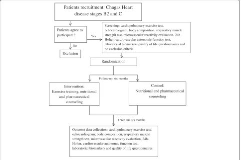
Trained research assistants or study investigators will conduct the evaluations in a quiet and private room. Ini-tial interviews will include a questionnaire to collect socio-demographic data (income, race, age, sex, and schooling), medical history (stage of CHD, comorbidities, medication, cardiac devices, and arrhythmias), functional class, and level of physical activity through the International Physical Activity Questionnaire-Short Form (IPAQ-SF). Evalua-tions of functional capacity (maximal progressive CPET), cardiac function (two-dimensional echocardiography), body composition (anthropometry and skinfolds), muscle respiratory strength (manovacuometry), microvascular reactivity (laser speckle flowmetry), cardiac rhythm abnor-malities (24-h Holter), autonomic function (active ortho-static stress test), laboratorial biomarkers (biochemical, inflammatory, and oxidative stress), and quality of life (Short Form-36 (SF36) and Minnesota Living with Heart Failure Questionnaire) will be performed at baseline, after 3 months, and at the end of follow-up (6 months). After initial evaluation, patients will be randomized to interven-tion or control groups as seen in Fig. 1.
Intervention
300 mg.dL−1 (16.7 mmol.L−1) unless they present with ketosis symptoms, or dehydration [15]. All training ses-sions will be performed in the morning, indoors, and with a controlled temperature environment under super-vision of medical staff.
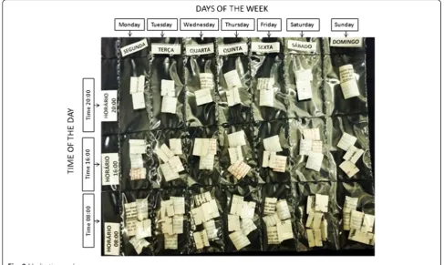
Patients of both groups will undergo regular med-ical appointments with the same cardiologist during the follow-up based on standard medical treatment of the Brazilian consensus on Chagas disease recommen-dations [3] and with other medical specialties if ne-cessary. In addition, nutritional and pharmaceutical counseling will be provided monthly for both groups. The nutritional counseling will consist of general guidance about healthy eating habits and will include how to reduce saturated fat and include poly and monounsaturated fatty acids, to consume more vita-mins, high-fiber carbohydrates, and to reduce the so-dium and water intake for patients with HF [16, 17]. Pharmacists will guide patients about medication usage, drug dosage, and compliance. In order to re-duce any bias due to medication noncompliance, all patients will receive monthly personalized packages according to the medical prescription. The pills will be organized by the time and days that the pills
should be taken (Fig. 2). The schedule of enrollment, interventions, and assessments of the PEACH study is seen in Fig. 3 and the Standard Protocol Items: Rec-ommendations for Interventional Trials (SPIRIT) checklist in Additional file 1.
Outcomes
These will be functional capacity, cardiac function, body composition, muscle respiratory strength, microvascular reactivity, cardiac rhythm abnormalities, autonomic function, biochemical, oxidative stress and inflammatory biomarkers, and quality of life will be assessed at base-line, 3 months, and 6 months of the study.
Cardiopulmonary exercise test
The primary outcome is functional-capacity-based on VO2 peak obtained by a CPET using the VO2000 gas
analyzer (MedGraphics®, St. Paul, MS, USA) with a com-puterized system Ergo PC Elite (Micromed®, Brasília, Brazil) and a treadmill (Inbramed®, Porto Alegre, Brazil). A blinded evaluator will perform an incremental exercise test using a ramp protocol, tailored to achieve a fatigue limited exercise duration of approximately 8 to 12 mi-nutes. [18]. The workloads will be based on age, gender,
Three and six months Follow-up: six months
Yes
No
Patients recruitment: Chagas Heart disease stages B2 and C
Patients agree to participate?
Exclusion
Screening: cardiopulmonary exercise test,
echocardiogram, body composition, respiratory muscle strength test, microvascular reactivity evaluation, 24h-Holter, cardiovascular autonomic function test, laboratorial biomarkers,quality of life questionnaires and no exclusion criteria.
Randomization
Intervention: Exercise training, nutritional
and pharmaceutical counseling
Control:
Nutritional and pharmaceutical counseling
Outcome data collection: cardiopulmonary exercise test, echocardiogram, body composition, respiratory muscle strength test, microvascular reactivity evaluation, 24h-Holter, cardiovascular autonomic function test, laboratorial biomarkers and quality of life questionnaires.
[image:4.595.57.541.89.411.2]height, and weight; adapted to each subject’s physical condition and effort tolerance. Gas and volume calibra-tions will be executed on the early morning of each test day. Pulmonary gas exchange will be analyzed breath-by-breath and averaged every 10 s. A 12-lead electrocar-diogram will monitor heart rhythm during CPET.
The CPET will be limited by symptoms through the subjective fatigue perception scale (Borg modified) ran-ging from 0 to 10, with 0 representing absence of fatigue and 10 maximum tolerated efforts. The examiner may interrupt the test in case of identification of any harmful hemodynamic response. The recovery phase will be active with walking in a pre-determined velocity of 2 km.h−1and 2 % of inclination.
The following CPET variables will be analyzed: max-imum achieved heart hate (HRmax); maxmax-imum achieved blood pressure (BPmax); respiratory exchange ratio (VCO2/VO2); oxygen consumption at peak of exercise
(VO2 peak); oxygen consumption at anaerobic threshold
(VO2 AT); oxygen pulse (O2 pulse); ventilation slope
equivalent to carbon dioxide production (slope VE/ VCO2); circulatory power (CP); presence of complex
ven-tricular arrhythmias (VA); functional aerobic impairment (FAI); and oxygen uptake efficiency slope (OUES) values.
The VO2 peak during exercise will be defined as the
greatest value during 30 s before and after maximum
effort. The VO2 AT will be determined by the point at
which expired carbon dioxide increases in a nonlinear fashion relative to the rate of oxygen consumption accord-ing to the V-slope method. Ergo PC Elite software will de-termine the other variables obtained on the CPET.
Before CPET, the rest electrocardiogram will be evalu-ated to define if the cardiac rhythm is acceptable to per-form the exam. The following markers will not be accepted: sustained ventricular tachycardia, second- and third-degree atrioventricular block, atrial or supraven-tricular tachycardia (more than 100 bpm).
Cardiac function
[image:5.595.54.542.89.380.2](A) diastolic filling velocities, E/A ratio, peak, and peak early (E’) diastolic myocardial velocities.
Body composition
The anthropometric evaluation will consist of measure-ments of body weight and height with minimal clothing and without shoes using a calibrated digital scale with coupled stadiometer. A ratio between weight (kg) and squared height (m) will determine body mass index (BMI), an important surrogate of nutritional status [22]. Two circumferences will be taken: the waist cir-cumference at the narrowest waist level and the hip circumference at the largest circumference around the buttocks [23].
A seven-site skinfold thickness protocol including chest, midaxillary, triceps, subscapular, abdomen, suprai-liac, and thigh sites will be used to evaluate body composition. Measurements will be taken twice on the right side of the body while standing in a relaxed position (Lange skinfold caliper, Beta Technology Inc., Cambridge, MD, USA). The average of each of these seven skinfold thickness will be summated and used to
estimate body composition using the Jackson and Pol-lock equation [24, 25].
Respiratory muscle strength
Respiratory muscle strength will be assessed by maximal inspiratory (MIP) and expiratory pressures (MEP) using a digital pressure manometer connected to a mouthpiece (MVD 3000®, Globalmed, Brazil). Patients will remain in a seated position with a nose clip. They will be requested to make a maximum inspiratory effort at residual vol-ume and a maximum expiratory effort at total lung cap-acity, sustaining it for 1 to 2 s [26]. Once the operator is satisfied, the maximum value of three maneuvers that vary by less than 20 % will be recorded [27].
Microvascular reactivity
A laser speckle contrast imaging system with a laser wavelength of 785 nm (PeriCam PSI system, Perimed, Järfälla, Sweden), coupled to iontophoresis of acetylcho-line and sodium nitroprusside, will noninvasively meas-ure real time cutaneous microvascular flow changes in the forearm [28, 29].
STUDY PERIOD
Enrolment Allocation Post-allocation
Close-out
TIMEPOINT January 2015 – July
2016
February 2015 – July 2016
m*1 m*2 m*3 m*4 m*5 m*6 January 2017
ENROLMENT:
Eligibility screen X
Informed consent X
Allocation X
INTERVENTIONS:
Exercise group
Exercise sessions
Nutritional and pharmaceutical
counseling X X X X X X
Control group
Exercise sessions
Nutritional and pharmaceutical
counseling X X X X X X
ASSESSMENTS:
[Baseline variables: Sociodemographic data; Medical history; Functional class]
X
[Outcome variables: Functional capacity, cardiac function, body composition, muscle respiratory strength, microvascular reactivity, rhythm abnormalities, autonomic function, biochemical, oxidative stress and inflammatory biomarkers, and quality of life]
X X X X
*m: month
[image:6.595.56.541.88.431.2]For the post-occlusive reactive hyperemia (PORH) test, arterial occlusion will be performed with suprasystolic pressure (50 mmHg above the systolic arterial pressure) using a sphygmomanometer applied to the arm of the subject over 3 min. Peak skin flow will be measured after pressure release.
Images will be analyzed using the manufacturer’s software (PIMSoft, Perimed, Järfälla, Sweden). The measurements of skin blood flow will be divided by the mean arterial pressure to yield the cutaneous vas-cular conductance (CVC) in arbitrary perfusion units (APU)/mmHg, to avoid interference of blood pressure levels on microvascular flow.
Cardiac rhythm abnormalities
Arrhythmias and heart rate variability (HRV) will be evaluated with a 24-h Holter (portable three-channel re-corder and analyzer; Cardio Light® and CardioSmart® 5.0, Cardio System, São Paulo, Brazil). Patients will be re-quested to maintain their normal daily activities during the exam. Standard frequency- and time-domain heart rate variability indexes will be measured and evaluated in patients who do not have artificial pacing or atrial fib-rillation rhythm. Only tracings of at least 18 h will be studied. Standard time-domain HRV indices (SDNN: standard deviation of normal-to-normal RR intervals; SDANN: standard deviation of the averages of normal-to-normal RR intervals in all 5-min segments of a 24-h recording; SDNNIDX: mean of the standard deviations of normal-to-normal RR intervals in all 5-min segments of a 24-h recording; rMSSD: root mean square of successive differences; and PNN >50: percentage of differences between adjacent normal-to-normal RR in-tervals that are greater than 50 ms) and frequency do-main (TP: total power; VLFP: very low-frequency power; LFP: low-frequency power; HFP: high-frequency power) will be calculated [30–32].
Cardiovascular autonomic function test
The active orthostatic stress test consists of the evalu-ation of the heart rate and blood pressure response ob-tained from orthostatic change. The patient will rest for 5 min in the supine position, which will be followed by a quick stand-up position (3 to 5 s). The electrocardio-gram will be digitally recorded (ErgoMet 13 V1.0.3.0 HW Heart Ware, Porto Alegre, Brazil) 10 s before the maneuver and will last until 40 s after. The RR intervals will be measured throughout the test period. The baseline average heart rate will be based on the 10 RR intervals immediately preceding the maneuver. The maximum RR at rest over the minimum RR after standup ratio will be calculated (index max:min RR) [33]. The blood pressure will be measured after 5 min at rest and the systolic blood pressure at 5 s after standing
with diastolic blood pressure collected within 5 s after recording systolic blood pressure immediately after standing up to evaluate postural hypotension. Patients with artificial pacing or atrial fibrillation rhythm during the exam will not be evaluated.
Laboratorial biomarkers
A laboratory accredited by the College of American Pathologists will perform biochemical measurements. Total cholesterol, high-density lipoprotein (HDL) chol-esterol, triacylglycerol, glucose, glycated hemoglobin, and the N-terminal of the prohormone brain natriuretic peptide (NT-proBNP) will be measured using Siemens Dimension® reagent cartridge with an intra- and inter-as-say coefficient of variation (CV) <5 %. The Friedewald equation, based on the triacylglycerol measures, will be used to determine low-density lipoprotein (LDL) terol and very low-density lipoprotein (VLDL) choles-terol concentrations [34].
Cytokine serum levels will be measured accordingly to the manufacturer’s instructions (EBioscience, San Diego, CA, USA). Antibodies specific for interferon gamma (IFN-γ), tumor necrosis factor (TNF), interleukin-beta 1 (IL-1β), interleukin-10 (IL-10), interleukin-4 (IL-4), interleukin-8 (IL-8), or monocyte chemotactic protein 1 (MCP-1) will be coated onto the 96-well ELISA micro-plate overnight. Washing solution will be added to each well three times. Standards and unknown samples will be pipetted into these wells and will be incubated for 2 h. After washing, a biotinylated (detection) antibody specific for the described cytokines will be added and in-cubated for 1 h. After washing, streptavidin-horseradish peroxidase will be added. After incubation for 30 min and washing to remove all unbound enzyme, color de-velopment solution will be added. Then, the plates will be read using a microplate reader (SpectraMax 190, Molecular Devices, Sunnyvale, CA, USA) at 450 nm. Oxidative stress will be accessed by two different meth-odologies: detection of serum carbonylated proteins and reduced/oxidized glutathione ratio. The oxidative modi-fied serum proteins will be detected after derivatization with 2,4-dinitrophenylhydrazine, through generation of dinitrophenylhydrazone, which will be analyzed in a spectrophotometer at 380 nm [35]. The reduced and ox-idized glutathione pool will be determined in patients’ sera using DetectX® Glutathione Fluorescent Detection Kit (Arbor Assays, Ann Arbor, MI, USA) as recom-mended by the manufacturer.
Quality of life
The SF-36 consists of 36 questions in eight different domains: general health, physical functioning, social functioning, mental health, physical role, emotional role, bodily pain, and vitality. Each of these dimensions range from 0 (worst possible health state) to 100 (best possible health state).
The Minnesota Living with Heart Failure Questionnaire has 21 questions about how the heart disease influences the lifestyle related to physical, psychological, and social areas. Each question’s responses range from 0 (none) to 5 (very much) and the maximum score is 105. In this ques-tionnaire, lower scores mean better quality of life.
Sample size
Considering a difference in peak oxygen intake of 2.9 ml.kg−1.min−1 with a standard deviation of 2.0 ml.kg−1.min−1[38], assuming anα= 0.05 andβ= 0.20, and increasing the sample size by 50 % accounting for losses to follow-up, a total of 30 patients (15 in the control group and 15 in the exercise group) will be included.
Randomization
A sequence will be computer-generated to randomly allocate 30 patients into two groups in a 1:1 ratio (WinPepi version 11). The sequence will be generated in blocks and by strata of CHD classification (B2 and C) by a single researcher not involved in recruitment. Opaque envelopes will be filled in sequentially to either control or exercise group. Block size will be blinded from inves-tigators involved in patients’recruitment.
Blinding
Given that exercise implies a behavioral intervention, it is not feasible to blind the patients. However, the evaluators will be blinded to the primary endpoint obtained by the CPET and the following secondary endpoints: microvascu-lar reactivity, cardiac rhythm abnormalities, cardiovascumicrovascu-lar autonomic function test, and laboratorial biomarkers. A blinded researcher will perform all data analysis.
Interim analysis and stopping rules
Three interim analyses are planned. The first will be conducted when the tenth volunteer completes 3 months of follow-up, the second when the twentieth volunteer completes 3 months of follow-up and the third when the last volunteer completes 3 months of follow-up.
Trial interruption for ethical reasons due to either posi-tive or negaposi-tive results exceeding expectations may be recommended by an independent committee. The pre-specified stopping rule is a difference of 50 % in VO2peak
between groups, serious adverse events twice as frequent in one of the groups as cardiovascular death, acute myo-cardial infarction, unstable angina, cardiopulmonary ar-rest, malignant ventricular arrhythmias, decompensated
HF, and stroke. All these estimates should have a signifi-cance level of 0.01 or less in any of the interim analyses.
Statistical analysis
Descriptive analysis will consist of mean and standard deviation for continuous variables and percentage for categorical variables. Skewness and Kurtosis testing will be performed to assess the normality of data which will be log-transformed in case of skewed distribution. Vari-ables that can change prognosis of the disease will be compared at baseline in relation to the exercise and con-trol groups. Longitudinal effects of exercise on primary and secondary outcomes will be evaluated through linear mixed models (LMM), which correlate with repeated measures over the time. LMM is an intention-to-treat analysis as it includes all observations of each one of the patients regardless of losses to follow-up or noncompli-ance to exercise protocol. The longitudinal analysis will be made by the treatment × time interaction, which esti-mates the rate of changes in the outcomes. Residual plots of all models will be examined and the likelihood-ratio test will be used to compare and select random intercept or random slope models.
The REDCap software will be used for data man-agement and the data analysis will be conducted by Stata 13.0 software. Statistical significance will be set at p< 0.05 for all analyses.
Discussion
Despite major advances in cardiovascular therapies, CHD still stands as an important cause of premature death in Latin America. Although the number of new cases of Chagas disease has decreased steadily since the late 1990s, many chronic cases are still part of routine care in public hospitals where patients with lower in-come have access to treatment. Moreover, decreased barriers to international travel and migration has led to an increase in migration of patients from Chagas disease-affected areas to nonendemic countries of North America and Europe. This globalization phenomenon transformed Chagas disease into a global medical chal-lenge [39, 40].
CHD treatment is based on trials that studied the effect of different drugs on ventricular dysfunction, survival, and quality of life in patients with cardiomyopa-thies from other etiologies [4, 41]. However, CHD has a specific autonomic imbalance, a different pattern of myocardial fibrosis associated with an inflammatory milieu generated by parasite and host defenses, an increased risk of complex arrhythmias and a known worse prognosis than cardiomyopathies from other etiologies [42, 43].
studies demonstrate that regular exercise is safe and associated with substantial benefits in patients with cardiovascular disease, mainly from ischemic etiology. Clinical adaptations to exercise training include improve-ments in functional capacity, enhanceimprove-ments in cardiac and vascular function, autonomic nervous system modulation, decreases in oxidative stress and low-grade inflammation, and improvements in lipid and glucose profiles [44, 45].
Despite these well-established benefits of exercise training in cardiac patients, there are few studies analyz-ing its effects on patients with CHD. Currently, only one randomized clinical trial [11] including 40 patients with CHD showed that exercise induced improvements in functional capacity and health-related quality of life. This study demonstrates that exercise is feasible, effective, and safe in patients with CHD but with restrictions as the indirect measurement of VO2 peak, the short-term
follow-up and the inclusion of patients in the early stages of CHD, which preclude a definite conclusion about the effects of exercise training in this population.
In the PEACH study, we will try to fill this knowledge gap and address the issue of whether patients with CHD have the same benefits promoted by exercise training in patients with cardiomyopathies from other etiologies. We hypothesize that exercise training will be safe and promote improvements in functional capacity and quality of life, as previously demonstrated by Lima et al. [11] and Fialho et al. [12] in a different sample of patients with CHD. Since an exacerbated inflammatory response is an important mechanism involved in the development of CHD [46] and because several studies have been demonstrating an im-portant anti-inflammatory property of exercise training [47], a decrease in the serum levels of pro-inflammatory cytokines and an increase in the serum levels of anti-in-flammatory cytokines in patients with CHD in the exercise group is expected. Although enhancements in autonomic function as results of exercise training are present in stud-ies with other cardiomyopathstud-ies [48], a recent article did not confirm this finding in patients with CHD [30]. We also hypothesize that cardiac function will improve by an increase in LVEF and an improvement in diastolic func-tion, as seen in coronary artery disease [49], and that exer-cise will improve body composition, respiratory strength, microvascular reactivity, and oxidative stress agents based on results from studies evaluating exercise in non-CHD patients [50, 51].
The benefits of exercise training that will be described by this study will set a new treatment strategy for CHD patients and that this strategy could be routinely in-cluded in clinical practice.
Trial status
Participants are currently being recruited.
Additional file
Additional file 1:SPIRIT checklist. (DOC 121 kb)
Acknowledgements
The authors wish to thank the Evandro Chagas National Institute of Infectious Diseases, the National Institute of Cardiology, and the Oswaldo Cruz Institute for clinical and laboratorial support.
Funding
This study was funded by the 6th Strategic Program for Support of Health from Oswaldo Cruz Foundation and National Council for Scientific and Technological Development (FIOCRUZ/CNPq). (No. 407742/2012-3).
Availability of data and materials
Not applicable.
Authors’contributions
FSNSM was involved in trial design, project development, manuscript drafting and review and will recruit, select, and collect clinical data from the patients. ASS was involved in trial design, manuscript drafting and review, acquisition of funding, and will collect clinical data from the patients. FCCS, RMS, HHV, ARC, JPB, EVT, ROP, FAL, AMHM, and PEAAB were involved in manuscript drafting or review and will collect clinical data from the patients. VLMP, PSS, SSX, MTH, and GMSS were involved in trial design and will collect clinical data from the patients. FMC will be involved in patient evaluations and will collect clinical data from the patients. MFFM was involved in trial design, manuscript drafting and review and will collect clinical data from the patients. All authors read and approved the final manuscript.
Authors’information
Not applicable.
Competing interests
The authors declare that they have no competing interests.
Consent for publication
Not applicable.
Ethical approval and consent to participate
The trial was approved by the Evandro Chagas National Institute of Infectious Diseases Research Ethics Committee (CAAE 38038914.6.0000.5262). All participants will read and sign a written informed consent and will be advised that they can decline to respond to any question or refuse to continue the research any time without compromising their treatment. All participants will receive travel allowance for the evaluations and exercise sessions provided by the project sponsor. Patients followed in the trial who sustain any harm from the intervention will be monitored and will have access to treatment until resolution of the clinical picture.
Author details
1Evandro Chagas National Institute of Infectious Diseases, Oswaldo Cruz
Foundation, Avenida Brasil 4365, Manguinhos, Rio de Janeiro 21040-360, Brazil.2National Institute of Cardiology, Rua das Laranjeiras 374, Laranjeiras,
Rio de Janeiro 22240-006, Brazil.3Physical Education and Sports Institute, State University of Rio de Janeiro, Rua São Francisco Xavier, 524, Maracanã, Rio de Janeiro 20550-900, Brazil.4Oswaldo Cruz Institute, Oswaldo Cruz Foundation, Avenida Brasil 4365, Manguinhos, Pavilhão Cardoso Fontes, Sala 64, Rio de Janeiro 21040-360, Brazil.
Received: 21 April 2016 Accepted: 13 August 2016
References
1. Bern C. Chagas’disease. N Engl J Med. 2015;373(5):456–66. 2. Rassi Jr A, Rassi A, Marin-Neto JA. Chagas disease. Lancet.
2010;375(9723):1388–402.
4. Ribeiro AL, Nunes MP, Teixeira MM, Rocha MO. Diagnosis and management of Chagas disease and cardiomyopathy. Nat Rev Cardiol. 2012;9(10):576–89. 5. Brush Jr JE, Handberg EM, Biga C, Birtcher KK, Bove AA, Casale PN, Clark MG,
Garson Jr A, Hines JL, Linderbaum JA, et al. 2015 ACC Health Policy Statement on Cardiovascular Team-Based Care and the Role of Advanced Practice Providers. J Am Coll Cardiol. 2015;65(19):2118–36.
6. Ades PA, Keteyian SJ, Balady GJ, Houston-Miller N, Kitzman DW, Mancini DM, Rich MW. Cardiac rehabilitation exercise and self-care for chronic heart failure. JACC Heart Fail. 2013;1(6):540–7.
7. McMurray JJ, Adamopoulos S, Anker SD, Auricchio A, Bohm M, Dickstein K, Falk V, Filippatos G, Fonseca C, Gomez-Sanchez MA, et al. ESC guidelines for the diagnosis and treatment of acute and chronic heart failure 2012: The Task Force for the Diagnosis and Treatment of Acute and Chronic Heart Failure 2012 of the European Society of Cardiology. Developed in collaboration with the Heart Failure Association (HFA) of the ESC. Eur J Heart Fail. 2012;14(8):803–69.
8. European Association of Cardiovascular Prevention, Rehabilitation Committee for Science G, Eacpr, Corra U, Piepoli MF, Carre F, Heuschmann P, Hoffmann U, Verschuren M, Halcox J, et al. Secondary prevention through cardiac
rehabilitation: physical activity counselling and exercise training: key components of the position paper from the Cardiac Rehabilitation Section of the European Association of Cardiovascular Prevention and Rehabilitation. Eur Heart J. 2010;31(16):1967–74.
9. Pina IL, Apstein CS, Balady GJ, Belardinelli R, Chaitman BR, Duscha BD, Fletcher BJ, Fleg JL, Myers JN, Sullivan MJ, et al. Exercise and heart failure: a statement from the American Heart Association Committee on exercise, rehabilitation, and prevention. Circulation. 2003;107(8):1210–25. 10. Bocchi EA. Exercise training in Chagas’cardiomyopathy: trials are welcome
for this neglected heart disease. Eur J Heart Fail. 2010;12(8):782–4. 11. Lima MM, Rocha MO, Nunes MC, Sousa L, Costa HS, Alencar MC, Britto RR,
Ribeiro AL. A randomized trial of the effects of exercise training in Chagas cardiomyopathy. Eur J Heart Fail. 2010;12(8):866–73.
12. Fialho PH, Tura BR, Sousa AS, Oliveira CR, Soares CC, Oliveira JR, Souza MV, Coelho MP, Souza FC, Cunha AB, et al. Effects of an exercise program on the functional capacity of patients with chronic Chagas’heart disease, evaluated by cardiopulmonary testing. Rev Soc Bras Med Trop. 2012;45(2):220–4. 13. Yancy CW, Jessup M, Bozkurt B, Butler J, Casey DE, Drazner MH, Fonarow
GC, Geraci SA, Horwich T, Januzzi JL, et al. 2013 ACCF/AHA guideline for the management of heart failure: a report of the American College of Cardiology Foundation/American Heart Association Task Force on practice guidelines. Circulation. 2013;128(16):e240–327.
14. Ponikowski P, Voors AA, Anker SD, Bueno H, Cleland JG, Coats AJ, Falk V, González-Juanatey JR, Harjola VP, Jankowska EA, et al. 2016 ESC Guidelines for the diagnosis and treatment of acute and chronic heart failure: The Task Force for the diagnosis and treatment of acute and chronic heart failure of the European Society of Cardiology (ESC). Developed with the special contribution of the Heart Failure Association (HFA) of the ESC. Eur J Heart Fail. 2016;18(8): 891–975.
15. Colberg SR, Albright AL, Blissmer BJ, Braun B, Chasan-Taber L, Fernhall B, Regensteiner JG, Rubin RR, Sigal RJ, Medicine ACoS, et al. Exercise and type 2 diabetes: American College of Sports Medicine and the American Diabetes Association: joint position statement. Exercise and type 2 diabetes. Med Sci Sports Exerc. 2010;42(12):2282–303.
16. daSaúde M. Dietary guidelines for the Brazilian population. 2nd ed. Brasília: Ministry of Health of Brazil; 2014.
17. Santos RD, Gagliardi AC, Xavier HT, Magnoni CD, Cassani R, Lottenberg AM, De Sociedade Brasileira C, Arpadi Faludi A, Geloneze B, Scherr C, et al. First guidelines on fat consumption and cardiovascular health. Arq Bras Cardiol. 2013;100(1 Suppl 3):1–40.
18. Balady GJ, Arena R, Sietsema K, Myers J, Coke L, Fletcher GF, Forman D, Franklin B, Guazzi M, Gulati M, et al. Clinician’s guide to cardiopulmonary exercise testing in adults: a scientific statement from the American Heart Association. Circulation. 2010;122(2):191–225.
19. Lang RM, Badano LP, Mor-Avi V, Afilalo J, Armstrong A, Ernande L, Flachskampf FA, Foster E, Goldstein SA, Kuznetsova T, et al.
Recommendations for cardiac chamber quantification by echocardiography in adults: an update from the American Society of Echocardiography and the European Association of Cardiovascular Imaging. J Am Soc Echocardiogr. 2015;28(1):1–39. e14.
20. Rudski LG, Lai WW, Afilalo J, Hua L, Handschumacher MD, Chandrasekaran K, Solomon SD, Louie EK, Schiller NB. Guidelines for the echocardiographic
assessment of the right heart in adults: a report from the American Society of Echocardiography endorsed by the European Association of
Echocardiography, a registered branch of the European Society of Cardiology, and the Canadian Society of Echocardiography. J Am Soc Echocardiogr. 2010;23(7):685–713. quiz 786–688.
21. Nagueh SF, Appleton CP, Gillebert TC, Marino PN, Oh JK, Smiseth OA, Waggoner AD, Flachskampf FA, Pellikka PA, Evangelista A.
Recommendations for the evaluation of left ventricular diastolic function by echocardiography. J Am Soc Echocardiogr. 2009;22(2):107–33.
22. Anjos LA. Body mass index (body mass.body height-2) as indicator of nutritional status in adults: review of the literature. Rev Saude Publica. 1992;26(6):431–6. 23. Lohman TG, Roche AF, Martorell R. Anthropometric standardization
reference manual. Illinois: Human Kinetics; 1988.
24. Jackson AS, Pollock ML, Ward A. Generalized equations for predicting body density of women. Med Sci Sports Exerc. 1980;12(3):175–81.
25. Jackson AS, Pollock ML. Generalized equations for predicting body density of men. Br J Nutr. 1978;40(3):497–504.
26. Caruso P, Albuquerque AL, Santana PV, Cardenas LZ, Ferreira JG, Prina E, Trevizan PF, Pereira MC, Iamonti V, Pletsch R, et al. Diagnostic methods to assess inspiratory and expiratory muscle strength. J Bras Pneumol. 2015;41(2):110–23.
27. American Thoracic Society/European Respiratory Society. ATS/ERS Statement on respiratory muscle testing. Am J Respir Crit Care Med. 2002;166(4):518–624. 28. Rousseau P, Mahe G, Haj-Yassin F, Durand S, Humeau A, Leftheriotis G,
Abraham P. Increasing the“region of interest”and“time of interest”, both reduce the variability of blood flow measurements using laser speckle contrast imaging. Microvasc Res. 2011;82(1):88–91.
29. Cordovil I, Huguenin G, Rosa G, Bello A, Kohler O, de Moraes R, Tibirica E. Evaluation of systemic microvascular endothelial function using laser speckle contrast imaging. Microvasc Res. 2012;83(3):376–9.
30. Nascimento BR, Lima MM, Nunes Mdo C, Alencar MC, Costa HS, Pinto Filho MM, Cota VE, Rocha MO, Ribeiro AL. Effects of exercise training on heart rate variability in Chagas heart disease. Arq Bras Cardiol. 2014;103(3):201–8. 31. Amaral Da Silva Souza MV, Santos Soares CC, Rega De Oliveira J, Rosa De
Oliveira C, Hargreaves Fialho P, Cunha DM, Cunha DM, Kopiler DA, Rangel Tura B, Batista Da Cunha A. Heart rate variability: analysis of time-domain indices in patients with chronic Chagas disease before and after an exercise program. Rev Port Cardiol. 2013;32(3):219–27.
32. Dreifus LS, Agarwal JB, Botvinick EH, Ferdinand KC, Fisch C, Fisher JD, Kennedy JW, Kerber RE, Lambert CR, Okike ON, et al. Heart rate variability for risk stratification of life-threatening arrhythmias. American College of Cardiology Cardiovascular Technology Assessment Committee. J Am Coll Cardiol. 1993;22(3):948–50.
33. Ribeiro AL, Ferreira LM, Oliveira E, Cruzeiro PC, Torres RM, Rocha MO. Active orthostatic stress and respiratory sinus arrhythmia in patients with Chagas’ disease with preserved left ventricular global systolic function. Arq Bras Cardiol. 2004;83(1):40–4. 35–49.
34. Friedwald WT, Levy RI, Fredrickson DS. Estimation of the concentration of low-density lipoprotein cholesterol in plasma, without use of preparative ultracentrifuge. Clin Clem. 1978;18:499–502.
35. Levine LR, Williams JA, Stadtman ER, Shacter E. Carbonyl assays for determination of oxidatively modified proteins. Methods Enzymol. 1994;233:346–57. 36. Ciconelli RM, Ferraz MB, Santos W, Meinão I, Quaresma MR. Brazilian
-Portuguese version of the SF-36. A reliable and valid quality of life outcome measure. Rev Bras Reumatol. 1999;39(3):143–50.
37. Carvalho VO, Guimaraes GV, Carrara D, Bacal F, Bocchi EA. Validation of the Portuguese version of the Minnesota Living with Heart Failure
Questionnaire. Arq Bras Cardiol. 2009;93(1):39–44.
38. Freyssin C, Verkindt C, Prieur F, Benaich P, Maunier S, Blanc P. Cardiac rehabilitation in chronic heart failure: effect of an 8-week, high-intensity interval training versus continuous training. Arch Phys Med Rehabil. 2012;93(8):1359–64.
39. Pinto Dias JC. Human Chagas disease and migration in the context of globalization: some particular aspects. J Trop Med. 2013;2013:789758. 40. Klein N, Hurwitz I, Durvasula R. Globalization of Chagas disease: a growing
concern in nonendemic countries. Epidemiol Res Int. 2012;2012(2012):1–13. 41. Bocchi EA, Braga FG, Ferreira SM, Rohde LE, Oliveira WA, Almeida DR, Moreira
Mda C, Bestetti RB, Bordignon S, Azevedo C, et al. III Brazilian Guidelines on Chronic Heart Failure. Arq Bras Cardiol. 2009;93(1 Suppl 1):3–70. 42. Cardoso J, Novaes M, Ochiai M, Regina K, Morgado P, Munhoz R,
in clinical and hemodynamic profile C. Arq Bras Cardiol. 2010;95(4): 518–23.
43. Rassi S, Barretto AC, Porto CC, Pereira CR, Calaca BW, Rassi DC. Survival and prognostic factors in systolic heart failure with recent symptom onset. Arq Bras Cardiol. 2005;84(4):309–13.
44. Piepoli MF, Corra U, Benzer W, Bjarnason-Wehrens B, Dendale P, Gaita D, McGee H, Mendes M, Niebauer J, Zwisler AD, et al. Secondary prevention through cardiac rehabilitation: from knowledge to implementation. A position paper from the Cardiac Rehabilitation Section of the European Association of Cardiovascular Prevention and Rehabilitation. Eur J Cardiovasc Prev Rehabil. 2010;17(1):1–17.
45. Haskell WL, Lee IM, Pate RR, Powell KE, Blair SN, Franklin BA, Macera CA, Heath GW, Thompson PD, Bauman A, et al. Physical activity and public health: updated recommendation for adults from the American College of Sports Medicine and the American Heart Association. Circulation. 2007;116(9):1081–93.
46. Keating SM, Deng X, Fernandes F, Cunha-Neto E, Ribeiro AL, Adesina B, Beyer AI, Contestable P, Custer B, Busch MP, et al. Inflammatory and cardiac biomarkers are differentially expressed in clinical stages of Chagas disease. Int J Cardiol. 2015;199:451–9.
47. Milani RV, Lavie CJ, Mehra MR. Reduction in C-reactive protein through cardiac rehabilitation and exercise training. J Am Coll Cardiol. 2004;43(6):1056–61. 48. Larsen AI, Gjesdal K, Hall C, Aukrust P, Aarsland T, Dickstein K. Effect of
exercise training in patients with heart failure: a pilot study on autonomic balance assessed by heart rate variability. Eur J Cardiovasc Prev Rehabil. 2004;11(2):162–7.
49. Zheng H, Luo M, Shen Y, Ma Y, Kang W. Effects of 6 months exercise training on ventricular remodelling and autonomic tone in patients with acute myocardial infarction and percutaneous coronary intervention. J Rehabil Med. 2008;40(9):776–9.
50. Tabet JY, Meurin P, Driss AB, Weber H, Renaud N, Grosdemouge A, Beauvais F, Cohen-Solal A. Benefits of exercise training in chronic heart failure. Arch Cardiovasc Dis. 2009;102(10):721–30.
51. Gielen S, Laughlin MH, O’Conner C, Duncker DJ. Exercise training in patients with heart disease: review of beneficial effects and clinical recommendations. Prog Cardiovasc Dis. 2015;57(4):347–55.
• We accept pre-submission inquiries
• Our selector tool helps you to find the most relevant journal
• We provide round the clock customer support
• Convenient online submission
• Thorough peer review
• Inclusion in PubMed and all major indexing services
• Maximum visibility for your research
Submit your manuscript at www.biomedcentral.com/submit