Effect of serum starvation and cell density in the
expression of VEGF, bFGF and Angiogenin, and also the
effect of VEGF up regulate in the expression of bFGF
and Angiogenin
Mohnad Abdalla , Wafa Ali Eltayb
School of Life Science, University of Science and Technology of China, PR China Email Address: [email protected]
Abstract- We investigated the effects of serum starvation and cell density expression of Vascular endothelial growth factor (VEGF), Basic fibroblast growth factor (bFGF) and Angiogenin by used PC3M cells, we demonstrated that low density increase the expression of VEGF and bFGF but no effects on Angiogenin in the mRNA levels, while no deferent in high density. However the VEGF and bFGF was decrease when the cells was starved 24h but no effects on angiogenin in the mRNA levels, also We transfected the VEGF gene in the sense orientation into PC3M cells, and obtained stable VEGF overexpressing transfectants. The bFGF and angiogenin was detected ,and we obtain the VEGF up regulate increase of bFGF expression level, and negative correlation was found between up regulate VEGF and angiogenin expression level.
Index Terms- PC3M cells, VEGF, bFGF, angiogenin, of serum starvation, cell density
I. INTRODUCTION
rostate cancer is a form of cancer that develops in the prostate, a gland in the male reproductive system. Most prostate cancers are slow growing; however, there are cases of aggressive prostate cancers. On the other hand, the more aggressive prostate cancers account for more cancer-related mortality than any other cancer except lung cancer(Siegel et al., 2011). Many factors, including genetics and diet, have been implicated in the development of prostate cancer(Djulbegovic et al., 2010).
Vascular endothelial growth factor (VEGF), also known as vascular permeability factor (VPF), is an important mediator of these angiogenic processes in both normal and diseased conditions. As one of the most important cytokines, VEGF is widely expressed by a number of human and animal tumors and has the ability to regulate most of the steps in the angiogenic signal cascade(Sutapa et al., 2009).
Basic fibroblast growth factor (bFGF), amember of a family of heparin binding multifunctional polypeptides, is one of the most potent angiogenic factors. It is highly expressed in a variety of tumor cells and tissues(Bikfaivi et al.,1997).
Angiogenin is localizes on chromosome 14q11, Its expression increases in various types of human cancers,
liver, kidney, prostate(Katona et al., 2005). Four aspects of ANG have been discovered to be necessary for the process of ANG-induced angiogenesis, including ribonuclease activity, basement membrane degradation, signaling transduction, and nuclear translocation(Xiangwei and Zhengping, 2008).
In recent years, the inhibitors of some angiogenic factors were expected to be used in tumor therapy, and themechanism of the interaction between angiogenic factors became important and appealing. The present study may be helpful in finding a potential therapeutic approach to human prostate cancer.
The single most important element that determined whether cancer cells were responsive to angiogenin or not was cell density. Other details such as the cell passage number, confluence status, attachment factors, and stimulation times had some influence, but were not as critical as cell density and starvation.
To investigate the effect of cell density in three gene the VEGF, bFGF and Angiogenin in prostate cancer. To investigate the effect of serum starvation in three gene the VEGF, bFGF and Angiogenin in prostate cancer. To further understand the biological role of VEGF,angiogenin and bFGF, we investigated the effect of VEGF up regulation on the expression of angiogenin, bFGF and PC3M cell growth.
II. MATERIALSANDMETHODS
Cell Culture
Cell Lines and Culture Conditions. The human Prostate cancer cell line PC-3M was obtained from the Northeast Normal University Lap. These cells were cultured and maintained in IMDM supplemented with 10% fetal bovine serum, 100 U/ml penicillin and100 µg/ml streptomycin and kept in a humidified atmosphere of 5% co2. They also use the Dimethyl sulfoxide
50µM for kept cells in -80. For induction of serum starvation, cells were grown to 100% confluence in IMDM and 10% fetal bovine serum, and the medium was then changed to serum-free medium. Cells were grown to 100% confluence to avoid any variations in VEGF, bFGF and Angiogenin expression that might occur when cells are grown to a lesser confluence, as demonstrated previously. a lesser confluence, as demonstrated previously. To determine the effects of serum starvation and high and low density, cells grown under these serum-free and high
ISSN 2250-3153
and low density conditions were harvested after various, time periods, and mRNA was obtained.
Transfection experiments
The day before transfection, PC3-M cells were seeded at a density of 3×10⁵ cell/cm2. twenty-four hour later, when cells reached 70-80% confluency, the decoy ODNs was transfected into PC-3M cells by LipofectaminTM2000 according to the manufacturer’s instructions. PC-3M cells were divided into two groups. VEGF decoy was transfected into first group, Control decoy was not transfected into second group, was used 1.6-2μg /50μl decoy DNA and 4-5μl/50μl lipofectamin were diluted into 50μl medium without serum. Then the above two were combinded and layed at room temperature for 20 mins. The complexes were added onto the cells cultured with serum-free medium. After 15-20 h, PC-3M cells were detected.
Reverse transcriptase-PCR (RT-PCR) Analysis
Expression of VEGF, bFGF and Angiogenin gene was determined by RT-PCR.Total. RNA was extracted using TRIzol Reagent(invitrogen). For oligonucleotide treated cells, RNA expression was performed 15 or 20 h after termination of oligonucleotide treatments.Two mcrogram of total RNA were reverse transcribed using 200 units of murine reverse transcriptase under recommended conditions. One-fourth of this cDNA was used as the template in a PCR using the following upstream and downstream primers:
VEGF sense primer 5, -ACATCTTCCAGGAGTACCCTGATGAG -3,
VEGF antisense primer 5,
-GCATTCACATTTGTTGTGCTGT-3,
A ngiogenin sense primer 5, CATCATGAGGAGACGGGG-3,
A sense antisense primer 5,- TCCAAGTGGACAGGTAAGCC-3,
bFGF sense primer 5,- ATGGCAGCCGGGAGCATCACC-3,
bFGF antisense primer 5,
-CACACACTCCTTTGATAGACACAA-3,
β-actin sense primer 5,- TGGGTCAGAAGGATTCCTATGT-3,
β-actin antisense primer 5,-
CAGCCTGGATAGCAACGTACA-3,
PCR for the detection of VEGF,BFGF and Angiogenin gene in field isolates for all reactions was done using Model Gene Amp® Chinese PCR system (Tiangen biotech) in the following conditions: one cycle at 94 ºC for 5 minutes; 28 cycles at 94 ºC for 30 second, 58 ºC for 30 second, and 72º C for 30 second; and a final extension at 72º C for10 minutes. This BFGF, but for Angiogenin gene was used 25 cycles, and for VEGF was used 26 cycle, For each series of samples, water was used β actin as Internal control. Each PCR product was run on an 1.5% gel electrophoresis at 100V for 30 min and the gel was scanned.
III. RESULT
studies were performed to examine effect of cell density and serum starvation in three gene the VEGF,bFGF and Angiogenin in prostate cancer, and the effect of up-regulated VEGF expiration in bFGF and Angiogenin expiration, two low density (1.5x10⁵ cells/well), high density (6x10⁵ cells/well), and 3x10⁵ was used as control in 6 well dish.
When cells were plated sparsely (1.5x10⁵ cells/well), there a significant increase in bFGF expression that occurred when decrease cells branched out to touch other cells via long this processes; this increase in bFGF levels remained as the cells became confluent. cells were plated high densities (6x10⁵ cells/well), high densities cells was present lower than lower densities cells for bFGF expression
Despite the dramatic changes between low densities and 24h in bFGF expression, but high and 0h expressed did not change significantly at densities of 6x10⁵-3x10⁵ cells per well.
Levels of the VEGF, bFGF and Angiogeninwere compared when cells were grown in replete medium vs. serum-starved conditions at 0 and 24h, only significant decrease in VEGF and bFGF was observed after cells were serum-starved for 24h, while the VEGF levels was increase at the low density. While Angiogenin expression in cells grown in serum-replete medium and serum-starved showed no significant difference.
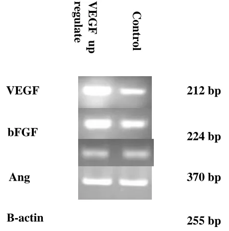
Using PCR, we genotyped the cells expression VEGF, bFGF, and angiogenin and found that , A strong correlation was found between up regulate VEGF and increase of bFGF expression level, and negative correlation was found between up regulate VEGF and angiogenin expression level.
IV. DISCUSSIONS
To test the hypothesis that constitutively active oncogenes or oncogene homologs can prevent the switch to a protective maintenance mode in response to starvation. Expression of VEGF, bFGF and Angiogenin mRNA by Serum Starvation, high and low density. We first performed differential stress to determine the effects of serum starvation, high and low density on VEGF, bFGF and Angiogenin expression. Our hypothesis was that the combination of these genetic manipulations with starvation, PC-3M cells were grown to 100% confluence in IMDM supplemented with 10% fetal bovine serum. The medium was then changed to serum-free medium, and the cells were harvested at intervals thereafter. and other cells it put in high and low density To determine the effects of these conditions on VEGF , bFGF and Angiogenin expression.
and low density (results not shown).but there is significant difference was found in the growth rates of the starved cells. This phenotypic change would presumably allow for motility and, eventually, wound contraction. Indeed, changes in motility induced by fibroblast growth factor have been demonstrated in cells at low cell density, while the same growth factor induces mitogenesis at higher cell density(Thomas et al., 1999).
When cells were plated sparsely (1.5x10⁵ cells/well), there a significant increase in bFGF expression that occurred when decrease cells branched out to touch other cells via long this processes; this increase in bFGF levels remained as the cells became confluent. cells were plated high densities (6x10⁵ cells/well), high densities cells was present lower than lower densities cells for bFGF expression. Same to Thomas et al., 1999.
Despite the dramatic changes between low densities and 24h in bFGF expression, but high and 0h expressed did not change significantly at densities of 6x10⁵-3x10⁵ cells per well.
Levels of the VEGF, bFGF and Angiogeninwere compared when cells were grown in replete medium vs. serum-starved conditions at 0 and 24h, only significant decrease in VEGF and bFGF was observed after cells were serum-starved for 24h, while the VEGF levels was increase at the low density. While Angiogenin expression in cells grown in serum-replete medium and serum-starved showed no significant difference. The mitogenic and proliferative activities of angiogenin are relatively low compared with those of bFGF and VEGF. These data also indicate that the endogenous angiogenin secreted by endothelial cells is neither necessary for nor inhibitory to the actions of bFGF and VEGF(Guo et al.,1997). Nuclear translocation of angiogenin in endothelial cells decreases as cell density increases and ceases when cells are confluent(Takanori et al., 2005).
We have examined Levels of the VEGF after 24h starvation, VEGF was decrease compare with control, while the VEGF levels was increase at the low density. Other researchers found that the expression of VEGF increased after the growth of tumor cells was inhibited. The inhibition of tumor cell growth by conventional forms of therapy such as irradiation or low dose chemotherapy led to an increase of VEGF expression(Polytarchou et al., 2004; Fersis et al., 2004). Gene expression studies comparing dense and sparse growing tumor cells have shown that VEGF gene expression increased in confluent slow growing H460 cells compared with sparse fast growing cells(Kuhn et al., 2004). VEGF is a key factor for angiogenesis in development, and lack of one allele of the VEGF gene causes embryonal death (Carmeliet et al., 1996).
Other researchers demonstrated that serum starvation induces increased VEGF expression in protein levels. Serum starvation has been shown variously to activate apoptosis pathways through NF-kB , to up-regulate IFN regulatory factor in cultured Swiss 3T3 cells, and to up-regulate expression of cyclins D and E in rat fibroblasts(Young et al., 1999).
A statistically significant correlation was found between the number of angiogenic factor. A strong correlation was found between VEGF and bFGF levels.
Angiogenin was independently correlated with VEGF. A negative significant correlation was found between VEGF levels and Angiogenin (Fig 3).
Yasuaki et al., 1999 demonstrate that bFGF induces VEGF mRNA expression (2.8-fold) and, thus, demonstrate a potential mechanistic explanation for the synergistic activity of VEGF and bFGF.
bFGF is not an endothelial cell-specific growth factor such as VEGF; it acts as a growth factor for many different cell types. bFGF levels seem to be important for the degree of tumor in our cells lines, because there is a positive correlation between bFGF and VEGF.
Human Prostate cancer is an angiogenesis dependent tumor. As VEGF, bFGF and Angiogenin is a potentially important an giogenic factor for Prostate cancer progression, we obtained stable VEGF over-expressing transfectants by transfecting the VEGF gene in the sense orientation into PC3M cells. A strong correlation was found between up regulate VEGF and increase of bFGF expression levels in mRNA levels, but there is no correlation between up regulate VEGF and Angiogenin in mRNA level.
In the present study, we have examined the effects of VEGF on the expression of bFGF and angiogenin in PC3M cells to further approach the interaction between VEGF, bFGF and angiogenin. Such results demonstrated that VEGF negatively affected the expression of angiogenin and positively affected the expression of bFGF in PC3M cells.
It has been reported that aFGF, bFGF, and FGF8 are co-localized in human prostate cancer(Dorkin et al., 1999). We have demonstrated that VEGF mRNA was affected in bFGF expression in PC3M cells. According to this result, we presumed that VEGF and bFGF may have a synergistic effect or that they may be in the same signal pathway in PC3m cell growth and progression.
bFGF activates stress-activated protein kinases (SAPK)/c-Jun N-terminal kinase (JNK) and p44/p42 mitogen-activated protein (MAP) to stimulate VEGF release, and bFGF-activated p38 MAPK negatively regulates the VEGF release(Yasuda et al., 2005). Angiogenin is also reported to activate SAPK/JNK(Xu et al., 2001). MAP kinase superfamily mediates intracellular signaling and plays a crucial role in cellular functions such as proliferation, differentiation, and cell death in a variety of cells(Widmann et al., 1999).
It has been reported that the 18-kDa bFGF interacts with a transcription factor in the nucleus and directly regulates rRNA transcription and ribosome biogenesis, a rate-limiting process in cell growth(Sheng et al., 2003; Dailey et al., 2005). bFGF first binds and activates high-affinity cell surface receptors, and after internalization it can translocate into the nucleus and nucleolus. Therefore, a change in bFGF expression levelmay affect the rRNA transcription and cell proliferation rate(Pederson, 1998; Claus et al., 2003; Xu et al., 2003; Sheng et al., 2004).
ISSN 2250-3153
interrelationships among other angiogenic factors are also unclear. How do these factors coordinate to induce angiogenesis? Are they simultaneously involved in all steps in the angiogenesis process or individually responsible for different steps? Are they interdependent or are their actions additive or synergistic? These questions will be studied in our future work, and we expect to answer some of these questions through our future investigations to guide the treatment of human Prostate cancer.
Multiple environmental stimuli important to tumor growth and metastasis may regulate the expression of VEGF mRNA. Some effects may be additive, whereas others may be mediated by common pathways. A better understanding of the signaling pathways activated in response to environmental stimuli may be important to therapeutic strategies that target angiogenesis of tumor cells.
REFERENCES
[1] Siegel, R, Ward E, Brawley O, Jemal A.. "Cancer statistics, 2011: the impact of eliminating socioeconomic and racial disparities on premature cancer deaths.". CA Cancer J Clin 61: 212–36.
[2] Djulbegovic M, Beyth RJ, Neuberger MM Stoffs TL, Vieweg J, Djulbegovic B, Dahm P. (2010). "Screening for prostate cancer: systematic review and meta-analysis of randomised controlled trials". BMJ 341: c4543. [3] Sutapa Sinha, Pawan Kumar Vohra, Resham Bhattacharya, Shamit Dutta, Shirshendu Sinha and Debabrata Mukhopadhyay. Dopamine regulates phosphorylation of VEGF receptor 2 by engaging Src-homology-2-domaincontaining protein tyrosine phosphatase 2. Journal of Cell Science 122, 3385-3392.(2009).
[4] Bikfaivi A, Klein S, Pintucci G, Rilkin DB: Biological roles of fibroblast growth factor-2. Endocr Rev, 18: 26-45, 1997.
[5] Katona TM, Neubauer BL, Iversen PW, Zhang S, Baldridge LA, Cheng L: Elevated expression of angiogenin in prostate cancer and its precursors. Clin Cancer Res, 11: 8358-8363, 2005.
[6] Xiangwei Gao and Zhengping Xu. Mechanisms of action of angiogenin. Biochim Biophys Sin (2008): 619-624.
[7] Thomas P. Richardson, Vickery Trinkaus-Randall, and Matthew A. Nugent. Regulation of Basic Fibroblast Growth Factor Binding and Activity by Cell Density and Heparan Sulfate. THE JOURNAL OF BIOLOGICAL CHEMISTRY. Vol. 274, No. 19, Issue of May 7, pp. 13534–13540, 1999. [8] GUO-FU HU, JAMES F. RIORDAN, AND BERT L. VALLEE. A putative
angiogenin receptor in angiogenin-responsive human endothelial cells. Natl. Acad. Sci. 94, pp. 2204–2209, March 1997.
[9] Takanori Tsuji, Yeqing Sun, Koji Kishimoto, Karen A. Olson, Shumei Liu, Saori Hirukawa, and Guo-fu Hu. Angiogenin Is Translocated to the Nucleus of HeLa Cells and Is Involved in Ribosomal RNA Transcription and Cell Proliferation. Cancer Res 2005;65:1352-1360.
[10] Polytarchou C, Gligoris T, Kardamakis D, Kotsaki E and Papadimitriou E: X-rays affect the expression of genes involved in angiogenesis. Anticancer Res 24: 2941-2945, 2004.
[11] Fersis N, Smyczek-Gargya B, Armeanu S, Gagulic E, Pantic L, Relakis K, Friedrich M and Wallwiener D: Changes in vascular enothelial growth factor (VEGF) after chemoendocrine therapy in breast cancer. Eur J Gynaecol Oncol 25: 45-50, 2004.
[12] Kuhn H, Bräunlich J, Hammerschmidt S and Wirtz H: Candidate genes upregulated in density dependent growth inhibition of lung cancer cells. Int J Oncol 25: 1481-1487, 2004.
[13] Carmeliet, P., Ferreira, V., Breier, G., Pollefeyt, S., Kieckens, L., Gertsenstein, M., Fahrig, M., Vandenhoeck, A., Harpal, K., Eberhardt, C., Declercq, C., Pawling, J., Moons, L., Collen, D., Risau, W., and Nagy, A. Abnormal blood vessel development and lethality in embryos lacking a single VEGF allele. Nature (Lond.), 380: 435–439, 1996.
[14] Young D. Jung, Kayo Nakano, Wenbiao Liu, Gary E. Gallick, and Lee M. Ellis. Extracellular Signal-regulated Kinase Activation Is Required for Up- Regulation of Vascular Endothelial Growth Factor by Serum Starvation in Human Colon Carcinoma Cells[J]. [CANCER RESEARCH, 1999: 59, 4804–4807.
[15] Yasuaki Hata, Susan L. Rook, and Lloyd Paul Aiello. Basic Fibroblast Growth Factor Induces Expression of VEGF Receptor KDR Through a Protein Kinase C and p44/p42 Mitogen Activated Protein Kinase– Dependent Pathway. DIABETES, VOL. 48, MAY 1999.
[16] Dorkin TJ, RobinsonMC,Marsh C,NealDE, LeungHY: aFGF immunoreactivity in prostate cancer and its co-localization with bFGF and FGF8. J Pathol, 189: 564–569, 1999.
[17] Yasuda E, Tokuda H, Ishisaki A, Hirade K, Kanno Y, Hanai Y, Nakamura N, Noda T, Katagiri Y, Kozawa O: PPAR-gamma ligands up-regulate basic fibroblast growth factor-induced VEGF release through amplifying SAPK/JNK activation in osteoblasts. Biochem Biophys Res Commun, 328: 137-143, 2005.
[18] Xu ZP,Monti DM, Hu GF: Angiogenin activates human umbilical artery smooth muscle cells. Biochem Biophys Res Commun, 285: 909–914, 2001. [19] Widmann C, Gibson S, Jarpe MB: Mitogen activated protein kinase:
conservation of a three kinase module from yeast to human. Physiol Rev, 79: 143–180, 1999.
[20] Sheng Z, Chang SB, Chirico WJ: Expression and purification of a biologically active basic fibroblast growth factor fusion protein. Protein Expr Purif, 27: 267-271, 2003.
[21] Dailey L, Ambrosetti D, Mansukhani A, Basilico C: Mechanisms underlying differential responses to FGF signaling. Cytokine Growth Factor Rev, 16: 233-247, 2005.
[22] Pederson T: Growth factors in the nucleolus? J Cell Biol, 143: 279-281, 1998.
[23] Claus P, Doring F, Gringel S, Ostermeyer FM, Fuhlrott J, Kraft T, Grothe C: Differential intranuclear localization of fibroblast growth factor-2 isoforms and specific interaction with the survival of motoneuron protein. J Biol Chem, 278: 479-485, 2003.
[24] Xu ZP, Tsuji T, Riordan JF, Hu GF: Identification and characterization of an angiogenin binding DNA sequence that stimulates luciferase reporter gene expression. Biochemistry, 42: 121-128, 2003.
[25] Sheng Z, Lewis JA, ChiricoWJ: Nuclear and nucleolar localization of 18 kDa fibroblast growth factor-2 is controlled by C-terminal signals. J Biol Chem, 279: 40153-40160, 2004.
AUTHORS
First Author – Mohnad Abdalla, School of Life Science, University of Science and Technology of China, PR China Email Address: [email protected].
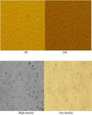
0h 24h
[image:5.612.152.461.65.450.2]High density low density
ISSN 2250-3153
[image:6.612.86.436.64.365.2]www.ijsrp.org
Figure 2 - The mRNA expression of basic fibroblast growth factor (bFGF), Vascular endothelial growth factor (VEGF), Angiogenin, and β-actin used as an internal control. In low and high density, starved cell
and control.
Figure 3 - The mRNA expression of Vascular endothelial growth factor (VEGF), basic fibroblast growth factor (bFGF), Angiogenin, and β-actin used as an internal control. In VEGF transfected cells and
control.