Original Article
Central venous-to-arterial partial pressure of carbon
dioxide difference indicates the prognosis of
cancer patients with sepsis
Yang Lu1,3*, Yang Yang2,3*, Xin He2,3, Peng Zhang4, Shangwen Dong4, Donghao Wang1, Wanhua Wang2,3
Departments of 1Intensive Care Unit, 2Anesthesia, Tianjin Medical University Cancer Institute and Hospital,
National Clinical Research Center for Cancer, Key Laboratory of Cancer Prevention and Therapy, Tianjin, PR China;
3Tianjin’s Clinical Research Center for Cancer, Tianjin 300060, PR China; 4Department of Cardiothoracic Surgery,
Tianjin Medical University General Hospital, Tianjin 300052, PR China. *Equal contributors and co-first authors.
Received March 10, 2017; Accepted September 10, 2017; Epub October 15, 2017; Published October 30, 2017
Abstract: This study aimed to investigate whether central venous-to-arterial partial pressure of carbon dioxide dif-ference (Pcv-aCO2) indicates the prognosis of cancer patients with sepsis post-operation. Total 157 cases of cancer patients with post-operation infection were enrolled, 127 cases were diagnosed with sepsis and received early capacity recovery. These patients were divided into two groups: Group A (Pcv-aCO2 < 6 mmHg) and B (Pcv-aCO2
≥ 6 mmHg). ROC curve analysis showed that three largest areas for ROC curve included Pcv-aCO2 (0.733), SOFA (0.768) and qSOFA (0.728). There was significant difference in survival between Group A and B (26.77±5.12 vs 19.60±12.13). Furthermore, we determined prognostic factors influencing the survival and identified Pcv-aCO2 as the significant factor of the survival by COX regression or logistic regression analysis. In conclusion, we added two indexes of qSOFA and SOFA for early detection and early diagnosis of sepsis with the introduction of the Sepsis-3.0 guidelines. The combination of Pcv-aCO2 with qSOFA and SOFA will help the diagnosis and prognosis of cancer
patients with sepsis.
Keywords: Central venous-to-arterial partial pressure of carbon dioxide difference, sepsis, cancer, capacity
recov-ery, sequential organ failure assessment score, quick SOFA
Introduction
After surgical operation and septic shock, sep-sis is one of the most common causes of death among patients with tumour. The rapid onset of
sepsis can sequentially lead to significant organ
failure over a short term, resulting in multiple organ dysfunction syndrome. Upon the admis-sion of sepsis patients to the Intensive Care Unit (ICU), hemodynamic monitoring is impor-tant to ensure the stability of the patient’s hemodynamic state, improve the oxygen supply of tissues and organs, enhance oxygen metab-olism, and reduce oxygen consumption. Early Goal Directed Therapy (EGDT) has been
regard-ed as standardizregard-ed early anti-shock fluid resus -citation therapy strategy [1-5]. However, owing to the diversity and complexity of the disease, a considerable part of septic shock patients have
high mortality rate and significant organ failure
and hemodynamic disorder after EGDT [6].
The sequential organ failure assessment (SOFA) and Quick SOFA (qSOFA) scores have been
in-troduced into the diagnostic criteria to improve the evaluation of early organ function in the early stage of shock [7]. Central venous-to-arte-rial partial pressure of carbon dioxide differ-ence (Pcv-aCO2) has been proposed as an indi-cator of cardiac index [8]. Therefore, we hypoth-esized that Pcv-aCO2 can be used to evaluate the effects of resuscitation therapy, organ per-fusion, and oxygen metabolism on patients with septic shock. This study aimed to explore the
value of Pcv-aCO2 for guiding fluid therapy and
evaluating the prognosis of patients with septic shock.
Subjects and methods
Subjects
sity Cancer Hospital intensive care unit (ICU) from January 2014 to December 2015, who had infection post-operation. They included 69
females and 88 males, their age was 65.89± 7.35 years. After sufficient assessment, 127
cases of patients were diagnosed with sepsis. The diagnostic criteria of abdominal infection included the gallbladder, biliary tract, liver, spl- een, pancreas, peritoneum, subphrenic organi-zations or other parts of tissues intra-abdomi-nal infections. The following standards are in- cluded in the diagnostic criteria: Standard One: Culture pathogens through surgery or from
abdominal puncture aspirate fluids. Standard Two: Infection verified by surgical or histological
examination or complicated with abdominal abscess post-operation. Standard Three: Two of the following symptoms or signs that can be explained by no other reason: fever, nausea, vomiting, abdominal pain or jaundice. In addi-tion, they should have one of the following con-ditions: Pathogens were cultured in a surgical drainage (closed suction, open and T tube drainages); Pathogens were found from surgery
or aspiration fluid by microscopy; Blood culture
positive or abnormal images were from special
health evaluation II (APACHEII) score > 15 po- ints; b. Hemodynamic abnormalities after sur-gery: systolic blood pressure < 90 mmHg, or a > 40 mmHg basic blood pressure decline, pulse pressure < 20 mmHg, urinary amount < 0.5 ml/ Kg/hr, heart rate > 100 beats/min, central ve- nous pressure (CVP) < 5 mmHg, blood lactate > 2.0 mmol/L, Central venous oxygen saturation
(ScvO2) < 60%.
Patients were excluded if they met the follow- ing exclusion criteria: Age < 18 years old; Shock status except septic shock; Chronic diseases (chronic obstructive pulmonary disease and chronic kidney disease) because of the acute exacerbation of organ dysfunction; Taking sali-cylic acid, biguanide hypoglycemic agents and other drugs that can affect blood lactic acid concentration; had undergone a broad spec-trum of antibiotic therapy previously; died in ICU or discharged from ICU within 24 hours.
Procedures
Among 157 patients, 127 were initially diag-nosed as sepsis, who received capacity recov-ery for 6 hours. 103 patients met EGDT stand-ard, and they were divided into two groups ba- sed on Pcv-aCO2 value: Group A (Pcv-aCO2 < 6
mmHg; 36 cases) and B (Pcv-aCO2 ≥ 6 mmHg;
67 cases). The differences of oxygen extraction (O2ext or O2ER) were compared in two groups at the time of the capacity recovery for 6 and 24 hours. COX regression analysis was per-formed to determine the relevant factors that may impact the survival of the patients.
The age of all patients, APACHE II score before treatment, heart rate (HR), mean arterial pres-Table 1. Comparison of general clinical data between Group A and B
Survival (day) Hemoglobin (g/L) Albumin (g/L) Cardiac index Age (years) BMI Group A 26.77±5.12 124.53±4.93 21.53±0.82 2.76±0.43 70.89±7.05 22.87±2.90
Group B 19.60±12.13* 124.75±5.23 21.63±1.07 2.75±0.52 68.79±7.96 22.24±2.76
t value 3.386 -0.206 -0.488 0.018 1.325 1.094
p value 0.001 0.837 0.626 0.986 0.188 0.277
[image:2.612.91.524.85.152.2]*p < 0.05 indicated significant difference.
Table 2. Comparison of other clinical data between Group A and B
Outcome Infection site Primary disease Primary disease classification classificationIncision Gender
Z value -3.108* -0.200 -0.861 -0.728 -1.222 -0.113
p value 0.002 0.842 0.389 0.466 0.222 0.910
*p < 0.05 indicated significant difference.
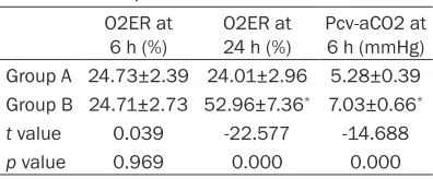
Table 3. Comparison of O2ER at 6 h and 24 h between Group A and B
O2ER at
6 h (%) O2ER at 24 h (%) Pcv-aCO2 at 6 h (mmHg) Group A 24.73±2.39 24.01±2.96 5.28±0.39
Group B 24.71±2.73 52.96±7.36* 7.03±0.66*
t value 0.039 -22.577 -14.688
p value 0.969 0.000 0.000
*p < 0.05 indicated significant difference.
examination such as ultra-sound, CT scan, magnetic resonance or abdominal X-ray examination.
[image:2.612.90.392.196.249.2] [image:2.612.90.288.300.382.2]sure (map) and systolic blood pressure (SBP), respiratory rate (RR), urine volume per hour, central venous pressure (CVP), blood lactic acid (LAC), central venous oxygen saturation (Scv-
O2), oxygenation index (PaO2/FiO2), serum cre -atinine, total bilirubin, platelet and Glasgow
[image:3.612.91.382.75.298.2]There were 36 patients in Group A (16 females, 20 males) and 67 patients in Group B (29 females, 38 males). The outcome included improved or death: 33 cases improved and 3 cases died in Group A; 45 cases improved and 22 cases died in Group B. The infected area
Figure 1. The ROC Curve of qSOFA, Pcv-aCO2 and other prognostic indicators. Pcv-aCO2 (0.733), SOFA (0.768), qSOFA (0.728), APACHII score (0.450), lactic
acid (0.520), ScvO2 (0.576), Age (0.423), and the largest ROC areas were
Pcv-aCO2, SOFA, qSOFA.
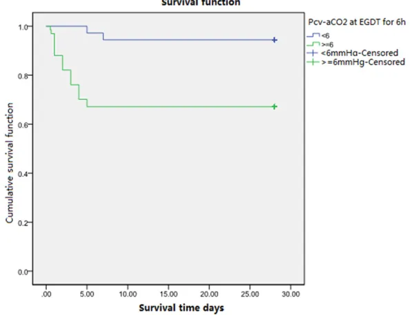
Figure 2. The survival curve analysis of Group A and B. The survival of patients in Group A was better than that of Group B (p=0.002).
Coma Score (GCS) were recorded. Oxygen metabo-lism index were calculated, including oxygen delivery (DO2), oxygen consumption (VO2), arterial oxygen con-tent (CaO2), central venous oxygen content (cvo2) and O2ER. Organ function
cor-relation index QSOFA were
calculated.
Statistical analysis
SPSS19.0 statistics soft-ware was used for data analysis. The measurement data were presented as
mean ± standard deviation (mean ± SD), and data
be-tween two groups were compared by independent sample t test. Spearman rank correlation analysis was performed. p < 0.05
indicated statistical
signifi-cance. Results
Clinical data of the two groups
The capacity resuscitation treatment was conducted early for 127 patients ini-tially diagnosed of sepsis, of which 103 patients met EGDT standards. We then divided 103 patients into Group A (Pcv-aCO2 < 6
mmHg) and B (Pcv-aCO2 ≥
6 mmHg) according to Pcv-aCO2 at 6 hours after EGDT.
[image:3.612.90.387.373.601.2]included the lungs, abdomen and unexplained: 27 for lungs, 8 for abdominal, 1 unexplained in Group A; 51 for lungs, 16 for abdominal cavity in Group B. The primary diseases were: 12 for lung cancer, 7 for esophageal cancer, 7 for gas-tric cancer, 2 for colorectal cancer, 1 for liver cancer, 6 for pancreatic cancer, 2 for gallblad-der cancer, 2 for renal cell carcinoma in Group A; 16 for lung cancer, 19 for esophageal can-cer, 9 for gastric cancan-cer, 5 for colorectal cancan-cer, 6 for liver cancer, 4 for pancreatic cancer, 2 for gallbladder cancer, 3 for kidney cancer, 2 for small intestine cancer, 1 for urinary tract tumor in Group B. In Group A, 12 had chest incision; 17 had abdominal incision; 7 had thoracic and abdominal combined incision. In Group B, 15 had chest incision; 32 had abdominal incision; 19 had thoracic and abdominal combined
inci-sion. The clinical data showed no significant dif -ferences between the two groups except for survival and outcome.
We compared oxygen metabolism indicators of capacity resuscitation for 6 and 24 hours
between the two groups. We found no signifi -cant difference in O2ER between Group A and B at EGDT for 6 hours, but O2ER of Group B
increased significantly compared to Group A at
EGDT for 24 hours (Table 3).
Correlation analysis of prognostic indicators
We analysed the correlation between Pcv-aCO2 and prognostic indicators. The areas under the
ROC were Pcv-aCO2 (0.733), SOFA (0.768), qSOFA (0.728), APACHII score (0.450), lactic
acid (0.520), ScvO2 (0.576), Age (0.423), and
the largest areas were Pcv-aCO2, SOFA and qSOFA (Figure 1).
Kaplan-Meier survival analysis showed that
the survival of patients in Group A was signifi -cantly better than that of Group B (Figure 2). The results of Cox regression survival analysis were: Pcv-aCO2 (p=0.020, B value =-2.369,
95.0% CI 0.013~0.692), SOFA before treat -ment (p=0.374, B value =0.088. 95.0% CI
0.899~1.326), qSOFA before treatment (p= 0.216. B values =0.615, 95.0% CI 0.698~ 4.897), APACHE II score (p=0.091, B value = 0.239, 95.0% CI 0.598~1.038), Hemoglobin (p=0.536, B value =-0.027, 95.0% CI 0.895~ 1.060), Albumin (p=0.370, B value =-0.241, 95.0% CI 0.464~1.331). The results of logistic regression analysis were: Pcv-aCO2 (p=0.012,
OR value =-3.293), SOFA before treatment (p=
0.235, OR value =0.167), qSOFA before treat -ment (p=0.333, OR value =0.645), APACHE II score (p=0.069, OR value =-0.356), Hemoglo- bin (p=0.480, OR value =-0.041), Albumin (p= 0.474, OR value =-0.237). Therefore, only
Pcv-aCO2 was the significant factor that affected
the survival (p < 0.05). Discussion
Sepsis is a common cause of death in critically
ill patients. For peritoneal cancer patients,
long-term poor nutritional status can cause hypoal-buminemia and decreased immune function after surgery, complicated with intra-abdominal infections. These conditions lead to continuous aggravation of abdominal infection, which can eventually result in septic shock. The mortality
rate of sepsis is 30-50%. Early identification
and evaluation of septic shock, the implemen-tation of capacity resusciimplemen-tation, improvement of tissue perfusion, and prevention of second-ary multiple organ dysfunction are essential to improve the prognosis. The mortality of patients
remains high even after fulfilling EGDT.
The standard of ScvO2 ≥ 70% separated part of
the patients from harmful hypoperfusion with
shock [9]. However, ScvO2 ≥ 70% does not pre -vent the progress of multiple organ dysfunction
[10]. Furthermore, continued high ScvO2 level
is even directly related to the high mortality rate in patients with shock [11, 12]. Notably, the combination of lactic acid and ScvO2 indi-cators determined the end point of capacity resuscitation, but they failed to achieve better results [13]. Pcv-aCO2 is the difference be- tween PcvCO2 and PaCO2, and its normal
range is 2-6 mmHg. Mixed venous blood flow
through the lungs, and gas exchange are excret-ed from the lungs. Increasexcret-ed Pcv-aCO2 can
reflect reduced blood flow because the dis -charge capacity of CO2 is very strong. Pv-aCO2 has a negative correlation with cardiac output (CO), thus hemodynamic status of a patient can be evaluated by Pcv-aCO2. Vallee et al. report-ed that Pcv-aCO2 > 6 mmHg may be a useful indicator of capacity resuscitation when ScvO2
> 70% [14]. Futier et al. showed that Pcv-aCO2
In this study, we selected 157 postoperative patients complicated with infections. According to the latest Sepsis-3.0 guideline, we chose
qSOFA and SOFA to screen the patients with
sepsis and 127 cases were diagnosed with sepsis and capacity resuscitation was immedi-ately carried out. According to the guidelines, the EGDT target for 103 patients is reached in six hours. The 103 patients were divided into
Group A aCO2 ≤ 6 mmHg) and B
(Pcv-aCO2 > 6 mmHg). We found that the survival of Group A was longer than that of Group B, and the overall patient outcome was better in Group
A than in Group B. Furthermore, we determined the relevant factors influencing the survival and identified Pcv-aCO2 as the significant fac -tor of the survival by COX regression or logistic regression analysis.
In summary, we added two index of qSOFA and SOFA for early detection and early diagnosis
of sepsis with the introduction of the Sepsis- 3.0 guidelines. We introduced Pcv-aCO2 as an important complement to traditional hemody-namic parameters to improve the treatment success rate of patients with sepsis. In the
clinical we can combine Pcv-aCO2 with qSOFA and SOFA to determine the diagnosis and prog -nosis of cancer patients with sepsis.
Acknowledgements
The study was supported by a grant from Science and technology fund of Tianjin Muni- cipal Health Bureau, China (No. 2013KZ091)
and the Science Foundation of Tianjin Medical
University, China (No. 2014KYQ10). Disclosure of conflict of interest
None.
Address correspondence to: Wanhua Wang, Depart- ment of Anesthesia, Tianjin Medical University Can- cer Institute and Hospital, National Clinical Research Center for Cancer, Key Laboratory of Cancer Prevention and Therapy, Tianjin, PR China; Tianjin’s Clinical Research Center for Cancer, Huan-Hu-Xi Road, Ti-Yuan-Bei, He Xi District, Tianjin 300060, PR China. Tel: 86-21-2334-0123; E-mail: Wanhua_ [email protected]
References
[1] Liu B, Ding X, and Yang J. Effect of early goal directed therapy in the treatment of severe
sepsis and/or septic shock. Curr Med Res Opin 2016; 32: 1773-1782.
[2] Rusconi AM, Bossi I, Lampard JG, Szava-Ko-vats M, Bellone A, and Lang E. Early goal-di-rected therapy vs usual care in the treatment of severe sepsis and septic shock: a system-atic review and meta-analysis. Intern Emerg Med 2015; 10: 731-743.
[3] O’Neill R, Morales J, and Jule M. Early goal-di-rected therapy (EGDT) for severe sepsis/septic shock: which components of treatment are
more difficult to implement in a
community-based emergency department? J Emerg Med 2012; 42: 503-510.
[4] Nguyen HB, Corbett SW, Menes K, Cho T, Daugharthy J, Klein W, and Wittlake WA. Early goal-directed therapy, corticosteroid, and re-combinant human activated protein C for the treatment of severe sepsis and septic shock in the emergency department. Acad Emerg Med 2006; 13: 109-113.
[5] Rivers E, Nguyen B, Havstad S, Ressler J, Muz-zin A, Knoblich B, Peterson E, Tomlanovich M; Early Goal-Directed Therapy Collaborative Group. Early goal-directed therapy in the treat-ment of severe sepsis and septic shock. N Engl J Med 2001; 345: 1368-1377.
[6] van Beest PA, Hofstra JJ, Schultz MJ, Boerma EC, Spronk PE, and Kuiper MA. The incidence of low venous oxygen saturation on admission to the intensive care unit: a multi-center obser-vational study in the Netherlands. Crit Care 2008; 12: R33.
[7] Singer M, Deutschman CS, Seymour CW, Shankar-Hari M, Annane D, Bauer M, Bellomo R, Bernard GR, Chiche JD, Coopersmith CM, Hotchkiss RS, Levy MM, Marshall JC, Martin GS, Opal SM, Rubenfeld GD, van der Poll T, Vin-cent JL, and Angus DC. The third international
consensus definitions for sepsis and septic
shock (sepsis-3). JAMA 2016; 315: 801-810. [8] Cuschieri J, Rivers EP, Donnino MW, Katilius M,
Jacobsen G, Nguyen HB, Pamukov N, and Horst HM. Central venous-arterial carbon diox-ide difference as an indicator of cardiac index. Intensive Care Med 2005; 31: 818-822. [9] Boulain T, Garot D, Vignon P, Lascarrou JB,
De-sachy A, Botoc V, Follin A, Frat JP, Bellec F, Que
-not JP, Mathonnet A, and Dequin PF. Preva -lence of low central venous oxygen saturation
in the first hours of intensive care unit admis -sion and associated mortality in septic shock patients: a prospective multicentre study. Crit Care 2014; 18: 609.
[11] Pope JV, Jones AE, Gaieski DF, Arnold RC,
Trzeciak S, and Shapiro NI. Multicenter study of central venous oxygen saturation (ScvO(2)) as a predictor of mortality in patients with sep-sis. Ann Emerg Med 2010; 55: 40-46.
[12] Textoris J, Fouché L, Wiramus S, Antonini F, Tho
S, Martin C, and Leone M. High central venous oxygen saturation in the latter stages of septic shock is associated with increased mortality. Crit Care 2011; 15: R176.
[13] Jones AE, Shapiro NI, Trzeciak S, Arnold RC, Claremont HA, and Kline JA. Lactate clearance vs central venous oxygen saturation as goals of early sepsis therapy: a randomized clinical trial. JAMA 2010; 303: 739-746.
[14] Vallee F, Vallet B, Mathe O, Parraguette J, Mari A, Silva S, Samii K, Fourcade O, and Genestal
M. Central venous-to-arterial carbon dioxide difference: an additional target for goal-direct-ed therapy in septic shock? Intensive Care Med 2008; 34: 2218-2225.
[15] Futier E, Robin E, Jabaudon M, Guerin R, Petit