Original Article
Decrease in serum levels of interleukin-1β, interleukin-5
and interleukin-9 after vitamin D repletion in patients
with chronic lymphocytic leukemia
Marcin Kubeczko1,2, Elżbieta Nowara1, Wojciech Spychałowicz2, Kamil Wdowiak2, Anna Mertas3, Wojciech
Król3, Tomasz Francuz2,4, Jerzy Chudek2,5, Jerzy Wojnar2
1Department of Clinical and Experimental Oncology, Maria Skłodowska-Curie Memorial Cancer Center and
Institute of Oncology, Gliwice Branch, Gliwice 44-400, Poland; 2Department of Internal Medicine and Oncological
Chemotherapy, School of Medicine in Katowice, Medical University of Silesia, Katowice 40-027, Poland; 3
Depart-ment of Microbiology and Immunology, School of Medicine with The Division of Dentistry in Zabrze, Medical University of Silesia, Zabrze 41-808, Poland; Departments of 4Biochemistry, 5Pathophysiology, School of Medicine
in Katowice, Medical University of Silesia, Katowice 40-752, Poland
Received December 20, 2015; Accepted February 27, 2016; Epub March 1, 2016; Published March 15, 2016
Abstract: Background: Vitamin D (VD) deficiency in chronic lymphocytic leukemia (CLL) is associated with a worse prognosis, a shorter time to start the treatment and the overall survival. VD deficiency is the first potentially modi-fiable prognostic factor in CLL that may interfere with the production of cytokines. However, there is the lack of studies concerning the VD repletion and its influence on interleukins in CLL patients. The aim of our prospective study was to assess the influence of cholecalciferol repletion on serum interleukins levels in CLL patients. Materials and methods: The study comprised 18 CLL patients. A six-month-long interventional study was conducted in CLL subjects with serum 25-OH-D3 concentrations at the level of <30 ng/ml. Patients were supplemented with cholecal-ciferol. Interleukins levels were assessed at the beginning of the study and after a six-month-long supplementation of cholecalciferol. Baseline measurements of interleukins were compared to those in apparently healthy controls. Results: CLL patients were characterized by significantly increased levels of IL-1β, IL-4, IL-5, IL-6, IL-9, IL-12, IL-13 and IL-17, in comparison with healthy controls. During the VD supplementation a decrease in IL-1β, IL-1ra, IL-5, IL-9 and IL-17 serum levels was found. The decrease in IL-1β, IL-5 and IL-9 was observed in the subgroup of CLL patients who were not receiving chemotherapy, while in IL-17 in CLL patients on chemotherapy. Conclusion: The VD repletion may exert favorable influence on interleukins levels in VD deficient CLL patients.
Keywords: Chronic lymphocytic leukemia, vitamin D, interleukin
Introduction
Chronic lymphocytic leukemia (CLL) is the most common type of leukemia in the western hemi -sphere and its incidence is ever-growing. CLL has been defined as the accumulation of mono -clonal neoplastic B lymphocytes [1] as a result of an imbalance between the proliferation and apoptosis of leukemic cells [2]. Diverse micro -environmental stimuli confer a growth advan -tage on these cells and extend their survival [3]. The most explicit manifestation of the impact of microenvironment on B-CLL is the rapid, spontaneous apoptosis of leukemic cells when cultured ex vivo [4]. Co-cultured stromal cells and certain cytokines can prevent this
spontaneous apoptosis [5]. Signals delivered by microenvironment do not only favor clonal expansion of leukemic cells but can also cause drug resistance [6]. Numerous soluble factors and cell-to-cell interactions involved in the sur-vival of neoplastic B-cells have been found [7]. Leukemic cells interact with several types of stromal cells, such as bone marrow stromal cells [8], nurse-like cells [9], T lymphocytes [10] and follicular dendritic cells [11]. Cytokines derived from stromal cells are well established factors, which support survival and growth in CLL cells [12].
-flammatory immune responses, triggered by binding with the IL-1 receptor (IL-1R) [13]. IL-1 receptor antagonist (IL-1ra), as a competitive inhibitor of IL-1 signaling, regulates activity of IL-1α and IL-1β [14]. IL-1β was found to be asso -ciated with the overall survival (OS) in CLL patients [15]. The recently found correlation between increased IL-1β concentration and sig -nificantly longer OS was somehow surprising, as the cytokine had been previously reported as a factor independently promoting in vitro CLL cell growth and survival [15]. IL-1β stimu -lates differentiation of naive T cells into Th17 cells [16], which secretes IL-17. Vitamin D insuf -ficient young adults were found to have higher serum concentrations of pro-inflammatory cytokines, such as IL-1β [17].
IL-5 is a T helper 2 (Th2) cytokine that stimu -lates normal B-cell growth and differentiation, as well as increases immunoglobulin secretion, particularly IgA [18]. IL-5 suppresses cell-medi -ated immune responses crucial for an effective antitumor activity [19], thus, increased concen-trations may support tumor growth. IL-5 is one of the best differentiators between CLL and healthy groups - in CLL levels of IL-5 are signifi -cantly higher [15]. Overexpression of IL-5 increases the risk of CLL transformation in mouse B cells [20]. The expansion of tran -scripts for IL-5 required the presence of CD5+ in CLL B [21]. Binding NFAT (nuclear factor of activated T cells) with an IL-5 promoter is cru -cial for IL-5 expression in human T cells [22]. In view of the significant role of NFAT in the tran -scriptional regulation of IL-5, agents which tar -get NFAT might be valuable clinically, particu-larly in those diseases where the over expression of IL-5 is associated with pathology. 1,25(OH)2D3 has been described to inhibit NFAT complex formation in activated normal T cells [23].
Th9 cells are a subset of Th cells that express IL-9 but no other Th cell lineage-specific cyto -kines [24]. IL-9 belongs to the IL-2 family, which is involved in the development and activation of lymphocytes. IL-9 binds with a heterodimeric receptor composed of the cytokine-specific α-chain (IL-9Rα) and the γ-chain [25] and acti -vates the JAK-STAT pathway [26]. IL-9 is a multi -functional Th2 cytokine that was shown to stim -ulate the proliferation of thymic lymphomas and inhibit dexamethasone-induced apoptosis
[27]. In CLL cytokine alterations, favoring regu -latory T cells (Tregs) and Th2 response with inhibition of Th1 differentiation, have been described [28]. The dysregulated expression of IL-9 can be identified in biopsies and serums from patients with various hematologic malig -nancies [29] and reveals an unfavorable course of the disease [30].
IL-17, a proinflammatory cytokine secreted pri -marily by CD4+ Th17 cells [16], can mediate both pro- and antitumor actions [31] and may impact CLL growth, survival, or both. Because the Th17 cells produce high amounts of IL-17A, most Th17-mediated effects are attributed to this cytokine [32]. IL-17 signals through IL-17R and activates mitogen-activated protein kinas-es and nuclear factor κB (NF-κB) [33]. IL-17 induces IL-6 and IL-8 [34]. A lack of Th17 cells might contribute to the suboptimal immune response in non-Hodgkin’s lymphoma [35]. Many of the IL-17 inflammatory actions can pri -marily benefit the host, but with the modify tumor microenvironment, IL-17 may promote tumor growth [31]. 1,25(OH)2D3 is a regulator of Th cells and has been shown to inhibit IL-17 secretion by the Th17 cells [36].
Additionally, higher concentrations of 25-OH-D3 were found to be associated with a reduced risk of CLL [46]. Living in an area with higher ambient UVR was found to be associated with a reduced risk of NHL, especially DLBCL and CLL [47]. Furthermore, after the vitamin D supple -mentation CLL remissions have been reported [48]. Shanafelt et al. revealed shorter OS and time-to-treatment (TTT) in CLL patients with vitamin D insufficiency [49]. Association of vita -min D deficiency with poor prognosis was con -firmed by Molica S. et al. [50] and Aref S. et al. [51]. Finally, vitamin D substitution enhances rituximab efficacy [52].
The aim of our prospective study was to assess the effect of cholecalciferol repletion on serum interleukins levels in CLL patients.
Materials and methods Patients and samples
Eighteen patients with CLL who were not treat -ed during the previous year and ten patients without CLL were enrolled between November 2013 and July 2015. The control group sub-jects compromised age-matched hospitalized patients, who met the same inclusion and exclusion criteria as the study group. The major exclusion criteria were malignancies, acute inflammatory diseases, history of renal, liver or heart failure and hypercalcemia. The protocol was approved by the Local Bioethics Committee and written informed consent was obtained from each participant. Blood specimens were collected after an 8h overnight fast during rou -tine visit. Serum and plasma samples were col-lected and stored in liquid nitrogen till assess -ment of the cytokines concentration. All laboratory tests (25-OH-D3, total calcium, ion-ized calcium, phosphate, alkaline phosphatase, creatinine, 24 h urine calcium excretion, intact parathormon (iPTH), complete blood count, IgG, IgA, IgM, LDH, β2-microglobulin, D-dimer) were repeated in the study group after 6 months of the VD supplementation. Secondary hyperparathyroidism was defined as iPTH level above manufacturer’s reference range (15-68.3 pg/ml) but with a normal concentration of ionized calcium.
The decision concerning the incorporation of chemotherapy or the “watchful waiting” strate -gy was made according to the International
Workshop on Chronic Lymphocytic Leukemia guidelines [53].
25-OH-D3 measurements
The serum 25-OH-D3 levels were measured on Cobas E422 Roche by electrochemilumines-cent immunoassay (ECLIA) with the inter-assay variability below 10.3%.
Serum cytokines measurements
Bio-Plex Pro™ Human Cytokine 27-plex Assay (Bio-Rad Laboratories Inc., Hercules, CA, USA) was used to measure cytokines in serum sam-ples. Cytokines were quantitated using the Bio-Plex 200 System based on xMAP suspension array technology (Bio-Rad Laboratories Inc., Hercules, CA, USA). All procedures were fol -lowed according to the manufacturer’s manual. Standard curves for each cytokine were creat -ed using standard solution with known concen-trations of recombinant human cytokine of interest and performed during the same run as the subjects’ serum analyses. Limits of detec -tion (LOD) for measured cytokines were as fol -lows: IL-1β - 0.6 pg/ml, IL-1ra - 5.5 pg/ml, IL-2 - 1.6 pg/ml, IL-4 - 0.7 pg/ml, IL-5 - 0.6 pg/ml, IL-6 - 2.6 pg/ml, IL-7 - 1.1 pg/ml, IL-9 - 2.5 pg/ml, IL-10 - 0.3 pg/ml, IL-12(p70) - 3.5 pg/ml, IL-13 - 0.7 pg/ml, IL-15 - 2.4 pg/ml, IL-17 - 3.3 pg/ml.
Intervention
VD deficiency was defined as a serum 25-OH-D3 level <30 ng/ml. Subjects in the study group were divided into 3 categories according to 25-OH-D3 levels: mild deficiency (20-30 ng/ml), moderate deficiency (10-19.9 ng/ml) and severe deficiency (<10 ng/ml). Patients with mild deficiency received 2000 IU/d of cholecal -ciferol, patients with moderate deficiency received 4000 IU/d and patients with severe deficiency received 6000 IU/d of cholecal-ciferol.
and prednisone in two patients (R-CVP), and bendamustine in two cases (R-B). One patient with 17p deletion after 4 cycles of R-CC was given idelalisib.
Safety issues
The two major concerns related with the VD supplementation: hypercalcemia and hypercal-ciuria were evaluated by quantification of ion -ized calcium and 24 h urine calcium excretion. Hypercalcemia was defined as ionized calcium levels above 1.35 mmol/l and hypercalciuria as urinary calcium excretion over 5 mmol/24 h, or urinary calcium-to-creatinine ratio higher than 0.4 mg/mg.
Statistical analysis
Differences in variables between the groups at the beginning of the study, as well as after 6
months into the supplementation were exam-ined by Mann-Whitney U test or Kruskal-Wallis one-way analysis of variance. The change of serum, plasma and urine examinations were compared using the Wilcoxon signed-rank test for paired data. The value of limit of detection (LOD) was used when interleukin measurement was lower than LOD. All analyses were per -formed using the statistical package Statistica, version 10 (StatSoft, Inc.). P<0.05 was consid -ered significant in all analyses.
Results
Characteristics of study subjects
[image:4.612.95.518.109.458.2]The characteristics of the subjects are shown in Table 1. Serum levels of IL-2, IL-7, IL-10 and IL-15 were below detection range in all controls and in 16, 15, 12 and 17 of the 18 CLL patients respectively and therefore were excluded from Table 1. Characteristics of the patients with CLL and patients in the control group. Interleukins con -centrations, age, BMI, β2-microblobulin and lymphocytes in bone marrow are presented as medians with an interquartile range (1-3 Q)
Characteristic CLL [N=18] Control [N=10] p value
Age, years 67 (63-73) 62.5 (57-66) 0.13
Male, n (%) 9 (50%) 4 (40%) 0.68
BMI, kg/m2 28 (25.3-30.8) 30.6 (28.0-34.0) 0.065
Concomitant diseases, n (%)
Type 2 diabetes 4 (22%) 1 (10%) 0.45
Coronary artery disease 6 (33%) 1 (10%) 0.19
Hypertension 12 (67%) 5 (50%) 0.41
Binet stage A, n (%) 7 (39%)
Binet stage B, n (%) 8 (44%)
Binet stage C, n (%) 3 (17%)
Genetics, n (%)
del 11q 4 (22%)
del 13q 9 (50%)
del 13q (as a sole mutation) 6 (33%)
del 17p 1 (6%)
Trisomy 12 2 (11%)
Previous chemotherapy 7 (39%)
Time from diagnosis, years 2 (0-7)
β2-microblobulin, mg/l 3.3 (2.6-5.2)
Lymphocyte in bone marrow, % 51 (41-77.5)
IL-1b, pg/ml 10.3 (9.6-11.5) 9.0 (8.0-10.0) <0.05
IL-1Ra, pg/ml 349.9 (333.2-424.5) 333.2 (291.5-374.8) 0.16
IL-4, pg/ml 9.6 (7.6-10.4) 7.7 (6.7-8.9) <0.05
IL-5, pg/ml 79.9 (71.7-90) 69.5 (60.3-79.4) <0.05
IL-6, pg/ml 2.6 (2.6-4.8) Below detection range in 11 pts 2.6 (2.6-2.6) Below detection range in 10 pts <0.05
IL-9, pg/ml 26.2 (12.9-43.1) 9.1 (2.5-22) <0.01
IL-12, pg/ml 9.6 (3.5-27.4) Below detection range in 10 pts 3.5 (3.5-3.5) Below detection range in 10 pts <0.05 IL-13, pg/ml 1.0 (0.7-6.7) Below detection range in 8 pts 0.7 (0.7-0.7) Below detection range in 9 pts <0.05
analysis. CLL patients were characterized by significantly increased levels of IL-1β, IL-4, IL-5, IL-6, IL-9, IL-12, IL-13 and IL-17.
After chemotherapy in group 2 five patients obtained a partial remission of the disease and three patients achieved a complete remission. In group 1 the disease remained stable. Vitamin D status and the cholecalciferol supplementation
In the group of the 18 analyzed CLL patients, only two patients had 25-OH-D3 level within the optimal range (30-80 ng/ml) and the remaining 16 subjects were enrolled into further interven -tion. Eight patients had mildly reduced (insuffi -cient) 25-OH-D3 level, seven moderate and one a severe deficiency. In total, almost 90% had suboptimal levels of 25-OH-D3. The mean 25-OH-D3 level at the baseline for the study subjects was 19.8 ± 6.9 ng/ml (range 7.6-29.7). Sixteen patients received cholecalciferol supplementation in a mean dose of 3125 ± 1218 IU. Supplementation resulted in a signifi -cant increase of 25-OH-D3 in study subjects (P<0.001), as well as in both subgroups (P<0.01). On completion of the study, the mean 25-OH-D3 level was 38.45 ± 14 ng/ml (range 18.9-70), one patient had a moderate VD defi -ciency. Four subjects (25%) had a mild VD defi -ciency and eleven (69%) had optimal VD levels.
Serum iPTH concentration significantly decr-eased (P<0.001) after 6 months of the VD sup -plementation and almost all patients (94%) achieved normal iPTH level. None of patients developed hypercalciuria, as well as hypercal-cemia during the study. Patients did not com-plain of constipation.
Changes of interleukins levels after the chole-calciferol repletion
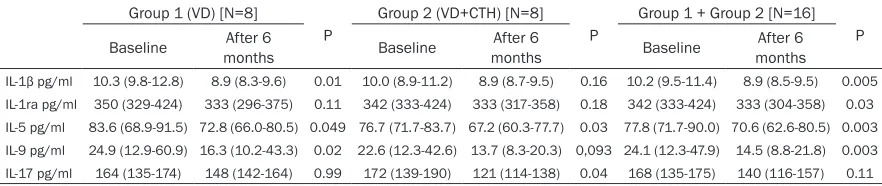
During the VD supplementation some signifi -cant changes (decreases) in IL-1β, IL-ra, IL-5, IL-9 and IL-17 serum levels were found (Table 2). The decrease of IL-1β, IL-5 and IL-9 was observed in CLL patients on the VD supplemen -tation exclusively, while of IL-17 in CLL patients on CTH and the VD supplementation. Changes in serum levels of IL-4, IL-6, IL-7, IL-10, IL-12, IL-13 and IL-15 were not statistically signifi-cant.
Discussion
[image:5.612.86.527.97.190.2]VD deficiency in CLL is associated with worse prognosis, shorter time to start treatment and the overall survival. It is the first potentially modifiable prognostic factor in CLL [51], which can modify the production of various cytokines that play an important role in the course of CLL [15]. In this prospective study we found that the VD repletion is associated with IL-1β level decrease. This is in agreement with the previ-ous report, which showed that a seasonal vari-ation in the VD status is associated with a decreased production of IL-1β during the sum -mer [54]. Prognostic significance of IL-1β remains a questionable issue: some research -ers found lower mRNA levels of IL-1β in CLL cells obtained from patients with progressive disease [55], whereas others [56] reported a higher expression of this cytokine in subjects with an unfavorable course of the disease. IL-1β was recently found to be correlated with positive prognostic markers and increased sur-vival [15]. Additionally, a reduced expression of IL-1β was found in CLL cells obtained from patients with shorter survival and at higher Rai stages [57]. On the other hand, the expression of IL-1β is increased in the presence of ZAP70 Table 2. Comparison of the baseline and a 6-month post supplementation characteristic in each group. Interleukins concentrations are presented as medians with an interquartile range (1-3 Q)
Group 1 (VD) [N=8]
P
Group 2 (VD+CTH) [N=8]
P
Group 1 + Group 2 [N=16]
P Baseline monthsAfter 6 Baseline monthsAfter 6 Baseline monthsAfter 6
IL-1β pg/ml 10.3 (9.8-12.8) 8.9 (8.3-9.6) 0.01 10.0 (8.9-11.2) 8.9 (8.7-9.5) 0.16 10.2 (9.5-11.4) 8.9 (8.5-9.5) 0.005
IL-1ra pg/ml 350 (329-424) 333 (296-375) 0.11 342 (333-424) 333 (317-358) 0.18 342 (333-424) 333 (304-358) 0.03
IL-5 pg/ml 83.6 (68.9-91.5) 72.8 (66.0-80.5) 0.049 76.7 (71.7-83.7) 67.2 (60.3-77.7) 0.03 77.8 (71.7-90.0) 70.6 (62.6-80.5) 0.003
IL-9 pg/ml 24.9 (12.9-60.9) 16.3 (10.2-43.3) 0.02 22.6 (12.3-42.6) 13.7 (8.3-20.3) 0,093 24.1 (12.3-47.9) 14.5 (8.8-21.8) 0.003
[58], which is a well-known marker of a poor prognosis. This inconsistency could illustrate a changeable expression of cytokines during the course of the disease, as well as both pro- and antitumor activities in diverse contexts [59]. The VD repletion was also found to suppress the production of IL-5. It is a Th2 cytokine over -expressed in CLL [60], which can support tumor growth by suppressing cell-mediated immune response [19], and is related with shorter TFT [15], and a worse response to lenalidomide [61]. However, in vitro studies revealed that IL-5 increases a spontaneous apoptosis of CLL cells [62]. Our results, which show that the VD reple-tion decreases IL-5, are consistent with the pre -vious in vitro findings concerning asthma [63] and may lead to a favorable effect on the course of CLL.
IL-9 was among the interleukins, which serum levels were increased in CLL and suppressed by the VD repletion. The increased serum IL-9 levels in CLL patients was previously found and was associated with adverse prognostic fac -tors, such as ZAP-70 expression, β2-micro-globulin expression, IGVH status and Rai & Binet classification [64]. The suppressive effect of VD on IL-9 levels was previously shown in vitro [63]. The beneficial consequences of IL-5 suppression related to the VD supplementation in CLL remains unknown.
We have demonstrated the increased levels of IL-17 in CLL patients, like some [65], but not all studies [60]. In general, higher IL-17 levels are considered beneficial. IL-17 activates immune cells and favors antitumor activity of immune system. It was shown, that low IL-17 levels cor -relate with a progression of CLL and a need of chemotherapy [60]. We have demonstrated that not the VD itself, but rather chemotherapy suppresses the production of IL-17, which is in line with the previous studies, demonstrating a decrease in Th17 cell number level after a ther -apy with rituximab [66] and chlorambucil [65], but not lenalidomide [67].
Furthermore, patients with CLL were found to have higher serum levels of IL-6, IL-12 [15], IL-1Ra, IL-13 and IL-15 [60], which is consistent with our findings. The levels of these interleu -kins did not change after the VD repletion, which is in agreement with other studies [68].
The results of our study demonstrate that the VD repletion in CLL patients may not only opti -mize musculoskeletal health [69], but also affect the function of the immune system (resulting in the suppression of the production of some interleukins) and potentially improve the clinical course of the disease. The VD defi -ciency can be easily determined by blood test-ing and treated accordtest-ing to the available rec-ommendations for general population. Large, multicenter, preferentially randomized studies are necessary to demonstrate the optimal VD levels in CLL patients.
The main limitation of the study was the num -ber of the enrolled patients, high variability of interleukins levels and the limited sensitivity of the method used in the detection of some inter -leukins. Further studies are needed to evaluate the effect of the cholecalciferol supplementa -tion on the levels of other interleukins.
In conclusion, the VD repletion may have a favorable effect on interleukins levels in VD deficient CLL patients.
Acknowledgements
The study was support by grant to M. K. from Medical University of Silesia (KNW-2-044/D/ 4/N). M. K. received a scholarship as part of the DoktoRIS-Scholarship Program for Inno-vative Silesia co-financed by the European Union under the European Social Fund.
Disclosure of conflict of interest
None.
Address correspondence to: Dr. Marcin Kubeczko, Department of Internal Medicine and Oncological Chemotherapy, School of Medicine in Katowice, Medical University of Silesia, Reymonta 8, Katowice 40-027, Poland. Tel: +48506333042; E-mail: [email protected]
References
[1] O’Brien S, del Giglio A, Keating M. Advances in the biology and treatment of B-cell chronic lym-phocytic leukemia. Blood 1995; 85: 307-18. [2] Chiorazzi N. Cell proliferation and death:
for-gotten features of chronic lymphocytic leuke-mia B cells. Best Pract Res Clin Haematol 2007; 20: 399-413.
Differ-ential effects on CLL cell survival exerted by different microenvironmental elements. Curr Top Microbiol Immunol 2005; 294: 135-45. [4] Collins RJ, Verschuer LA, Harmon BV, Prentice
RL, Pope JH, Kerr JF. Spontaneous pro-grammed death (apoptosis) of B-chronic lym-phocytic leukaemia cells following their culture in vitro. Br J Haematol 1989; 71: 343-50. [5] Plander M, Seegers S, Ugocsai P,
Diermeier-Daucher S, Iványi J, Schmitz G, Hofstädter F, Schwarz S, Orsó E, Knüchel R, Brockhoff G. Different proliferative and survival capacity of CLL-cells in a newly established in vitro model for pseudofollicles. Leukemia 2009; 23: 2118-28.
[6] Kay NE, Shanafelt TD, Strege AK, Lee YK, Bone ND, Raza A. Bone biopsy derived marrow stro-mal elements rescue chronic lymphocytic leu-kemia B-cells from spontaneous and drug in-duced cell death and facilitates an ‘angiogenic switch. Leuk Res 2007; 31: 899-906.
[7] Munk Pedersen I, Reed J. Microenvironmental interactions and survival of CLL B-cells. Leuk Lymphoma 2004; 45: 2365-72.
[8] Lagneaux L, Delforge A, Bron D, De Bruyn C, Stryckmans P. Chronic lymphocytic leukemic B cells but not normal B cells are rescued from apoptosis by contact with normal bone marrow stromal cells. Blood 1998; 91: 2387-96. [9] Burger JA, Tsukada N, Burger M, Zvaifler NJ,
Dell›Aquila M, Kipps TJ. Blood-derived nurse-like cells protect chronic lymphocytic leukemia B cells from spontaneous apoptosis through stromal cell-derived factor-1. Blood 2000; 96: 2655-63.
[10] Bagnara D, Kaufman MS, Calissano C, Marsilio S, Patten PE, Simone R, Chum P, Yan XJ, Allen SL, Kolitz JE, Baskar S, Rader C, Mellstedt H, Rabbani H, Lee A, Gregersen PK, Rai KR, Chi-orazzi N. A novel adoptive transfer model of chronic lymphocytic leukemia suggests a key role for T lymphocytes in the disease. Blood 2011; 117: 5463-72.
[11] Pedersen IM, Kitada S, Leoni LM, Zapata JM, Karras JG, Tsukada N, Kipps TJ, Choi YS, Ben-nett F, Reed JC. Protection of CLL B cells by a follicular dendritic cell line is dependent on in-duction of Mcl-1. Blood 2002; 100: 1795-801. [12] Ghamlouch H, Ouled-Haddou H, Damaj G, Roy-er B, GublRoy-er B, Marolleau JP. A combination of cytokines rescues highly purified leukemic CLL B-cells from spontaneous apoptosis in vitro. PLoS One 2013; 8: e60370.
[13] Dinarello CA. Interleukin-1 in the pathogenesis and treatment of inflammatory diseases. Blood 2011; 117: 3720-32.
[14] Hoeft B, Becker N, Deeg E, Beckmann L, Niet-ers A. Joint effect between regular use of non-steroidal anti-inflammatory drugs, variants in
inflammatory genes and risk of lymphoma. Cancer Causes Control 2008; 19: 163-73. [15] Yan XJ, Dozmorov I, Li W, Yancopoulos S, Sison
C, Centola M, Jain P, Allen SL, Kolitz JE, Rai KR, Chiorazzi N, Sherry B. Identification of out-come-correlated cytokine clusters in chronic lymphocytic leukemia. Blood 2011; 18: 5201-10.
[16] Korn T, Bettelli E, Oukka M, Kuchroo VK. IL-17 and Th17 Cells. Annu Rev Immunol 2009; 27: 485-517.
[17] Barker T, Martins TB, Hill HR, Kjeldsberg CR, Dixon BM, Schneider ED, Henriksen VT, Weav-er LK. Circulating pro-inflammatory cytokines are elevated and peak power output correlates with 25-hydroxyvitamin D in vitamin D insuffi-cient adults. Eur J Appl Physiol 2013; 113: 1523-34.
[18] Takatsu K, Kouro T, Nagai Y. Interleukin 5 in the link between the innate and acquired im-mune response. Adv Immunol 2009; 101: 191-236.
[19] Lucey DR, Clerici M, Shearer GM. Type 1 and type 2 cytokine dysregulation in human infec-tious, neoplastic, and inflammatory diseases. Clin Microbiol Rev 1996; 9: 532-62.
[20] Wen X, Zhang D, Kikuchi Y, Jiang Y, Nakamura K, Xiu Y, Tsurui H, Takahashi K, Abe M, Ohtsuji M, Nishimura H, Takatsu K, Shirai T, Hirose S. Transgene-mediated hyper-expression of IL-5 inhibits autoimmune disease but increases the risk of B cell chronic lymphocytic leukemia in a model of murine lupus. Eur J Immunol 2004; 34: 2740-9.
[21] Garaud S, Morva A, Lemoine S, Hillion S, Bor-dron A, Pers JO, Berthou C, Mageed RA, Ren-audineau Y, Youinou P. CD5 promotes IL-10 production in chronic lymphocytic leukemia B cells through STAT3 and NFAT2 activation. J Im-munol 2011; 186: 4835-44.
[22] De Boer ML, Mordvinov VA, Thomas MA, Sand-erson CJ. Role of nuclear factor of activated T cells (NFAT) in the expression of interleukin-5 and other cytokines involved in the regulation of hemopoetic cells. Int J Biochem Cell Biol 1999; 31: 1221-36.
[23] Takeuchi A, Reddy GS, Kobayashi T, Okano T, Park J, Sharma S. Nuclear factor of activated T cells (NFAT) as a molecular target for 1alpha, 25-dihydroxyvitamin D3-mediated effects. J Immunol 1998; 160: 209-18.
[24] Veldhoen M, Uyttenhove C, van Snick J, Helm-by H, Westendorf A, Buer J, Martin B, Wilhelm C, Stockinger B. Transforming growth factor-beta ‘reprograms’ the differentiation of T help-er 2 cells and promotes an inthelp-erleukin 9-pro-ducing subset. Nat Immunol 2008; 9: 1341-6. [25] Hornakova T, Staerk J, Royer Y, Flex E, Tartag-
JC. Acute lymphoblastic leukemia-associated JAK1 mutants activate the Janus kinase/STAT pathway via interleukin-9 receptor alpha ho-modimers. J Biol Chem 2009; 284: 6773-81. [26] Demoulin JB, Van Roost E, Stevens M, Groner
B, Renauld JC. Distinct roles for STAT1, STAT3, and STAT5 in differentiation gene induction and apoptosis inhibition by interleukin-9. J Biol Chem 1999; 274: 25855-61.
[27] Renauld JC, Vink A, Louahed J, Van Snick J. In-terleukin-9 is a major anti-apoptotic factor for thymic lymphomas. Blood 1995; 85: 1300-5. [28] Christopoulos P, Pfeifer D, Bartholomé K, Follo
M, Timmer J, Fisch P, Veelken H. Definition and characterization of the systemic T-cell dysregu-lation in untreated indolent B-cell lymphoma and very early CLL. Blood 2011; 117: 3836-46. [29] Merz H, Houssiau FA, Orscheschek K, Renauld
JC, Fliedner A, Herin M, Noel H, Kadin M, Muel-ler-Hermelink HK, Van Snick J, et al. Interleu-kin-9 expression in human malignant lympho-mas: unique association with Hodgkin’s dis- ease and large cell anaplastic lymphoma. Blood 1991; 78: 1311-7.
[30] Fischer M, Bijman M, Molin D, Cormont F, Uyt-tenhove C, van Snick J, Sundström C, Enblad G, Nilsson G. Increased serum levels of inter-leukin-9 correlate to negative prognostic fac-tors in Hodgkin’s lymphoma. Leukemia 2003; 17: 2513-6.
[31] Maniati E, Soper R, Hagemann T. Up for Mis-chief? IL-17/Th17 in the tumour microenviron-ment. Oncogene 2010; 29: 5653-62.
[32] Murugaiyan G, Saha B. Protumor vs antitumor functions of IL-17. J Immunol 2009; 183: 4169-75.
[33] Shalom-Barak T, Quach J, Lotz M. Interleukin-17-induced gene expression in articular chon-drocytes is associated with activation of mito-gen-activated protein kinases and NF-kappaB. J Biol Chem 1998; 273: 27467-73.
[34] Tartour E, Fossiez F, Joyeux I, Galinha A, Gey A, Claret E, Sastre-Garau X, Couturier J, Mosseri V, Vives V, Banchereau J, Fridman WH, Wi-jdenes J, Lebecque S, Sautès-Fridman C. Inter-leukin 17, a T-cell-derived cytokine, promotes tumorigenicity of human cervical tumors in nude mice. Cancer Res 1999; 59: 3698-704. [35] Yang ZZ, Novak AJ, Ziesmer SC, Witzig TE,
An-sell SM. Malignant B cells skew the balance of regulatory T cells and TH17 cells in B-cell non-Hodgkin’s lymphoma. Cancer Res 2009; 69: 5522-30.
[36] Cantorna MT, Snyder L, Lin YD, Yang L. Vitamin D and 1,25(OH)2D regulation of T cells. Nutri-ents 2015; 7: 011-21.
[37] Anderson JL, May HT, Horne BD, Bair TL, Hall NL, Carlquist JF, Lappé DL, Muhlestein JB; In-termountain Heart Collaborative (IHC) Study
Group. Relation of vitamin D deficiency to car-diovascular risk factors, disease status, and incident events in a general healthcare popu-lation. Am J Cardiol 2010; 106: 963-8. [38] Hayes CE, Hubler SL, Moore JR, Barta LE,
Pras-ka CE, Nashold FE. Vitamin D actions on CD4(+) T Cells in autoimmune disease. Front Immunol 2015; 6: 100.
[39] Gröber U, Spitz J, Reichrath J, Kisters K, Holick MF. Vitamin D: Update 2013: From rickets pro-phylaxis to general preventive healthcare. Der-matoendocrinol 2013; 5: 331-47.
[40] Haussler MR, Whitfield GK, Haussler CA, Hsieh JC, Thompson PD, Selznick SH, Dominguez CE, Jurutka PW. The nuclear vitamin D receptor: biological and molecular regulatory properties revealed. J Bone Miner Res 1998; 13: 325-49. [41] Provvedini DM, Tsoukas CD, Deftos LJ, Manola-gas SC. 1,25-dihydroxyvitamin D3 receptors in human leukocytes. Science 1983; 221: 1181-3.
[42] Veldman CM, Cantorna MT, DeLuca HF. Expres-sion of 1,25-dihydroxyvitamin D(3) receptor in the immune system. Arch Biochem Biophys 2000; 374: 334-8.
[43] van Etten E, Mathieu C. Immunoregulation by 1,25-dihydroxyvitamin D3: basic concepts. J Steroid Biochem Mol Biol 2005; 97: 93-101. [44] Mathieu C, Adorini L. The coming of age of
1,25-dihydroxyvitamin D(3) analogs as immu-nomodulatory agents. Trends Mol Med 2002; 8: 174-9.
[45] Palmer MT, Lee YK, Maynard CL, Oliver JR, Bikle DD, Jetten AM, Weaver CT. Lineage-spe-cific effects of 1,25-dihydroxyvitamin D(3) on the development of effector CD4 T cells. J Biol Chem 2011; 86: 997-1004.
[46] Łuczyńska A, Kaaks R, Rohrmann S, Becker S, Linseisen J, Buijsse B, Overvad K, Trichopoulou A, Valanou E, Barmpitsioti A, Masala G, Agnoli C,Tumino R, Panico S, Bueno-de-Mesquita HB, van Duijnhoven FJ, Peeters PH, Vermeulen R, Weiderpass E, Brustad M, Skeie G, González CA, Jakszyn P, Quirós JR, Sánchez MJ, Huerta JM, Ardanaz E, Melin B, Johansson AS, Alm- quist M, Malm J, Khaw KT, Wareham N, Travis RC, Fedirko V, Romieu I, Jenab M, Gallo V, Riboli E, Vineis P, Nieters A. Plasma 25-hydro- xyvitamin D concentration and lymphoma risk: results of the European Prospective Investi- gation into Cancer and Nutrition. Am J Clin Nutr 2013; 98: 827-38.
[48] Arlet JB, Callens C, Hermine O, Darnige L, Ma-cintyre E, Pouchot J, Capron L. Chronic lympho-cytic leukaemia responsive to vitamin D ad-ministration Br J Haematol 2012; 156: 148-9. [49] Shanafelt TD, Drake MT, Maurer MJ, Allmer C,
Rabe KG, Slager SL, Weiner GJ, Call TG, Link BK, Zent CS, Kay NE, Hanson CA, Witzig TE, Cerhan JR. Vitamin D insufficiency and progno-sis in chronic lymphocytic leukemia. Blood 2011; 117: 1492-8.
[50] Molica S, Digiesi G, Antenucci A, Levato L, Mi-rabelli R, Molica M, Gentile M, Giannarelli D, Sperduti I, Morabito F, Conti L. Vitamin D insuf-ficiency predicts time to first treatment (TFT) in early chronic lymphocytic leukemia (CLL). Leuk Res 2012; 36: 443-7.
[51] Aref S, Ibrahim L, Azmy E. Prognostic impact of serum 25-hydroxivitamin D [25(OH)D] concen-trations in patients with lymphoid malignan-cies. Hematology 2013; 18: 20-5.
[52] Bittenbring JT, Neumann F, Altmann B, Achen-bach M, Reichrath J, Ziepert M, Geisel J, Regitz E, Held G, Pfreundschuh M.Vitamin D deficien-cy impairs rituximab-mediated cellular deficien- cytotox-icity and outcome of patients with diffuse large B-cell lymphoma treated with but not without rituximab. J Clin Oncol 2014; 32: 3242-8. [53] Hallek M, Cheson BD, Catovsky D,
Caligaris-Cappio F, Dighiero G, Döhner H, Hillmen P, Ke-ating MJ, Montserrat E, Rai KR, Kipps TJ; Inter-nationalWorkshop on Chronic Lymphocytic Leukemia. Guidelines for the diagnosis and treatment of chronic lymphocytic leukemia: a report from the International Workshop on Chronic Lymphocytic Leukemia updating the National Cancer Institute-Working Group 1996 guidelines. Blood 2008; 111: 5446-56. [54] Khoo AL, Chai LY, Koenen HJ, Sweep FC,
Joosten I, Netea MG, van der Ven AJ. Regula-tion of cytokine responses by seasonality of vitamin D status in healthy individuals. Clin Exp Immunol 2011; 164: 72-9.
[55] Aguilar-Santelises M, Magnusson R, Svenson SB, Loftenius A, Andersson B, Mellstedt H, Jondal M. Expression of interleukin-1 alpha, interleukin-1 beta and interleukin-6 in chronic B lymphocytic leukaemia (B-CLL) cells from pa-tients at different stages of disease progres-sion. 1991; 84: 422-8.
[56] di Celle PF, Carbone A, Marchis D, Zhou D, Soz-zani S, Zupo S, Pini M, Mantovani A, Foa R. Cy-tokine gene expression in B-cell chronic lym-phocytic leukemia: evidence of constitutive interleukin-8 (IL-8) mRNA expression and se-cretion of biologically active IL-8 protein. Blood 1994; 84: 220-8.
[57] Stratowa C, Löffler G, Lichter P, Stilgenbauer S, Haberl P, Schweifer N, Döhner H, Wilgenbus KK. CDNA microarray gene expression analysis
of B-cell chronic lymphocytic leukemia propos-es potential new prognostic markers involved in lymphocyte trafficking. Int J Cancer 2001; 91: 474-80.
[58] Pede V, Rombout A, Vermeire J, Naessens E, Vanderstraeten H, Philippé J, Verhasselt B. Ex-pression of ZAP70 in chronic lymphocytic leu-kaemia activates NF-κB signalling. Br J Haema-tol 2013; 163: 621-30.
[59] Rodriguez-Vita J, Lawrence T. The resolution of inflammation and cancer. Cytokine Growth Factor Rev 2010; 21: 61-5.
[60] Karmali R, Paganessi LA, Frank RR, Jagan S, Larson ML, Venugopal P, Gregory SA, Christo-pherson KW 2nd. Aggressive disease defined by cytogenetics is associated with cytokine dysregulation in CLL/SLL patients. J Leukoc Biol 2013; 93: 161-70.
[61] Chanan-Khan AA, Chitta K, Ersing N, Paulus A, Masood A, Sher T, Swaika A, Wallace PK, Mashtare TL Jr, Wilding G, Lee K, Czuczman MS, Borrello I, Bangia N. Biological effects and clinical significance of lenalidomide-induced tumour flare reaction in patients with chronic lymphocytic leukaemia: in vivo evidence of im-mune activation and antitumour response. Br J Haematol 2011; 155: 457-67.
[62] Mainou-Fowler T, Craig VA, Copplestone JA, Hamon MD, Prentice AG. Interleukin-5 (IL-5) increases spontaneous apoptosis of B-cell chronic lymphocytic leukemia cells in vitro in-dependently of bcl-2 expression and is inhibit-ed by IL-4. Blood 1994; 84: 2297-304. [63] Keating P, Munim A, Hartmann JX. Effect of
vi-tamin D on T-helper type 9 polarized human memory cells in chronic persistent asthma. Ann Allergy Asthma Immunol 2014; 112: 154-62.
[64] Chen N, Lv X, Li P, Lu K, Wang X. Role of high expression of IL-9 in prognosis of CLL. Int J Clin Exp Pathol 2014; 7: 716-21.
[65] Tang D, Niu Q, Jiang N, Li J, Zheng Q, Jia Y. In-creased frequencies of Th17 in the peripheral blood of patients with chronic lymphocytic leu-kemia: A one year follow-up. Pak J Med Sci 2014; 30: 1128-33.
[66] van de Veerdonk FL, Lauwerys B, Marijnissen RJ, Timmermans K, Di Padova F, Koenders MI, Gutierrez-Roelens I, Durez P, Netea MG, van der Meer JW, van den Berg WB, Joosten LA. The anti-CD20 antibody rituximab reduces the Th17 cell response. Arthritis Rheum 2011; 63: 1507-16.
[68] Jorde R, Sneve M, Torjesen PA, Figenschau Y, Gøransson LG, Omdal R. No effect of supple-mentation with cholecalciferol on cytokines and markers of inflammation in overweight and obese subjects. Cytokine 2010; 50: 175-80.
[69] Płudowski P, Karczmarewicz E, Bayer M, Carter G, Chlebna-Sokół D, Czech-Kowalska J, Dębski R, Decsi T, Dobrzańska A, Franek E, Głuszko P,Grant WB, Holick MF, Yankovskaya L, Konstantynowicz J, Książyk JB, Księżopolska-Orłowska K, Lewiński A, Litwin M, Lohner S,