Original Article
Increased expression of long non-coding RNA
HOXC-AS1 associates with the malignant
status and poor prognosis in glioma
Zhen-Hua Chen1*, Hong-Kang Hu2*, Cheng-Yin Lu2*, Dong-Mei Wang2, Yi Chen2, Jun Qian2, Wei Sun2, Xiao-Jun Wu2, Guo-Han Hu2, Lei Jiang2
1Department of Neurosurgery, The Second Affiliated Hospital of Nantong University, Nantong, People’s Republic
of China; 2Department of Neurosurgery, Changzheng Hospital, Second Military Medical University, Shanghai,
People’s Republic of China. *Equal contributors.
Received December 16, 2015; Accepted May 4, 2016; Epub April 15, 2018; Published April 30, 2018
Abstract: Long non-coding RNA (lncRNA), which is longer than 200 nucleotides, is a type of RNA without the function of encoding proteins. Growing evidence in recent years indicates that lncRNAs are novel regulators in cancerigen-esis and progression. However, little is known about the therapeutic significance of lncRNAs in glioma. In this study, we focused on a typical lncRNA HOXC cluster antisense RNA 1 (HOXC-AS1) with unknown function and detected its expression in glioma and normal brain tissues, in an attempt to confirm the role of HOXC-AS1 in the pathogenesis of glioma and explore the relationship between HOXC-AS1 expression and the clinicopathological features of glioma patients. The results showed that the expression level of HOXC-AS1 was increased significantly in high-grade glioma tissues (WHO grade III-IV) compared with that in low-grade glioma tissues (WHO grade I-II) (P<0.01) and normal brain tissues (P<0.05). In addition, HOXC-AS1 expression was not significantly correlated with age (<50 vs. ≥50, P=0.170), gender (male vs. female, P=0.467), tumor size (<5 cm vs. ≥5 cm; P=0.052), and KPS (<70 vs. ≥70, P=0.661). The overall survival (OS) of glioma patients was significantly associated with the expression of HOXC-AS1 (P<0.001). Multivariate regression analysis showed that increased HOXC-AS1 expression was an independent risk factor for poor prognosis of glioma patients (P=0.039). Taken together, HOXC-AS1 may play an important role in the progression and prognosis of glioma, and may prove to be a latent biomarker and therapeutic target for glioma.
Keywords: Long noncoding RNA, HOXC-AS1, prognosis, glioma, tumor marker
Introduction
Glioma constitutes the most common and deadliest primary malignant brain tumor, accounting for 50-60% of all brain tumors [1]. According to the 2007 WHO classification of gliomas [2], they can be classified into grade I-IV according to their degree of malignancy. Although great advances have been made in the conventional treatments for glioma includ-ing surgery, radiotherapy and chemotherapy in recent years, patient outcomes remain unfavor-able [3, 4]. The characteristics of progressive proliferation and diffuse invasion of glioma may directly result in the overall poor prognosis of patients with glioma. The currently available histopathological classification systems have undoubtedly provided a precious basis for de- fining the clinical assessment of groups of pa-
tients and predicting the clinical behavior of the corresponding cancer as the guideline for treatment [2]. However, studies in recent years suggest that these criteria alone may not be able to fully evaluate the prognosis of glioma patients [5]. Thus, there is an urgent need to identify new potential biomarkers to predict the prognosis of glioma patients more accurately, and find new targets for cancer therapy.
that is associated with many tumors, including promoting glioma cell invasion by directly regu-lating miR-675 expression [11]. Some lncRNAs, such as HOTAIR (Hox transcript antisense inter-genic RNA), have been considered to be signs of various cancers [12]. However, the relation-ship between most lncRNAs and cancerigene-sis remains unclear.
We previously performed microarrays with the glioma specimens and found that HOXC-AS1 was aberrantly expressed in glioma [13]. HOXC-AS1 (ENST00000505700) is an lncRNA whose function has never been described. In the pres-ent study, we first detected the expression level of HOXC-AS1 in the glioma tissue and normal brain tissue to determine the role of HOXC-AS1 in the pathogenesis of gliomas, and then ana-lyzed the relationship between HOXC-AS1 expression and clinicopathological features of glioma including the survival time of patients. It was found that the expression level of HOXC-AS1 in high-grade glioma tissues was signifi-cantly higher than that in low-grade glioma tis-sues and normal brain tistis-sues. In addition, the relatively higher HOXC-AS1 expression was sig-nificantly related to the malignant status and poor prognosis of patients with glioma.
Materials and methods
Clinical samples
This study was approved by the Specialty Com- mittee on Ethics of Biomedicine Research of the Second Military Medical University (Shang- hai, China). Informed consent was obtained from all patients concerned.
Forty-four glioma tissues were selected, and the pathological information was identified according to the WHO classification by experi-enced clinical pathologists. Six samples of normal brain tissues were obtained from six patients sustaining severe head trauma, for whom partial resection of the normal brain was required for decompression during surgery. None of the patients had received chemothera-py or radiotherachemothera-py before resection. All the samples were resected from primary surgery, and the specimens were put into liquid nitro- gen for real-time polymerase chain reaction (PCR). The treatment was carried out accord- ing to the National Comprehensive Cancer Net- work (NCCN) guideline in all glioma patients included in this study. Clinical follow-up was
available for all patients. Overall survival (OS) time of the patients was calculated from the date of initial surgery to the date of patient death.
RNA isolation and quantitative real-time PCR
Total RNA was extracted from the tissue with Trizol reagent (Invitrogen, Carlsbad, CA, USA) according to the manufacturer’s instructions. After purification, complementary DNA (cDNA) was synthesized from 1 μg total RNA using the Prime Script RT Master Mix (Takara). The primers (Sangon) were designed as follows: for human HOXC-AS1, the forward primer was 5’-CCATCTCTGCGACACTTCC-3’ and the reverse primer was 5’-AGCTACTTGCCCACGACC-3’. For human GAPDH, the forward primer was 5’-GG- GAAACTGTGGCGTGAT-3’ and the reverse pri- mer was 5’-GAGTGGGTGTCGCTGTTGA-3. Real-time PCR was conducted by SYBR Premix Ex TaqTM II (Takara) on 7900HT (Applied Biosys- tems). Change in expression level was calculat-ed by quantitative analysis in triplicate using the comparative cycle threshold method. The raw data of target lncRNA were normalized to GAPDH.
Statistical analysis
All data were analyzed using SPSS version 21.0 and GraphPad 5.0 software. Data are expressed as mean ± SD. One-way analysis of variance (ANOVA) was used to test for differences between the glioma and normal brain tissues in all groups, and a least significant difference post-hoc test was used to obtain individual P values followed by ANOVA. The chi-square test was used to examine the relationship between HOXC-AS1 expression level and the clinicopath-ologic features. Survival curves were plotted using the Kaplan-Meier method and compared using the log-rank test. The Cox multivariate proportional hazards model was used to ana-lyze the significance of survival variables. Differences were considered statistically sig-nificant when the p value was <0.05.
Results
HOXC-AS1 up-regulation in high-grade glioma tissues
quantitative real-time PCR. It was found that HOXC-AS1 expression was significantly higher in the glioma tissues compared with that in the normal brain tissues (Figure 1A). HOXC-AS1 expression was significantly up-regulated in the high-grade glioma tissues compared with that in the normal brain tissues (P<0.05) and low-grade glioma tissues (P<0.01) (Figure 1B), while there was no significant difference in HOXC-AS1 expression between the low-grade glioma and normal brain tissues.
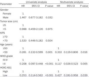
[image:3.612.91.373.73.215.2]grade and age were also significantly correlated with the patient outcomes (Figure 2B and 2C). Univariate analysis identified three prognostic factors: age (<50 or ≥50), WHO grade (I-II or III-IV), and HOXC-AS1 expression. The other clini-copathological characteristics (gender, tumor size and KPS) were not statistically significant prognostic factors. Multivariate analysis of the prognostic factors confirmed that high HOXC-AS1 expression was an independent predictor of poor survival in glioma patients (P=0.039), in Figure 1. Quantitative real-time PCR analysis of HOXC-AS1. A. HOXC-AS1
ex-pression was significantly higher in the glioma tissues compared with that in the normal brain tissues. B. HOXC-AS1 expression was significantly higher in the high-grade glioma tissues compared with that in the normal brain tis-sues and low-grade glioma tistis-sues. NBT: normal brain tissue, LGG: low-grade glioma, HGG: high-grade glioma, ns; P>0.05, *P<0.05, **P<0.01.
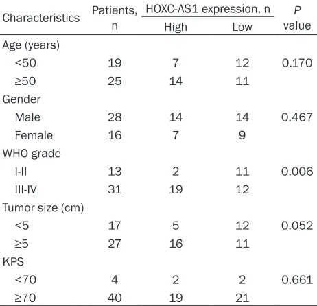
Table 1. HOXC-AS1 expression and clinicopathological features of human gliomas
Characteristics Patients, n HOXC-AS1 expression, n valueP
High Low
Age (years)
<50 19 7 12 0.170
≥50 25 14 11
Gender
Male 28 14 14 0.467
Female 16 7 9
WHO grade
I-II 13 2 11 0.006
III-IV 31 19 12
Tumor size (cm)
<5 17 5 12 0.052
≥5 27 16 11
KPS
<70 4 2 2 0.661
≥70 40 19 21
KPS: Karnofsky performance score.
Correlation between HOXC-AS1 expression and clini-copathological features in patients with glioma
We next identified the corre- lation between HOXC-AS1 ex- pression and the clinicopath-ological features of glioma. The glioma tissues were divid-ed as high-expression group (n=21) and low-expression group (n=23), based on the median expression level of all gliomas (mean expression value 1568.208). As summa-rized in Table 1, HOXC-AS1 was significantly associated with WHO grade (I-II vs. III-IV, P=0.006). However, no signi- ficant association was observed between HOXC-AS1 expression and other clinicopa- thological parameters, including age (<50 vs. ≥50, P=0.170), gender (male vs. fe- male, P=0.467), tumor size (<5 cm vs. ≥5 cm, P=0.052), and karnofsky performance score (KPS) (<70 vs. ≥70, P=0.661). High HOXC-AS1 expression indicates poor prognosis
[image:3.612.91.322.333.556.2]addition to age (P=0.016) and WHO grade (P=0.005) (Table 2).
Discussion
It is widely accepted that genetic information is expressed as a protein. However, almost 98% human genomes do not code for proteins [14], and their function remains largely unknown. Research in recent years has focused more attention on noncoding genes, especially lncRNAs. More evidence has revealed that lncRNAs are generally transcribed in eukaryotic
tocellular carcinoma (HCC). Additionally, the down-regulation of lncRNA-LET was found to be related to hypoxia-induced cancer cell invasion [21]. These examples confirmed that lncRNAs could be used as candidate targets for cancer therapy.
[image:4.612.92.523.75.203.2]LncRNAs are of great importance to gliomas [22, 23]. LncRNA metastasis-associated lung adenocarcinoma transcript 1 (MALAT1) has been reported to be significantly increased in glioma tissues compared with paired adja-cent normal brain tissues. Additionally, lncRNA Figure 2. Kaplan-Meier curves for overall survival by HOXC-AS1 expression (A), WHO grade (B), age (C).
Table 2. Univariate and multivariate Cox regression analyses of overall survival
Parameter Univariate analysis Multivariate analysis
HR 95% CI P value HR 95% CI P value
Gender
Female 1
Male 1.467 0.677-3.182 0.332
Tumor size (cm)
≥5 1
<5 0.988 0.459-2.126 0.975 KPS
≥70 1
<70 1.520 0.446-5.182 0.504 Age (years)
≥50 1 1
<50 0.281 0.132-0.599 0.001 0.310 0.119-0.806 0.016 WHO grade
III-IV 1 1
I-II 0.208 0.097-0.448 <0.001 0.117 0.026-0.523 0.005 HOXC-AS1
High 1 1
Low 0.253 0.114-0.562 <0.001 0.427 0.191-0.958 0.039
HR: Hazard ratio, 95% CI: 95% confidence interval, KPS: Karnofsky performance score.
[image:4.612.92.395.271.534.2]hepa-MALAT1 over-expression was markedly associ-ated with poor prognosis of glioma patients [24]. Yao et al [25] reported that lncRNA X-inactive specific transcript (XIST) expression was up-regulated in glioma tissues and glio-blastoma stem cells, and knockdown of XIST expression exerted a tumor-suppressive func-tion by reducing cell proliferafunc-tion, invasion and migration as well as inducing apoptosis. The mechanism study showed that miR-152 medi-ated the tumor-suppressive effect that XIST knockdown produced. Guo et al [26] found that long intergenic noncoding RNA POU3F3 (linc-POU3F3) might affect the development and progression of glioma by altering the expres-sion of POU3F3. However, our understanding about the concrete mechanism of lncRNAs in the pathogenesis of glioma is far behind other solid tumors, and more lncRNAs associated with glioma need to be found, especially as gli-oma specific bigli-omarkers.
In this study, we found that HOXC-AS1 was over-expressed in high-grade glioma tissues compared with that in low-grade glioma and normal brain tissues, suggesting that it might play an important role in the development of glioma. Besides, we showed that HOXC-AS1 expression in glioma was negatively correlated with OS of glioma patients, and that OS in glio-ma patients with high HOXC-AS1 expression was relatively short. In addition, multivariate Cox regression analysis showed that HOXC-AS1 over-expression was an independent indicator of poor prognosis in glioma patients. Some pre-vious studies reported that age was another important factor affecting the survival time of glioma patients [27, 28], and our results may provide more favorable evidence for them. Yet, the accuracy and reliability of this study may be affected by the small sample size and the lack of the normal brain tissue around the glioma. In conclusion, our data offer convincing evi-dence that the increased expression of HOXC-AS1 may predict unfavorable prognosis in glio-ma patients, indicating that HOXC-AS1 glio-may play a crucial role in promoting the progression of glioma and be a potential target for the treat-ment of this disease. However, the particular mechanism by which HOXC-AS1 is up-regulated in glioma is not clear. More studies are needed to verify the role of HOXC-AS1 as a reliable clini-cal predictor of the outcome for glioma patients in the future.
Acknowledgements
This study was financially supported by the National Natural Science Foundation of China (No. 81270038).
Disclosure of conflict of interest
None.
Address correspondence to: Guo-Han Hu and Lei Jiang, Department of Neurosurgery, Changzheng Hospital, Second Military Medical University, No. 415, Fengyang Road, Shanghai 200003, People’s Republic of China. Tel: +86-21-81885673; E-mail: [email protected] (GHH); Tel: +86-21-81885- 680; E-mail: [email protected] (LJ)
References
[1] Ohgaki H and Kleihues P. Epidemiology and etiology of gliomas. Acta Neuropathol 2005; 109: 93-108.
[2] Louis DN, Ohgaki H, Wiestler OD, Cavenee WK, Burger PC, Jouvet A, Scheithauer BW and Kleihues P. The 2007 WHO classification of tu-mours of the central nervous system. Acta Neuropathol 2007; 114: 97-109.
[3] Taylor LP. Diagnosis, treatment, and prognosis of glioma: five new things. Neurology 2010; 75: S28-S32.
[4] Omuro A and DeAngelis LM. Glioblastoma and other malignant gliomas: a clinical review. JAMA 2013; 310: 1842-1850.
[5] Johnson DR and Galanis E. Incorporation of Prognostic and Predictive Factors Into Glioma Clinical Trials. Curr Oncol Rep 2013; 15: 56-63.
[6] Ponting CP, Oliver PL and Reik W. Evolution and functions of long noncoding RNAs. Cell 2009; 136: 629-641.
[7] Caley DP, Pink RC, Trujillano D and Carter DR. Long noncoding RNAs, chromatin, and devel-opment. ScientificWorldJournal 2010; 10: 90-102.
[8] Spizzo R, Almeida MI, Colombatti A and Calin GA. Long non-coding RNAs and cancer: a new frontier of translational research? Oncogene 2012; 31: 4577-4587.
[9] Gibb EA, Brown CJ and Lam WL. The functional role of long non-coding RNA in human carcino-mas. Mol Cancer 2011; 10: 38.
[10] Wapinski O and Chang HY. Long noncoding RNAs and human disease. Trends Cell Biol 2011; 21: 354-361.
[12] Bhan A and Mandal SS. LncRNA HOTAIR: A master regulator of chromatin dynamics and cancer. Biochim Biophys Acta 2015; 1856: 151-164.
[13] Chen Y, Wu JJ, Lin XB, Bao Y, Chen ZH, Zhang CR, Cai Z, Zhou JY, Ding MH, Wu XJ, Sun W, Qian J, Zhang L, Jiang L and Hu GH. Differential lncRNA expression profiles in recurrent glio-mas compared with primary glioglio-mas identified by microarray analysis. Int J Clin Exp Med 2015; 8: 5033-5043.
[14] Ponting CP and Belgard TG. Transcribed dark matter: meaning or myth? Hum Mol Genet 2010; 19: R162-168.
[15] Tsai MC, Manor O, Wan Y, Mosammaparast N, Wang JK, Lan F, Shi Y, Segal E and Chang HY. Long Noncoding RNA as Modular Scaffold of Histone Modification Complexes. Science 2010; 329: 689-693.
[16] Ling H, Spizzo R, Atlasi Y, Nicoloso M, Shimizu M, Redis RS, Nishida N, Gafa R, Song J, Guo Z, Ivan C, Barbarotto E, De Vries I, Zhang X, Ferracin M, Churchman M, van Galen JF, Beverloo BH, Shariati M, Haderk F, Estecio MR, Garcia-Manero G, Patijn GA, Gotley DC, Bhardwaj V, Shureiqi I, Sen S, Multani AS, Welsh J, Yamamoto K, Taniguchi I, Song MA, Gallinger S, Casey G, Thibodeau SN, Le Marchand L, Tiirikainen M, Mani SA, Zhang W, Davuluri RV, Mimori K, Mori M, Sieuwerts AM, Martens JW, Tomlinson I, Negrini M, Berindan-Neagoe I, Foekens JA, Hamilton SR, Lanza G, Kopetz S, Fodde R and Calin GA. CCAT2, a nov-el noncoding RNA mapping to 8q24, underlies metastatic progression and chromosomal in-stability in colon cancer. Genome Res 2013; 23: 1446-1461.
[17] Sun M, Liu XH, Wang KM, Nie FQ, Kong R, Yang JS, Xia R, Xu TP, Jin FY, Liu ZJ, Chen JF, Zhang EB, De W and Wang ZX. Downregulation of BRAF activated non-coding RNA is associated with poor prognosis for non-small cell lung can-cer and promotes metastasis by affecting epi-thelial-mesenchymal transition. Mol Cancer 2014; 13: 68.
[18] Zhou C, Ye L, Jiang C, Bai J, Chi Y and Zhang H. Long noncoding RNA HOTAIR, a hypoxia-induc-ible factor-1alpha activated driver of malignan-cy, enhances hypoxic cancer cell proliferation, migration, and invasion in non-small cell lung cancer. Tumour Biol 2015; 36: 9179-9188. [19] Gupta RA, Shah N, Wang KC, Kim J, Horlings
HM, Wong DJ, Tsai MC, Hung T, Argani P, Rinn JL, Wang Y, Brzoska P, Kong B, Li R, West RB, van de Vijver MJ, Sukumar S and Chang HY. Long non-coding RNA HOTAIR reprograms chromatin state to promote cancer metastasis. Nature 2010; 464: 1071-1076.
[20] Yang F, Zhang L, Huo XS, Yuan JH, Xu D, Yuan SX, Zhu N, Zhou WP, Yang GS, Wang YZ, Shang JL, Gao CF, Zhang FR, Wang F, Sun SH. Long non-coding RNA high expressed in hepatocel-lular carcinoma (lncRNA-HEIH) facilitates tu-mor growth through enhancer of zeste homo-log 2. Hepatohomo-logy 2011; 54: 1679-1689. [21] Yang F, Huo XS, Yuan SX, Zhang L, Zhou WP,
Wang F and Sun SH. Repression of the long noncoding RNA-LET by histone deacetylase 3 contributes to hypoxia-mediated metastasis. Mol Cell 2013; 49: 1083-1096.
[22] Sun YZ, Wang Z and Zhou D. Long non-coding RNAs as potential biomarkers and therapeutic targets for gliomas. Med Hypotheses 2013; 81: 319-321.
[23] Bian EB, Li J, Xie YS, Zong G, Li J and Zhao B. LncRNAs: new players in gliomas, with special emphasis on the interaction of lncRNAs With EZH2. J Cell Physiol 2015; 230: 496-503. [24] Ma KX, Wang HJ, Li XR, Li T, Su G, Yang P and
Wu JW. Long noncoding RNA MALAT1 associ-ates with the malignant status and poor prog-nosis in glioma. Tumour Biol 2015; 36: 3355-3359.
[25] Yao Y, Ma J, Xue Y, Wang P, Li Z, Liu J, Chen L, Xi Z, Teng H, Wang Z, Li Z and Liu Y. Knockdown of long non-coding RNA XIST exerts tumor-sup-pressive functions in human glioblastoma stem cells by up-regulating miR-152. Cancer Lett 2015; 359: 75-86.
[26] Guo H, Wu L, Yang Q, Ye M and Zhu X. Functional linc-POU3F3 is overexpressed and contributes to tumorigenesis in glioma. Gene 2015; 554: 114-119.
[27] Shaw E, Arusell R, Scheithauer B, O’Fallon J, O’Neill B, Dinapoli R, Nelson D, Earle J, Jones C, Cascino T, Nichols D, Ivnik R, Hellman R, Curran W and Abrams R. Prospective random-ized trial of low- versus high-dose radiation therapy in adults with supratentorial low-grade glioma: initial report of a North Central Cancer Treatment Group/Radiation Therapy Oncology Group/Eastern Cooperative Oncology Group study. J Clin Oncol 2002; 20: 2267-2276. [28] Carson KA, Grossman SA, Fisher JD and Shaw