organic papers
Acta Cryst.(2005). E61, o1721–o1723 doi:10.1107/S1600536805014650 Stashet al. C
10H13N5O4
o1721
Acta Crystallographica Section E Structure Reports Online
ISSN 1600-5368
1-[4,5-Bis(hydroxymethyl)-1,2,3-triazol-1-yl-methyl]thymine
Adam I. Stash,a* Valery E. Zavodnik,a Sergei G. Zavgorodny,b Galina V. Gurskayacand Vladimir G. Tsirelsond
aKarpov Institute of Physical Chemistry,
10 Vorontsovo Pole, 105064 Moscow, Russia,
b
Shemyakin-Ovchinnikov Institute of Bioorganic Chemistry, Russian Academy of Sciences, 16/10 Miklukho-Maklaya Street, 117871 Moscow, Russia,cEngelhardt Institute of Molecular Biology, Russian Academy of Sciences, 32 Vavilova Street, 119991 Moscow, Russia, anddMendeleev University of Chemical
Technology, 9 Miusskaya Square, 125047 Moscow, Russia
Correspondence e-mail: [email protected]
Key indicators
Single-crystal X-ray study T= 293 K
Mean(C–C) = 0.002 A˚ Rfactor = 0.030 wRfactor = 0.086
Data-to-parameter ratio = 13.9
For details of how these key indicators were automatically derived from the article, see http://journals.iucr.org/e.
#2005 International Union of Crystallography Printed in Great Britain – all rights reserved
The title compound, C10H13N5O4, can be classed as both a
homo-C-nucleoside and an analogue of nucleosides possessing a 1,2,3-triazole ring instead of a furanose residue, some derivatives of which are potent antiviral agents. The mutual molecular arrangement of the five- and six-membered cyclic residues, as well as their disposition relative to the bridging –CH2– group, has been analyzed. In the crystal structure,
intermolecular N—H O and O—H O hydrogen bonds form an infinite three-dimensional molecular network.
Comment
In recent years, the synthesis of nucleoside analogues bearing different heterocyclic residues instead of the furanose ring has attracted considerable attention, since some of their deriva-tives are potent antiviral agents. A versatile method of constructing five-membered heterocycles possessing variable substituents is the 1,3-dipolar cycloaddition reaction (Xianget al., 1996; Adamset al., 1998, and references therein), yielding compounds such as the title compound, (I). Furthermore, compound (I) can also be classed as a homo-C-nucleoside analogue (Doboszewski, 1997; Sallam & Townsend, 1998). The spatial arrangement of the heterocycles in (I) (Fig. 1) has now been analysed and the results are presented here.
The dihedral angle between the planes of the six-membered pyrimidine ring (N1/C2/N3/C4–C6) and the five-membered 1,2,3-triazole ring (N10–N30/C40/C50) is 77.0 (1). The dihedral angles between the N1/C10/N10plane and the pyrimidine and triazole rings are 82.2 (1)and 41.8 (1), respectively.
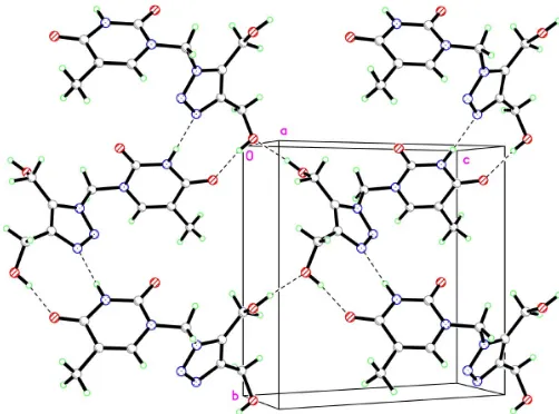
In the crystal structure, the molecules of (I) form a spiral along the 21 axis, with neighbouring molecules in the spiral
connected by N3—H3 N30i
and O40 0—H400 O4ii
hydrogen bonds, while O500—H500 O400iii
hydrogen bonds link neigh-bouring spirals, forming an infinite three-dimensional mol-ecular network (Table 1, Fig. 2; symmetry codes as in Table 1).
Experimental
Compound (I) was obtained by the general procedure of cyclo-addition of the azide dipole to alkyne dipolarophiles. 1-Methylthio-methylthymine (Zavgorodny et al., 1997) was converted to 1-azidomethylthymine by the procedure developed earlier forO,S -acetals (Zavgorodnyet al., 2000), in 87% yield. This azido synthon gives the title compound in 88% yield by heating with 2-butyne-1,4-diol in methanol (m.p. 455–456 K). Crystals of (I) for X-ray analysis were prepared from a saturated solution of (I) in methanol, to which several drops of hexane had been added, kept at 277 K. Full details of the synthesis and characterization of (I) will be published elsewhere.
Crystal data
C10H13N5O4
Mr= 267.25
Monoclinic,P21
a= 4.516 (1) A˚
b= 11.618 (2) A˚
c= 11.481 (2) A˚ = 91.43 (2) V= 602.2 (2) A˚3
Z= 2
Dx= 1.474 Mg m
3 MoKradiation Cell parameters from 24
reflections = 12.3–14.2
= 0.12 mm1
T= 293 (2) K Prism, colourless 0.320.300.12 mm
Data collection
Enraf–Nonius CAD-4 diffractometer !/2scans
Absorption correction: none 8497 measured reflections 2746 independent reflections 2350 reflections withI> 2(I)
Rint= 0.016
max= 35.0
h= 0!7
k=18!18
l=18!18 3 standard reflections
frequency: 60 min intensity decay: 0.3%
Refinement
Refinement onF2
R[F2> 2(F2)] = 0.030
wR(F2) = 0.086
S= 1.04 2746 reflections 197 parameters
H atoms treated by a mixture of independent and constrained refinement
w= 1/[2(F
o2) + (0.0712P)2] whereP= (Fo2+ 2Fc2)/3 (/)max= 0.001
max= 0.24 e A˚
3 min=0.21 e A˚
3
Table 1
Hydrogen-bond geometry (A˚ ,).
D—H A D—H H A D A D—H A
N3—H3 N30i
0.90 (2) 2.00 (2) 2.8989 (12) 177 (2) O40 0
—H40 0
O4ii
0.81 (2) 1.94 (2) 2.7137 (15) 159 (2) O50 0—H50 0 O40 0iii
0.89 (1) 1.80 (1) 2.6863 (12) 175 (1)
Symmetry codes: (i) xþ1;y1
2;zþ1; (ii) xþ1;yþ 1
2;zþ1; (iii)
x;y1 2;z.
All H atoms were located in difference syntheses and refined isotropically. In the refinement, atoms H40 0, H50 0, H3 and H6 were fully refined, while for the others,Uiso(H) values were refined with
thexyzcoordinates constrained. The C—H bond lengths are in the range 0.92 (2)–0.98 (2) A˚ , N—H bond lengths are 0.904 (2) A˚ and O—H bond lengths are in the range 0.83 (2)–0.89 (1) A˚ .
Data collection:CAD-4/PC Software (Enraf–Nonius, 1993); cell refinement: CAD-4/PC Software; data reduction: CAD-4/PC Soft-ware; program(s) used to solve structure: SHELXS97 (Sheldrick, 1997a); program(s) used to refine structure:SHELXL97(Sheldrick, 1997a); molecular graphics: XP in SHELXTL (Sheldrick, 1997b); software used to prepare material for publication: CIFTAB97
(Sheldrick, 1997a) andSHELXL97.
This work was supported by the Russian Foundation for Basic Research, Grant No. 04-03-33053, and the President Grant No. 1781.2003.4 for support of the Leading Russian Scientific Schools.
References
Adams, D. R., Boyd, A. S. F., Ferguson, R., Grierson, D. S. & Monneret, C. (1998).Nucleosides Nucleotides,17, 1053–1075.
Doboszewski, B. (1997).Nucleosides Nucleotides,16, 1049–1052.
Enraf–Nonius (1993).CAD-4/PC Software. Version 1.2. Enraf–Nonius, Delft, The Netherlands.
Sallam, M. A. E. & Townsend, L. B. (1998).Nucleosides Nucleotides,17, 1215– 1229.
Sheldrick, G. M. (1997a).SHELXS97,SHELXL97and CIFTAB. Release 97-2. University of Go¨ttingen, Germany.
organic papers
o1722
Stashet al. C [image:2.610.313.564.70.256.2]10H13N5O4 Acta Cryst.(2005). E61, o1721–o1723
Figure 1
The molecular structure of (I), showing the atom-labelling scheme. Displacement ellipsoids are drawn at the 50% probability level and H
atoms are drawn as spheres of arbitrary radii. Figure 2
[image:2.610.46.300.74.231.2]Sheldrick, G. M. (1997b). SHELXTL. Version 5.10. Bruker AXS Inc., Madison, Wisconsin, USA.
Xiang, Y., Chen, J., Shinazi, R. F. & Zhao, K. (1996).Biomed. Chem. Lett.6, 1051–1054.
Zavgorodny, S. G., Malyshev, A. A., Konstantinova, I. D., Kuznetsov, S. A. & Miroshnikov, A. I. (1997).Bioorg. Khim.23, 69–71. (In Russian). Zavgorodny, S. G., Pechenov, A. E., Shvets, V. I. & Miroshnikov, A. I. (2000).
Nucleosides Nucleotides Nucleic Acids,19, 1977–1991.
organic papers
Acta Cryst.(2005). E61, o1721–o1723 Stashet al. C
supporting information
sup-1
Acta Cryst. (2005). E61, o1721–o1723
supporting information
Acta Cryst. (2005). E61, o1721–o1723 [https://doi.org/10.1107/S1600536805014650]
1-[4,5-Bis(hydroxymethyl)-1,2,3-triazol-1-ylmethyl]thymine
Adam I. Stash, Valery E. Zavodnik, Sergei G. Zavgorodny, Galina V. Gurskaya and Vladimir G.
Tsirelson
1-[Bis(4,5-hydroxymethyl)-1,2,3-triazol-1-ylmethyl]thymine
Crystal data
C10H13N5O4 Mr = 267.25
Monoclinic, P21
Hall symbol: P 2yb
a = 4.516 (1) Å
b = 11.618 (2) Å
c = 11.481 (2) Å
β = 91.43 (2)°
V = 602.2 (2) Å3 Z = 2
F(000) = 280
Dx = 1.474 Mg m−3
Mo Kα radiation, λ = 0.71073 Å Cell parameters from 24 reflections
θ = 12.3–14.2°
µ = 0.12 mm−1 T = 293 K Prism, colourless 0.32 × 0.30 × 0.12 mm
Data collection
Enraf–Nonius CAD-4 diffractometer
Radiation source: fine-focus sealed tube
β-filter′ monochromator
ω/2θ scans
8497 measured reflections 2746 independent reflections 2350 reflections with I > 2σ(I)
Rint = 0.016
θmax = 35.0°, θmin = 1.8°
h = 0→7
k = −18→18
l = −18→18
3 standard reflections every 60 min intensity decay: 0.3%
Refinement
Refinement on F2
Least-squares matrix: full
R[F2 > 2σ(F2)] = 0.030 wR(F2) = 0.086 S = 1.04 2746 reflections 197 parameters 1 restraint
Primary atom site location: structure-invariant direct methods
Secondary atom site location: difference Fourier map
Hydrogen site location: difference Fourier map H atoms treated by a mixture of independent
and constrained refinement
w = 1/[σ2(Fo2) + (0.0712P)2]
where P = (Fo2 + 2Fc2)/3
(Δ/σ)max = 0.001
Δρmax = 0.24 e Å−3
supporting information
sup-2
Acta Cryst. (2005). E61, o1721–o1723 Special details
Geometry. All e.s.d.'s (except the e.s.d. in the dihedral angle between two l.s. planes) are estimated using the full covariance matrix. The cell e.s.d.'s are taken into account individually in the estimation of e.s.d.'s in distances, angles and torsion angles; correlations between e.s.d.'s in cell parameters are only
used when they are defined by crystal symmetry. An approximate (isotropic) treatment of cell e.s.d.'s is used for estimating e.s.d.'s involving l.s. planes.
Refinement. Refinement of F2 against ALL reflections. The weighted R-factor wR and goodness of fit S are based on F2,
conventional R-factors R are based on F, with F set to zero for negative F2. The threshold expression of F2 > σ(F2) is used
only for calculating R-factors(gt) etc. and is not relevant to the choice of reflections for refinement. R-factors based on F2
are statistically about twice as large as those based on F, and R- factors based on ALL data will be even larger.
Fractional atomic coordinates and isotropic or equivalent isotropic displacement parameters (Å2)
x y z Uiso*/Ueq
O2 0.2831 (2) 0.02525 (8) 0.45490 (7) 0.04333 (17)
O4 0.2803 (3) 0.14152 (9) 0.83244 (8) 0.0549 (2)
N1 −0.03431 (18) 0.16888 (7) 0.50471 (7) 0.03122 (14)
C2 0.18000 (19) 0.08706 (7) 0.52957 (7) 0.03025 (15)
N3 0.27080 (18) 0.08302 (7) 0.64428 (7) 0.03328 (15)
C4 0.1798 (2) 0.15398 (8) 0.73250 (8) 0.03582 (18)
C5 −0.0315 (3) 0.24269 (8) 0.69914 (9) 0.03724 (17)
C6 −0.1265 (2) 0.24643 (8) 0.58771 (8) 0.03460 (16)
C7 −0.1315 (4) 0.32579 (14) 0.78901 (13) 0.0610 (4)
C1′ −0.1378 (2) 0.17923 (10) 0.38560 (7) 0.03575 (18)
N1′ 0.06362 (16) 0.25123 (7) 0.31885 (6) 0.02950 (13)
N2′ 0.1660 (2) 0.34927 (8) 0.36632 (7) 0.03868 (18)
N3′ 0.3252 (2) 0.39986 (8) 0.28728 (7) 0.03699 (16)
C5′ 0.15171 (17) 0.23879 (7) 0.20745 (7) 0.02686 (14)
C4′ 0.32007 (19) 0.33556 (8) 0.18843 (7) 0.02970 (15)
C5′′ 0.0819 (2) 0.13764 (9) 0.13279 (9) 0.03698 (19)
O5′′ −0.22467 (18) 0.11452 (10) 0.13752 (9) 0.0500 (2)
C4′′ 0.4755 (2) 0.37334 (11) 0.08132 (9) 0.0407 (2)
O4′′ 0.3870 (3) 0.48514 (10) 0.04720 (8) 0.0545 (2)
H3 0.402 (4) 0.0279 (17) 0.6657 (15) 0.040 (4)*
H6 −0.260 (5) 0.300 (2) 0.560 (2) 0.060 (6)*
H4′′ 0.452 (5) 0.531 (2) 0.095 (2) 0.059 (5)*
H5′′ −0.2699 (15) 0.0686 (7) 0.0780 (6) 0.045 (4)*
H71 −0.2254 0.2769 0.8462 0.106 (11)*
H72 0.0246 0.3691 0.8217 0.146 (17)*
H73 −0.2683 0.3849 0.7643 0.078 (7)*
H11′ −0.1349 0.1028 0.3504 0.050 (4)*
H12′ −0.3347 0.2120 0.3842 0.045 (4)*
H41′′ 0.4090 0.3251 0.0160 0.060 (6)*
H42′′ 0.6840 0.3665 0.0826 0.044 (4)*
H51′′ 0.1843 0.0706 0.1620 0.065 (6)*
supporting information
sup-3
Acta Cryst. (2005). E61, o1721–o1723 Atomic displacement parameters (Å2)
U11 U22 U33 U12 U13 U23
O2 0.0578 (4) 0.0411 (4) 0.0313 (3) 0.0068 (3) 0.0076 (3) −0.0072 (3)
O4 0.0859 (7) 0.0517 (5) 0.0267 (3) 0.0101 (5) −0.0101 (4) −0.0039 (3)
N1 0.0378 (3) 0.0329 (3) 0.0230 (2) −0.0003 (3) 0.0012 (2) 0.0021 (2)
C2 0.0380 (3) 0.0283 (3) 0.0247 (3) −0.0019 (3) 0.0034 (3) −0.0008 (3)
N3 0.0433 (4) 0.0305 (3) 0.0260 (3) 0.0049 (3) −0.0012 (3) −0.0007 (2)
C4 0.0504 (5) 0.0320 (4) 0.0250 (3) −0.0017 (4) 0.0013 (3) −0.0014 (3)
C5 0.0513 (5) 0.0302 (4) 0.0305 (4) 0.0027 (3) 0.0060 (3) −0.0043 (3)
C6 0.0413 (4) 0.0306 (4) 0.0321 (4) 0.0041 (3) 0.0047 (3) 0.0016 (3)
C7 0.0855 (10) 0.0497 (7) 0.0481 (6) 0.0144 (7) 0.0093 (6) −0.0192 (6)
C1′ 0.0355 (4) 0.0458 (5) 0.0258 (3) −0.0096 (3) −0.0023 (3) 0.0055 (3)
N1′ 0.0333 (3) 0.0329 (3) 0.0223 (2) −0.0040 (3) −0.0002 (2) 0.0009 (2)
N2′ 0.0484 (4) 0.0401 (4) 0.0276 (3) −0.0121 (3) 0.0039 (3) −0.0057 (3)
N3′ 0.0459 (4) 0.0343 (4) 0.0309 (3) −0.0096 (3) 0.0027 (3) −0.0020 (3)
C5′ 0.0298 (3) 0.0282 (3) 0.0224 (3) 0.0018 (3) −0.0017 (2) 0.0009 (2)
C4′ 0.0336 (3) 0.0304 (3) 0.0250 (3) 0.0003 (3) −0.0003 (2) 0.0041 (3)
C5′′ 0.0385 (4) 0.0360 (4) 0.0365 (4) −0.0010 (3) 0.0036 (3) −0.0110 (3)
O5′′ 0.0371 (3) 0.0655 (6) 0.0471 (4) −0.0062 (3) −0.0024 (3) −0.0293 (4)
C4′′ 0.0471 (5) 0.0442 (5) 0.0310 (4) −0.0007 (4) 0.0060 (3) 0.0100 (4)
O4′′ 0.0804 (6) 0.0467 (5) 0.0355 (4) −0.0062 (5) −0.0147 (4) 0.0183 (4)
Geometric parameters (Å, º)
O2—C2 1.2196 (11) C1′—H12′ 0.9671
O4—C4 1.2320 (13) N1′—N2′ 1.3400 (11)
N1—C2 1.3813 (12) N1′—C5′ 1.3566 (10)
N1—C6 1.3830 (12) N2′—N3′ 1.3108 (12)
N1—C1′ 1.4391 (12) N3′—C4′ 1.3584 (12)
C2—N3 1.3704 (11) C5′—C4′ 1.3777 (12)
N3—C4 1.3767 (12) C5′—C5′′ 1.4837 (12)
N3—H3 0.904 (19) C4′—C4′′ 1.4968 (13)
C4—C5 1.4493 (15) C5′′—O5′′ 1.4125 (13)
C5—C6 1.3399 (14) C5′′—H51′′ 0.9618
C5—C7 1.4915 (15) C5′′—H52′′ 0.9574
C6—H6 0.92 (3) O5′′—H5′′ 0.887 (7)
C7—H71 0.9731 C4′′—O4′′ 1.4116 (15)
C7—H72 0.9370 C4′′—H41′′ 0.9776
C7—H73 0.9613 C4′′—H42′′ 0.9446
C1′—N1′ 1.4661 (12) O4′′—H4′′ 0.81 (2)
C1′—H11′ 0.9757
C2—N1—C6 121.82 (8) N1′—C1′—H12′ 110.4
C2—N1—C1′ 117.58 (8) H11′—C1′—H12′ 111.9
C6—N1—C1′ 120.27 (8) N2′—N1′—C5′ 111.58 (7)
O2—C2—N3 123.03 (9) N2′—N1′—C1′ 118.98 (8)
supporting information
sup-4
Acta Cryst. (2005). E61, o1721–o1723
N3—C2—N1 114.42 (8) N3′—N2′—N1′ 106.80 (7)
C2—N3—C4 126.79 (8) N2′—N3′—C4′ 109.49 (8)
C2—N3—H3 117.7 (11) N1′—C5′—C4′ 103.80 (7)
C4—N3—H3 115.5 (11) N1′—C5′—C5′′ 124.46 (8)
O4—C4—N3 120.33 (10) C4′—C5′—C5′′ 131.68 (8)
O4—C4—C5 123.69 (10) N3′—C4′—C5′ 108.32 (8)
N3—C4—C5 115.98 (8) N3′—C4′—C4′′ 121.78 (9)
C6—C5—C4 117.93 (8) C5′—C4′—C4′′ 129.89 (9)
C6—C5—C7 122.97 (11) O5′′—C5′′—C5′ 108.84 (8)
C4—C5—C7 119.10 (11) O5′′—C5′′—H51′′ 107.2
C5—C6—N1 122.82 (9) C5′—C5′′—H51′′ 110.3
C5—C6—H6 123.2 (15) O5′′—C5′′—H52′′ 111.9
N1—C6—H6 114.0 (15) C5′—C5′′—H52′′ 109.8
C5—C7—H71 103.5 H51′′—C5′′—H52′′ 108.7
C5—C7—H72 112.7 C5′′—O5′′—H5′′ 107.0 (4)
H71—C7—H72 112.3 O4′′—C4′′—C4′ 111.25 (10)
C5—C7—H73 117.5 O4′′—C4′′—H41′′ 103.6
H71—C7—H73 109.2 C4′—C4′′—H41′′ 108.7
H72—C7—H73 101.9 O4′′—C4′′—H42′′ 110.9
N1—C1′—N1′ 110.70 (7) C4′—C4′′—H42′′ 116.8
N1—C1′—H11′ 108.1 H41′′—C4′′—H42′′ 104.5
N1′—C1′—H11′ 106.7 C4′′—O4′′—H4′′ 108.8 (16)
N1—C1′—H12′ 109.0
Hydrogen-bond geometry (Å, º)
D—H···A D—H H···A D···A D—H···A
N3—H3···N3′i 0.904 (19) 1.996 (19) 2.8989 (12) 176.9 (16)
O4′′—H4′′···O4ii 0.81 (2) 1.94 (2) 2.7137 (15) 159 (2)
O5′′—H5′′···O4′′iii 0.887 (7) 1.802 (7) 2.6863 (12) 174.7 (7)