Protein Structure Refinement
Algorithms
!
!
!
Thesis by
Mohsen Chitsaz
!
In Partial Fulfillment of the Requirements for the
degree of
PhD in Molecular Biophysics and Minor in Computer
Science
!
!
!
!
CALIFORNIA INSTITUTE OF TECHNOLOGY
Pasadena, California
2014
Acknowledgements
!
I would like to express my special appreciation and thanks to my advisor Professor
Dr. Stephen L. Mayo, for being a tremendous mentor for me. I would like to thank
you for encouraging my research and for allowing me to grow as a research
scien-tist. Your advice on both research as well as on my career have been priceless. I
will always look back and appreciate the chance that I was given to have you as
my mentor. I would also like to thank Professor William A. Goddard, Professor
Pamela J. Bjorkman, and Professor Chris Umans for serving as my committee
members. I would also like to thank Dr. Marie Arie for her help with writing
manu-scripts and her comments on the organization of the text of this thesis. I also would
like to thank Rhonda DiGuisto and everyone else in Mayo Lab. I also would like to
express my great appreciation and thanks to the graduate office, Dean Joseph
Shepherd and Ms. Natalie Gilmore for their invaluable kindness and help with the
students in tough times. I would also like to thank Daniel Yoder and ISP for their
great help throughout my programs.
Words cannot express how grateful I am to my parents for all of the sacrifices that
they have made to provide me with the opportunity to be able to focus on
develop-ing myself scientifically and professionally, and explore and investigate my
re-search ideas, and pursue my education. Your prayers and inspirations for me was
what made me succeed thus far. I would also like to thank my siblings and my
fam-ily for all their support, help, inspirations and encouragements. I also would like to
greatly thank all my friends: Alireza Mohammad Karim, Bernardo Sosa Padilla
Babakhani, Masoud Farivar, Asghar Aryanfar, Vanessa Heckman, and Gokjan
Karakus all of whom have made my residence at Caltech very memorable,
!
!
Abstract
Protein structure prediction has remained a major challenge in structural biology
for more than half a century. Accelerated and cost efficient sequencing
technolo-gies have allowed researchers to sequence new organisms and discover new
pro-tein sequences. Novel propro-tein structure prediction technologies will allow
re-searchers to study the structure of proteins and to determine their roles in the
un-derlying biology processes and develop novel therapeutics.
Difficulty of the problem stems from two folds: (a) describing the energy landscape
that corresponds to the protein structure, commonly referred to as force field
prob-lem; and (b) sampling of the energy landscape, trying to find the lowest energy
configuration that is hypothesized to be the native state of the structure in solution.
The two problems are interweaved and they have to be solved simultaneously.
This thesis is composed of three major contributions. In the first chapter we
de-scribe a novel high-resolution protein structure refinement algorithm called GRID.
In the second chapter we present REMC
GRID
, an algorithm for generation of low
energy decoy sets. In the third chapter, we present a machine learning approach
to ranking decoys by incorporating coarse-grain features of protein structures.
!
!
Figures and Tables
Figure 1-1. Schematic showing the GRID algorithm
...
24
!
Figure 1-2. Post-refinement analysis of GRID moves
...
25
!
Figure 1-3. Performance of GRID vs. Backrub
...
26
!
Figure 1-4. Backbone RMSD with respect to starting crystal structure
...
27
!
Figure 1-S1. Energy improvement following refinement with GRID
...
28
!
Figure 1-S2. GRID moves plotted as a function of residue number
...
29-31
!
Table 1-1. Proteins used as test set for structure refinement
...
32
!
Figure 2-1. REMC
GRIDalgorithm psedocode
...
43
!
Figure 2-2. The energy improvement of 10 structures using REMC
GRID...
44
!
Figure 2-3. REMC
GRIDenergy improvement compared to GRID
...
45
!
Figure 2-4. The ratio of energy improvement of REMC
GRIDvs. GRID
...
46
!
Figure 2-5. Computational effort comparison of REMC
GRIDand GRID
...
47
!
Figure 2-6. Backbone rmsds of 80 replicas
...
48
!
Figure 2-7. Backbone rmsds of decoys (pdbID: 1pga)
...
49
!
Figure 2-8. Backbone rmsd vs. energy of decoys generated
...
50
!
Figure 2-9. Backbone rmsd distribution of decoy sets generated by REMC
GRID...
51
!
Figure 2-10. Comparison of Rosetta decoys and REMC
GRID...
52
!
Table 2-1. Ten high-resolution structures used in testing REMC
GRID...
53
!
Figure 3-1. Coarse-grained model of protein structures
...
62
!
Figure 3-2(A,B,C). Energy model for the coarse-grained model proposed in Chapter 3
...
63-65
!
Figure 3-3. Pairwise energies among amino acid groups
...
66
!
Figure 3-4. Backbone rmsd distribution of decoys generated by REMC
GRID...
67
!
Figure 3-5. Comparison of three different classifiers
...
68
!
Figure 3-S1. Coarse-grained score vs. backbone RMSD (pdbID: 1fkb)
...
69
!
Figure 3-S2. Comparison of coarse-grained score vs. minimum and average rmsd
...
70
!
Figure 3-S3. Protein structure description in the coarse-grained model
...
71
!
Figure 3-S4. Modeling the scoring function using sigmoid functions
...
72
!
Figure 3-S5. Sidechain-sidechain energy term
...
73
!
Figure 3-S6. Backbone-backbone energy term
...
74
!
Figure 3-S7. Sidechain-backbone energy term
...
75
!
Figure 3-S8. Gradient of the energy function
...
76
!
Figure 3-S9. Coarse-grained model, energy function, and the gradient formulation
...
77
!
!
Table 3-1. Amino acids categorization into 7 groups
...
79
!
Table 3-2. Pairwise energies that are used in the coarse-grained scoring function
...
80
!
Table 3-3. Comparison of three different classifiers trained on decoys
...
81
!!
Nomenclature
!
RMSD:
Root Mean Square Deviation
GRID:
A high-resolution refinement algorithm
REMC
GRID:
Replica Exchange Monte Carlo GRID
AU:
Arbitrary Units
!
SVM:
Support Vector Machine
!
PDB:
Protein Data Bank
!
Residue:
an amino acid in the context of a protein structure
!
ϕ
:
Dihedral angle formed on the backbone of a residue by C, N, C-alpha, C atoms
!
ψ
:
Dihedral angle formed on the backbone of a residue by N, C-alpha, C, N atoms
!
Side-chain:
Chemical group attached to the backbone of the protein, determining the type of amino acid
!
Conformation:
3-dimensional state of a protein structure in Cartesian space
!
Forcefield:
a scoring function that takes a conformation of a protein and assigns energy to it
!
Decoy:
a conformation of a protein, typically, perturbed state of a protein structure
!
!
TABLE OF CONTENTS
!
Acknowledgements
...
ii
!
Abstract
...
iv
!
Figures and Tables
...
v
!
Nomenclature
...
vii
!
Table of Contents
...
viii
!
Chapter 1: GRID Algorithm
...
9
!
Chapter 2: REMC
GRID
Algorithm
...
33
!
Chapter 3: Coarse-grained Scoring and Machine Learning
...
54
!
Appendix A: Header file for the algorithms
...
83
!
Appendix B: Main code for the algorithms
...
88
!
!
Chapter 1
!
!
GRID: A High-Resolution Protein Structure Refinement Algorithm
!
The text of this chapter is adapted from a published review article that was co-authored
with Professor Stephen L. Mayo
Chitsaz M, Mayo SL.,
J Comput Chem
2013;
34
:445–450.
!
!
Abstract
The energy-based refinement of protein structures generated by fold prediction
algo-rithms to atomic-level accuracy remains a major challenge in structural biology.
Energy-based refinement is mainly dependent on two components: (1) sufficiently accurate force
fields, and (2) efficient conformational space search algorithms. Focusing on the latter, we
developed a high-resolution refinement algorithm called GRID. It takes a
three-dimen-sional protein structure as input and, employing an all-atom force field, attempts to
im-prove the energy of the structure by systematically perturbing backbone dihedrals and
side chain rotamer conformations. We compare GRID to Backrub, a stochastic algorithm
that has been shown to predict a significant fraction of the conformational changes that
occur with point mutations. We applied GRID and Backrub to 10 high-resolution (
≤
2.8 Å)
crystal structures from the Protein Data Bank and measured the energy improvements
obtained and the computation times required to achieve them. GRID resulted in energy
improvements that were significantly better than those attained by Backrub while
expend-ing about the same amount of computational resources. GRID resulted in relaxed
struc-tures that had slightly higher backbone RMSDs compared to Backrub relative to the
start-ing crystal structures. The average RMSD was 0.25 ± 0.02 Å for GRID versus 0.14 ± 0.04
Å for Backrub. These relatively minor deviations indicate that both algorithms generate
structures that retain their original topologies, as expected given the nature of the
algo-rithms.
Introduction
Although much progress has been made in recent years, the ability to predict the
three-dimensional structure of a protein from its amino acid sequence remains a major
chal-lenge in structural biology. Good prediction techniques would allow us to obtain the
struc-tures of novel protein sequences without having to determine them experimentally and
would be particularly useful for membrane and other proteins whose structures are
exper-imentally difficult to obtain or are highly diverged from the set of proteins with existing
structures
1
. The knowledge obtained from accurate protein structure prediction could
im-prove our understanding of how proteins function, aid in the discovery of proteins with
new and improved properties, and facilitate the development of new drugs
2, 3
. Many
bio-logical applications, including studies on enzymatic activity, virtual ligand screening,
struc-ture-based protein function prediction, and strucstruc-ture-based drug design require
knowl-edge of protein structures at atomic resolution
1
. In addition, the expansion of
high-throughput genome sequencing projects has led to a continually widening gap between
protein sequence data and the number of solved structures. Thus, there is a significant
need for computational techniques that can model protein structures with atomic accuracy
comparable to experimental methods.
Protein structure prediction methods can be broadly classified into three main
cate-gories: comparative modeling, threading, and template-free modeling
4
. In comparative
modeling, the protein structure is built by using sequence alignments to find the best
matching evolutionarily related protein that has a solved structure (the template)
4
. Most
structures constructed using this approach have a root mean square deviation (RMSD) of
pro-teins, threading can be used, which matches the target protein directly to the sequences
of solved three-dimensional structures. Threading methods often identify correct
tem-plates and provide models with an RMSD of 2-6 Å, with inaccuracies occurring mainly in
the loop regions
6
. For targets that have no structurally-related solved proteins (< 30%
se-quence similarity), template-free modeling methods are used; that is, the model is built
from scratch
5, 7
. Successful prediction using template-free modeling is limited to small
pro-teins (< 120 residues), with accuracy in the range of 4-8 Å
8
. Much effort has been
ex-pended in the last several decades to achieve the current levels of success in each of
these three categories. However, the resulting models are still low-resolution and further
refinement is needed in order to achieve the atomic-level accuracy required for most
bio-logical applications. Refinement of low-resolution structures to higher resolution models
therefore remains a major challenge in the field of protein structure prediction
1, 4, 9-11
.
Recognition of the importance of refinement is reflected by the addition of a refinement
category in the eighth Critical Assessment of Techniques for Protein Structure Prediction
(CASP8)
12
.
Approaches to high-resolution refinement have included molecular mechanics energy
minimization and Monte Carlo methods including replica exchange Monte Carlo
1, 13-16
.
Knowledge-based statistically derived potentials have also been incorporated into these
techniques
17
. Recently, a flexible backbone modeling algorithm was applied
18-20
. All these
methods have attained some degree of success; however, none has emerged as clearly
superior
13, 14, 16
. Current methods require improvements in terms of computational
effi-ciency and/or accuracy
18, 20-23
. For those approaches that generate an ensemble
effi-cient in processing many structures in a reasonable amount of time. In addition, the
re-fined structure must accurately reflect the naturally folded state. Because the minimum
energy configuration is considered to be the best estimate of native structure
24,25
,
high-resolution refinement methods must be able to efficiently find lower energy configurations.
The energy improvement attained relative to the starting structure is a measure of a
method's progress towards a model consistent with the experimental data, assuming an
accurate energy function. Thus, refinement procedures typically employ multiple
refine-ment cycles in which the energy of a low-resolution structure is iteratively reduced. The
energy of the structure is calculated based on molecular mechanics force fields that
de-scribe the underlying physics and chemistry of protein molecules
14, 16
. Knowledge-based
force fields are also commonly used
26
.
To address the problems of computational efficiency and accuracy, we devised a
high-resolution refinement algorithm called GRID. The method sequentially applies
perturba-tions to a structure's
φ
and
ψ
angles and side chain rotamers, accepting perturbations
that produce the most improvement in the energy of the structure. Unlike many
refine-ment methods that rely on stochastic techniques, GRID has been tuned to use a single
pass search strategy and therefore requires less computational effort. We compared
GRID to Backrub
27
, a flexible backbone modeling method that is able to predict most of
the small conformational changes associated with point mutations
18, 20
. GRID and
Back-rub were applied to a set of 10 high-resolution crystal structures from the PDB, and the
algorithms were assessed in terms of energy improvement and computational efficiency.
Computational Methods
GRID refinement algorithm
GRID is a refinement method that takes a three-dimensional protein structure as input
and, employing an all-atom force field, attempts to improve the energy of the structure
us-ing a move set that allows for backbone and side chain flexibility (Fig. 1). Three degrees
of freedom are considered:
φ
and
ψ
angle perturbations and side-chain rotamer
optimiza-tion. The algorithm starts from the first residue (N terminus) and evaluates all
combina-tions of
Δφ
,
Δψ
, and rotamer, given a specified maximum perturbation angle and precision
level (perturbation angle step size).
Δφ
and
Δψ
perturbations are applied to the C-terminal
protein segment. The continuous space of
Δφ
,
Δψ
perturbations is broken down into
dis-crete grid points around the current
φ
and
ψ
angles of the residue. Similarly, a rotamer
library is used to define a discrete set of rotamers for the residue. For each combination
of
Δφ
,
Δψ
, and rotamer available for a given residue, the algorithm perturbs the structure
and calculates the energy of the trial configuration (the rest of the molecule is treated as a
rigid body). This information is used to fill out a three-dimensional matrix that reflects the
discretized energy space for the residue. The move that results in the lowest energy is
then applied to the structure before proceeding to the next residue. GRID marches
for-ward from N to C terminus, sequentially calculating the energy matrix and applying the
lowest energy move on each of the residues. When the last residue is reached, the
algo-rithm can terminate or return to the N terminus for a subsequent iteration.
Note that although the moves are restricted to local perturbations relative to a single
residue, the perturbation selected influences the entire structure. Thus, relatively minor
of the protein. In order to further improve the energies, at the end of the procedure, the
algorithm walks through the structure once (from N to C terminus) and performs four
steps of local conjugate gradient energy minimization on each residue while keeping the
rest of the structure fixed in Cartesian space.
!
GRID implementation and parameterization
The GRID refinement algorithm was developed in C++ and incorporated into the Triad
protein design software package
33
. The Rosetta force field with default parameters
28
was
used to calculate the energy of the protein conformations. Rotamers were selected from a
backbone-dependent side-chain rotamer library
29
.
As noted above, GRID takes as input four main parameters: (1) the maximum
pertur-bation angle (
Δφ
,
Δψ
) for each iteration, (2) the precision level (step size for
Δφ
and
Δψ
)
for each iteration, (3) the rotamer library used for each iteration, and (4) the number of
it-erations. GRID was run on 2-10 test proteins using several combinations of these
para-meters to determine the optimal protocol for refinement. Values for maximum
Δφ
and
Δψ
ranged from ±0.1° to ±8.0° and precision levels ranged from 0.1° to 8.0°, forming
Δφ
×
Δψ
matrices containing from 9 to 441 grid points; the number of iterations ranged from 1 to
10. The same set of rotamers (from the Dunbrack backbone-dependent rotamer library
29
)
was used in every case.
!
Backrub algorithm
The Backrub concept was developed by the Richardson lab
27
and was implemented by
that ultra-high-resolution crystal structures often show alternate conformations in which
subtle local backbone motion is coupled to much larger two-state changes in side-chain
conformation
18, 20, 21
. This “backrub” motion supplies a common type of local plasticity in
protein backbones. A refinement algorithm based on the Backrub concept performs
10,000 steps of Monte Carlo simulation using Metropolis acceptance criteria. In every
step, Backrub employs two types of moves with equal probability: (1) it replaces a
side-chain rotamer (randomly chosen from a specified rotamer library) at a randomly chosen
residue position, or (2) it performs a Backrub move in which a randomly chosen 2- or
3-residue fragment is rotated about the axis defined by the C
α
atoms of the first and the last
residue of the fragment. The bond angles extending from the C
α
atoms of the first and the
last residues are then optimized using a bond angle potential. We used the Rosetta
ver-sion 3.1 implementation of Backrub, with parameters as described in Friedland et al
18
.
!
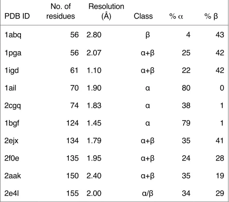
Test sets and assessment criteria
GRID and Backrub were initially tested on 10 single-chain high-resolution (
≤
2.8 Å) crystal
structures from the PDB representing proteins of various sizes and folds (
α
,
β
,
α
+
β
, and
α
/
β
) (Table 1). Two criteria were used to compare the algorithms: (1) energy improvement
(relative to that of the starting structure), and (2) computation time. The validity of the first
criterion is based on the assumption that the minimum energy configuration is the best
estimate of the native state
24, 32
and that the degree of energy improvement attained
re-flects the method's success in moving toward the native form and, perhaps more
impor-tantly, allowing selection of the best structural models from an ensemble of predicted
!
Results
GRID parameterization: smaller perturbations of
φ and
ψ allow more global
struc-ture refinement
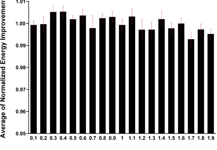
Initial trials on 2–10 test proteins indicated that refinements done using a single iteration
and the lowest precision level, a 3
×
3
Δφ
×
Δψ
matrix, produced energy improvements
that were at least 70% of the values obtained with larger matrices (11
×
11) and multiple
iterations (up to 10). These initial studies also showed that perturbations of
φ
and
ψ
greater than ± 2.0° caused no significant energy improvement over GRID refinements
performed using smaller perturbations. More extensive tests were performed using
small-er psmall-erturbation angles that ranged from ± 0.1° to ± 1.9°, and all of these resulted in the
same level of energy improvement (Fig. S1). Completely eliminating
φ
and
ψ
perturba-tions (i.e., sidechain rotamer optimization alone) was less effective at energy
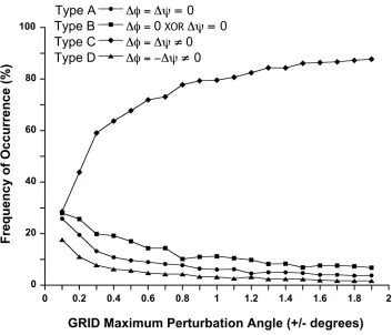
improve-ment (Fig. S2). We then conducted a post-refineimprove-ment analysis of the moves that had
been applied to achieve the final refined structures (Fig. 2). The moves were divided into
four types that differ in their effects on protein structure: type A, the backbone is not
per-turbed (
Δφ
= 0,
Δψ
= 0); type B, only one of the dihedrals is perturbed (
Δφ
= 0,
Δψ
≠
0) or
(
Δφ
≠
0,
Δψ
= 0); type C, both dihedrals are perturbed by the same amount in the same
direction (
Δφ
=
Δψ
); and type D, both dihedrals are perturbed by the same amount, but in
opposite directions (
Δφ
=
−Δψ
). Side chain rotamer optimization was allowed in all
cas-es. The frequency of occurrence of each of these different types of moves was then
cal-culated. As seen in Figure 2, move type A (no backbone perturbation) became
ap-plied. The next most common move was type B, followed by type D, then type C; this
trend was consistent at all perturbation angles tested. At the smallest perturbation angle
(0.1°), the type A move (no backbone perturbation) was no longer dominant and occurred
at about the same frequency as types B and D (~27%).
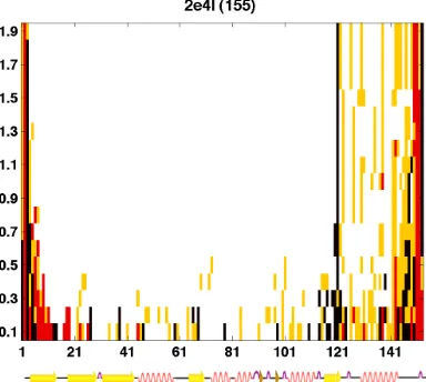
In choosing the parameters for refinement with GRID, one of the criteria was that the
perturbations be fairly evenly dispersed throughout the protein structure and not restricted
to specific regions. We found that using the smallest perturbation (± 0.1°) for
Δφ
and
Δψ
gave the best results (i.e., the frequency of dihedral angle change was similar for all
residues), whereas with larger perturbations, most of the moves caused no backbone
movement (only the rotamer was changed), and moves that did perturb the backbone
were restricted to residues near the termini (Fig. S3). These results led us to choose a
maximum perturbation angle of ± 0.1°. To minimize computation time, the lowest values
were also used for the number of iterations and for the precision level. The final GRID
re-finement parameters are therefore as follows: maximum perturbation angle (
Δφ
,
Δψ
) = ± 0.1°, precision level = 0.1°, and iterations = 1, with rotamers from the Dunbrack
backbone-dependent rotamer library
29
.
!
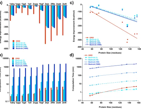
GRID outperforms Backrub
As shown in Figure 3a, refinement with GRID resulted in structures with energy
improve-ments ranging from ~100 to ~600 kcal/mol. In every case, these values were significantly
larger than those obtained with a single trajectory of Backrub. Increasing the number of
Backrub trajectories to 10, 100, or 1000 caused minimal additional improvement. The four
pro-cedure has a significant impact on the resulting energies, increasing the overall energy
improvement by about a factor of 2 (Fig. S4). The computation time expended by GRID
was comparable to that required for one trajectory of Backrub (Fig. 3b); run times ranged
from 45 sec to about 6 minutes, averaging ~3 minutes on a single CPU core (Intel Xeon
at 2.33 GHz). As expected, computational effort increased linearly with the number of
Backrub trajectories, with 1000 trajectories taking ~2400 minutes for the largest protein in
the test set (155 residues).
Plots of energy improvement versus protein size show an enhanced effect (greater
improvement) as protein size increases; this trend is seen for both algorithms, but is more
pronounced for GRID (Fig. 3c). As expected, computation time scales linearly with the
number of residues for both Backrub and GRID (Fig. 3d). Taken together, these results
demonstrate that GRID outperforms Backrub for high-resolution structure refinement,
achieving substantially greater energy improvements in every case while expending
com-parable computational effort.
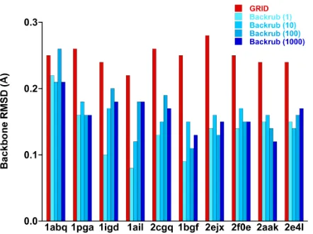
Calculation of the backbone RMSD of the final refined structure with respect to the
starting crystal structure for each of the ten proteins shows that GRID produces
some-what larger deviations than Backrub; the average RMSD was 0.25 ± 0.02 Å for GRID
ver-sus 0.14 ± 0.04 Å for Backrub (Fig. 4). These relatively minor deviations indicate that both
algorithms generate refined structures that retain their original topologies.
An implementation of GRID that uses random residue order for applying perturbations
was also run and found to produce nearly identical results to the sequential N terminus to
C terminus version described above (Fig. S5).
Discussion
Our results indicate that the GRID algorithm is more effective in reducing the energy of
structures than the Backrub algorithm: applying the same all-atom force field, GRID
con-sistently yields lower energy configurations. Although stochastic algorithms have been
successful in refining small proteins (< 85 residues), refinement of larger proteins has
proven to be particularly difficult
3
. In contrast, GRID was effective on all the proteins
test-ed, which included structures up to 155 residues. GRID also achieves these
improve-ments with relatively little computational resources (equivalent to one trajectory of
Back-rub), thus making it feasible to use on even larger proteins. This level of computational
efficiency is essential for the last stage of protein structure prediction, since low-resolution
prediction techniques typically produce a large number of structures that must be refined
in order to select the best structural model. GRID's high performance capabilities stem
from using a strategy that is straightforward, but effective.
To achieve structures that are closer to the native state, refinement methods must be
able to find lower energy configurations. This can be problematic for algorithms that use
local perturbations to achieve refinement, as they are more likely to fall into local minima.
It is possible that GRID minimizes this problem because it applies local perturbations that
are propagated down the whole protein chain. These perturbations, which are effectively
more global in nature, have a higher probability of explicitly moving the structure out of a
local minimum. Thus, GRID may allow for a more thorough exploration of the energy
landscape, making it easier to find lower energy structures.
Acknowledgments
This work was supported by the Defense Advanced Research Projects Agency (DARPA)
and a Department of Defense National Security Science and Engineering Faculty
Fellow-ship (S.L.M).
I would like to thank Marie Ary for her comments and assistance in writing
the manuscript.
!
References
!
[1]
G. Chopra, C. M. Summa, M. Levitt,
Proc. Natl. Acad. Sci. USA
2008
,
105
,
20239-20244.
[2]
D. Baker, A. Sali,
Science
2001
,
294
, 93-96.
[3]
P. Bradley, K. M. S. Misura, D. Baker,
Science
2005
,
309
, 1868-1871.
[4]
Y. Zhang,
Curr. Opin. Struct. Biol.
2009
,
19
, 145-155.
[5]
V. Mariani, F. Kiefer, T. Schmidt, J. Haas, T. Schwede,
Proteins
2011
,
79 Suppl 10
,
37-58.
[6]
R. Jauch, H. C. Yeo, P. R. Kolatkar, N. D. Clarke,
Proteins
2007
,
69 Suppl 8
,
57-67.
[7]
J. Xu, J. Peng, F. Zhao,
Proteins
2009
,
77 Suppl 9
, 133-137.
[8]
J. Kopp, L. Bordoli, J. N. Battey, F. Kiefer, T. Schwede,
Proteins
2007
,
69 Suppl 8
,
38-56.
[9]
I. H. Park, V. Gangupomu, J. Wagner, A. Jain, N. Vaidehi,
J. Phys. Chem. B
2012
,
[10] O. Graña, D. Baker, R. M. MacCallum, J. Meiler, M. Punta, B. Rost, M. L. Tress, A.
Valencia,
Proteins
2005
,
61 Suppl 7
, 214-224.
[11]
C. Venclovas, A. Zemla, K. Fidelis, J. Moult,
Proteins
2003
,
53 Suppl 6
, 585-595.
[12] J. L. MacCallum, L. Hua, M. J. Schnieders, V. S. Pande, M. P. Jacobson, K. A. Dill,
Proteins
2009
,
77 Suppl 9
, 66-80.
[13] S. Kannan, M. Zacharias,
Proteins
2007
,
66
, 697-706.
[14] S. Kannan, M. Zacharias,
J. Struc. Biol.
2009
,
166
, 288-294.
[15] M. Levitt, S. Lifson,
J. Mol. Biol.
1969
,
46
, 269-279.
[16] A. Shmygelska, M. Levitt,
Proc. Natl. Acad. Sci. USA
2009
,
106
, 1415-1420.
[17] C. M. Summa, M. Levitt,
Proc. Natl. Acad. Sci. USA
2007
,
104
, 3177-3182.
[18] G. D. Friedland, A. J. Linares, C. A. Smith, T. Kortemme,
J. Mol. Biol.
2008
,
380
,
757-774.
[19] M. S. Lin, T. Head-Gordon,
J. Comput. Chem.
2011
,
32
, 709-717.
[20] C. A. Smith, T. Kortemme,
J. Mol. Biol.
2008
,
380
, 742-756.
[21] T. E. Creighton,
Biochem. J.
1990
,
270
, 1-16.
[22] K. A. Dill,
Biochemistry
1990
,
29
, 7133-7155.
[23] H. Venselaar, R. P. Joosten, B. Vroling, C. A. B. Baakman, M. L. Hekkelman, E.
Krieger, G. Vriend,
Eur. Biophys. J.
2010
,
39
, 551-563.
[24] G. I. Makhatadze, P. L. Privalov,
Adv. Protein. Chem.
1995
,
47
, 307-425.
[25] O. B. Ptitsyn,
Adv. Protein. Chem.
1995
,
47
, 83-229.
[27] I. W. Davis, W. B. Arendall, D. C. Richardson, J. S. Richardson,
Structure
2006
,
14
, 265-274.
[28] C. A. Rohl, C. E. Strauss, K. M. Misura, D. Baker,
Methods Enzymol.
2004
,
383
,
66-93.
[29] R. L. Dunbrack, F. E. Cohen,
Protein Sci.
1997
,
6
, 1661-1681.
[30] E. L. Humphris, T. Kortemme,
Structure
2008
,
16
, 1777-1788.
[31] I. Georgiev, D. Keedy, J. S. Richardson, D. C. Richardson, B. R. Donald,
Bioinfor-matics
2008
,
24
, i196-204.
[32] C. B. Anfinsen,
Science
1973
,
181
, 223-230.
[33] Protabit, www.protabit.com.
[34] W. Kabsch, C. Sander,
Biopolymers
1983
,
22
, 2577-2637.
!
!
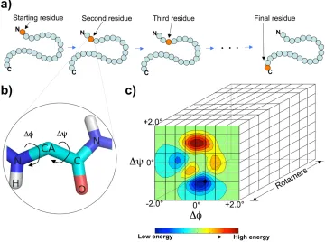
[image:25.612.126.486.106.374.2]!
Figure 1
.
Schematic showing the GRID algorithm. a) In each iteration, GRID starts from the N-terminal
residue and marches forward to the C terminus, sequentially applying the lowest energy move on the
structure before proceeding to the next residue. b) For each residue, GRID evaluates all the possible
perturbations (combinations of
Δφ
,
Δψ
, and rotamer) given a specified maximum perturbation angle and
precision level, then applies the move that results in the lowest energy. c) Three-dimensional energy
ma-trix (grid) that corresponds to all the possible perturbations (
Δφ
,
Δψ
, and rotamer) relative to the current
configuration of a residue. In this example, the maximum perturbation angle for
Δφ
and
Δψ
is ± 2.0°, the
precision level is 0.5°, and 8 rotamers are specified from the backbone dependent rotamer library.
!!
!
Figure 2.
Post-refinement analysis of GRID moves for different maximum dihedral angle
perturbations.
A single iteration of GRID was run on each of the 10 test proteins using
max-imum perturbation angles ranging from ± 0.1° to ± 1.9°; the precision level was always set to
form the smallest
Δφ
×
Δψ
matrix (9 grid points). GRID moves were divided into four types:
no perturbation of the backbone (type A), perturbation of
φ
or
ψ
, but not both (type B),
per-turbation of both
φ
and
ψ
by the same amount in the same direction (type C), and
perturba-tion of both
φ
and
ψ
by the same amount, but in opposite directions (type D). The frequency
of occurrence of each of the four different types of moves was calculated; points are the
average for the 10 test proteins.
[image:26.612.130.483.100.402.2]!
!
!
[image:27.612.77.537.98.454.2]!
Figure 3.
Performance of GRID versus Backrub in refining 10 high-resolution crystal structures from the PDB. Energy
improvement (a) and computation time (b) are shown for each of the 10 proteins. For Backrub, which is a stochastic
algorithm, values are given for 1, 10, 100, and 1000 trajectories. Energy improvement (c) and computation time (d) are
also plotted as a function of protein size.
!
!
[image:28.612.201.422.102.267.2]#
Figure 4.
Backbone RMSD with respect to starting crystal
structure following refinement with GRID and Backrub. Both
algorithms keep the refined structures close to their original
topologies.
!!
!
!
Figure S1.
Energy improvement following refinement with GRID using maximum dihedral angle
perturbations ranging from ± 0.1° to ± 1.9°; a single iteration was performed and the precision level
was set to produce a 9-point
Δφ
×
Δψ
matrix (3
×
3). For each of the 10 proteins in the test set, the
energy improvement (kcal/mol) was averaged over all the perturbation angles and normalized.
Av-erages of these normalized values for all 10 proteins ± S.E. are shown.
[image:29.612.130.485.117.363.2]!
!
!
!
!
!
!
!
!
!
!
!
!
!
!
!
!
!
!
!
!
!
!
!
!
!
!
!
!
[image:32.612.93.285.169.341.2]!
!
Figure S2.
GRID moves plotted as a function of residue number (x axis) and maximum dihedral angle perturbation (y
axis) for each of the 10 proteins tested (PDB ID and length of protein are given at top of each graph). The precision
level was always set to form the smallest
Δφ
×
Δψ
matrix (9 grid points) and a single iteration was performed. The four
move types are as follows: type A (white), type B (yellow), type C (red), and type D (black). At larger perturbation
an-gles, moves that cause no backbone movement (type A) become increasingly dominant, and moves that actually
per-turb the backbone (types B–D) are restricted to residues near the termini. Smaller perper-turbation angles allow movement
to occur more uniformly throughout the entire protein structure.
!
!
[image:33.612.72.307.128.335.2]!
!
!
!
Table 1.
Proteins used as test set for structure refinement.
PDB ID
No. of
residues
Resolution
(Å)
Class
%
α
% β
1abq
56 2.80
β
4
43
1pga
56 2.07
α+β
25
42
1igd
61 1.10
α+β
22
42
1ail
70 1.90
α
80
0
2cgq
74 1.83
α
38
1
1bgf
124 1.45
α
79
1
2ejx
134 1.79
α+β
35
41
2f0e
135 1.95
α+β
24
28
2aak
150 2.40
α+β
35
19
2e4l
155 2.00
α/β
34
29
a
Classifications based on Dictionary of Secondary Structure of
!
!
!
!
Chapter 2
!
!
REMC
GRID
: A Sampling Algorithm for Protein Structure
Abstract
We present replica exchange Monte Carlo (REMC)
GRID
, an algorithm that incorporates the
core strategies of GRID
1
, a previously developed refinement algorithm, with REMC
sam-pling. GRID uses a deterministic approach to high-resolution refinement, improving the
energy of a protein structure by systematically perturbing the backbone dihedrals and
side-chain conformations. In this study, REMC
GRID
was used for two purposes: (1)
struc-ture refinement, and (2) generation of decoy strucstruc-tures. In both applications, REMC
GRID
takes a protein structure as input and generates a conformationally diverse set of
low-en-ergy perturbed structures. One can specify the level of dihedral angle perturbations
ap-plied and the probability of accepting a perturbed conformation using
θ
max
and kT
f
para-meters. For refinement, we used 10 high-resolution (
≤
2.8
Å
) crystal structures from the
Protein Data Bank (PDB)
2
. The energy improvements obtained and the computational
effort required to achieve them were compared for REMC
GRID
and GRID alone. When
longer compute times are allowed, REMC
GRID
results in energy improvements that are
significantly better than those attained by GRID. For decoy generation, we used 59
high-resolution (
≤
2.8
Å
) crystal structures from the PDB of proteins ranging in size from 50-150
residues. We applied REMC
GRID
with 25 different parameter sets specifying different
φ
/
ψ
perturbation angles and acceptance probabilities to generate 472,000 decoys. We
com-puted the backbone RMSD of each decoy with respect to its crystal structure and found
that REMC
GRID
can generate a uniform distribution of perturbed structures covering a
broad range of RMSDs (0–10+ Å) that is independent of protein size and class. These
characteristics (uniformity and broadness) are important for the proper performance of
Introduction
Computational modeling of protein structures continues to be essential for advancing
research in biology and drug discovery, especially given that experimental structures for
many biologically important targets are difficult to obtain. Although investigators have
fo-cused efforts on protein structure prediction for over half a century, the accurate and
reli-able prediction of protein structures still remains a major challenge
3
. A successful
struc-ture prediction algorithm must solve two problems: (1) inaccuracies in the force field, and
(2) inefficient sampling of the conformational space
4
. The two problems are inter-related;
for example, one needs an efficient sampling algorithm in order to validate improvements
in a force field
5
. Several groups have worked on developing force fields
6-15
, and creating
better sampling methods
16-22
.
One of the major problems with current force fields is that they are too sensitive
6,23
.
Small perturbations in the configuration of a structure can result in large changes in its
energy. For instance, a few atomic clashes can result in an unfavorable energy for the
en-tire structure, even though the overall fold might be very close to the native state. This
im-poses ruggedness on the energy landscape and makes it hard for sampling algorithms to
search the conformational space and find the lowest energy states of the structure without
getting trapped in local minima. Coarse-grained approaches aim to decrease the
sensitiv-ity of current force fields by using a simpler model to describe proteins and their
inter-atomic interactions
24
. To test the validity of any coarse-grained model, one needs a decoy
set that covers a broad range of RMSDs so that the model’s ability to properly rank the
decoys can be tested. We therefore set out to create a decoy-generating algorithm that
RMSD’s for proteins of different sizes and classes. We were able to achieve this by
in-corporating a previously developed deterministic refinement algorithm (GRID) with replica
exchange Monte Carlo sampling in a novel algorithm called REMC
GRID
.
Results and Discussion
REMC
GRID
algorithm
REMC
GRID
is a sampling algorithm that takes a three-dimensional protein structure as
in-put and, employing an all-atom force field, perturbs the structure using a move set that
allows for backbone and side chain flexibility (see Fig. 1). Three degrees of freedom are
considered:
φ
and
ψ
dihedral perturbations and side-chain conformation. The algorithm
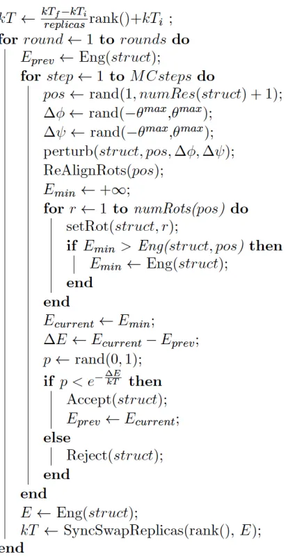
runs multiple Monte Carlo simulations (replicas) in parallel at different temperatures. After
a specified number of steps of Monte Carlo simulation, replicas join and stochastically
swap configurations based on their energies and the temperatures they are running at
according to the Metropolis-Hastings criterion
25
(see Fig. 6). Each step of Monte Carlo
simulation consists of four stages: (1) picking a residue at random, (2) perturbing
φ
and
ψ
backbone dihedrals of that residue, given a specified maximum perturbation angle range,
(3) scanning through all the possible side chain conformations (from a predefined rotamer
library) and picking the one that results in the lowest energy for the perturbed structure,
and (4) accepting or rejecting the move based on the Metropolis acceptance criterion,
given a specified temperature. A rotamer library is used to define a discrete set of
confor-mations for each residue. Although the moves are restricted to local perturbations relative
to a single residue, the perturbation selected influences the entire structure. Thus,
rela-tively minor local changes can propagate down the chain resulting in large shifts in the
REMC
GRID
Improves Structure Refinement
REMC
GRID
was used to refine a set of 10 high-resolution (
≤
2.8
Å
) crystal structures from
the PDB (Table 1). We calculated the energy improvements attained and the compute
times required to achieve them for REMC
GRID
and for GRID alone. Given enough
compu-tational resources, REMC
GRID
results in structures with significantly better energies than
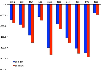
GRID alone (Fig. 2 and Fig. 3). As seen in a plot showing the ratio of computational
re-sources required for REMC
GRID
to match the level of energy improvement obtained by
GRID (see Fig. 4), with longer compute times (number of moves) REMC
GRID
achieves
en-ergy improvements that are up to ~15%
better than GRID alone. Since REMC
GRID
runs
multiple trajectories in parallel and the compute time of each trajectory is comparable to
that of GRID (GRID takes ~1-2 minutes, since it is deterministic and therefore just runs
once), on a large cluster, REMC
GRID
can get this 15% improvement with an overall
com-pute time that is ~3-fold that of GRID; i.e., in ~2-6 minutes. Low score decoys generated
REMC
GRID
Generates Uniform Distributions of Diversely Perturbed Decoys
We used REMC
GRID
to generate a set of decoys based on 59 high-resolution (
≤
2.8
Å
)
crystal structures from the PDB. These 59 structures included proteins ranging in size
from 50-150 residues and is the same dataset used by Das et al. for decoy generation
with Rosetta
26
. We applied REMC
GRID
with 25 different parameter sets specifying 5
dif-ferent
φ
/
ψ
perturbation angles and 5 different acceptance probabilities. For each pair of
parameters, we generated 320 decoys for each of the 59 proteins, for a total of 472,000
decoys. We found that smaller perturbations of
φ
and
ψ
result in smaller deviations in
structure. Moreover, higher simulation temperatures caused the acceptance of more
moves that worsen the energy of the structure, resulting in more diverse and more
per-turbed decoy sets. The parameter set with the largest
φ
/
ψ
perturbations and the least
re-stricting kT
f
values resulted in the most perturbed decoy structures. These decoys
cov-ered the broadest range of RMSD’s (including 10+ Å) and showed uniform broadness
across protein size. These characteristics (uniformity and broadness) are important for
the proper performance of machine learning algorithms. We planned to use this decoy set
to train a machine learning classifier (SVM), which would be used as a scoring function
for structure prediction (see Chapter 3).
REMC
GRID
generates more uniform decoys than Rosetta
In generating decoys, Rosetta starts from an unfolded polypeptide chain and after several
rounds of full structure prediction, produces a set of low energy conformations
27
. In
con-trast, REMC
GRID
starts from the crystal structure and systematically and gradually
decoy structures. REMC
GRID
uses an all-atom force field to ensure that structures with
se-vere clashes are not generated.
We compared our most perturbed REMC
GRID
decoys (those produced with the
largest
φ
/
ψ
perturbations and the least restricting kT
f
values) with those obtained using
Rosetta for broadness (range of backbone RMSDs) and uniformity (distribution of RMSDs
across different protein structures). The REMC
GRID
decoys are very uniform: the median
value is ~1 Å and is very close to 3 percentile value, and the most perturbed structures
(98 percentile) rarely surpass 12 Å RMSD (Fig. 10). In contrast, the Rosetta decoys are
quite varied: the median typically ranges from 5-10 Å, and the 98 percentile values are
much higher, with some decoys reaching > 30 Å RMSD. REMC
GRID
decoys
show
ade-quate broadness (0 to 10+ Å) and have distributions that are fairly consistent, whereas
Rosetta decoys show very large differences in their distributions. Thus, REMC
GRID
can
generate a uniform distribution of perturbed structures covering a broad range of RMSDs
that is independent of protein size and class.
Methods
REMC
GRID
implementation and parameterization
The REMC
GRID
algorithm was developed in C++ and incorporated into the phoenix protein
design software package. Standard Message Passing Interface (MPI) was used for
multi-processor parallelization. The phoenix force field with default parameters was used to
calculate the energy of the protein conformations. Side-chain conformations were
select-ed from the Dunbrack backbone-dependent rotamer library
28
.
REMC
GRID
takes as input six main parameters: (1) the maximum perturbation
swapping configurations, (4) number of rounds of running Monte Carlo simulations, (5)
lowest temperature replica, and (6) highest temperature replica. Values for maximum
Δϕ
and
Δψ
ranged from ± 2° to ± 10°; the number of parallel replicas was 80; the number of
Monte Carlo steps before swapping configuration was 100; number of rounds of Monte
Carlo was 20; lowest temperature was kT
i
= 0.5; and highest temperature ranged from
kT
f
= 1 to kT
f
= 9. The final set of parameters was obtained after trying REMC
GRID
with
several different values for each parameter and computing the RMSDs of the resulting
decoys.
Acknowledgments
This work was supported by the Defense Advanced Research Projects Agency (DARPA)
and a Department of Defense National Security Science and Engineering Faculty
Fellow-ship (S.L.M).
!
References
!
1. Chitsaz, M.; Mayo, S. L. J Comput Chem 2013, 34(6), 445-450.
2. Bernstein, F. C.; Koetzle, T. F.; Williams, G. J.; Meyer, E. F., Jr.; Brice, M. D.; Rodgers,
J. R.; Kennard, O.; Shimanouchi, T.; Tasumi, M. Eur J Biochem 1977, 80(2), 319-324.
3. Dill, K. A.; MacCallum, J. L. Science 2012, 338(6110), 1042-1046.
4. Summa, C. M.; Levitt, M. Proc Natl Acad Sci U S A 2007, 104(9), 3177-3182.
5. Best, R. B.; Zhu, X.; Shim, J.; Lopes, P. E.; Mittal, J.; Feig, M.; Mackerell, A. D., Jr. J
Chem Theory Comput 2012, 8(9), 3257-3273.
7. Vanommeslaeghe, K.; Hatcher, E.; Acharya, C.; Kundu, S.; Zhong, S.; Shim, J.;
Dari-an, E.; Guvench, O.; Lopes, P.; Vorobyov, I.; Mackerell, A. D., Jr. J Comput Chem 2010,
31(4), 671-690.
8. Hsieh, M. J.; Luo, R. J Phys Chem B 2010, 114(8), 2886-2893.
9. Marrink, S. J.; Risselada, H. J.; Yefimov, S.; Tieleman, D. P.; de Vries, A. H. J Phys
Chem B 2007, 111(27), 7812-7824.
10. Periole, X.; Marrink, S. J. Methods Mol Biol 2013, 924, 533-565.
11. Yesylevskyy, S. O.; Schafer, L. V.; Sengupta, D.; Marrink, S. J. PLoS Comput Biol
2010, 6(6), e1000810.
12. Li, X.; Wu, X.; Wu, G. J Theor Biol 2014.
13. Berhanu, W. M.; Jiang, P.; Hansmann, U. H. Phys Rev E Stat Nonlin Soft Matter Phys
2013, 87(1), 014701.
14. Vendruscolo, M. J Biol Phys 2001, 27(2-3), 205-215.
15. Koga, N.; Takada, S. J Mol Biol 2001, 313(1), 171-180.
16.
Daidone, I.; Amadei, A.; Roccatano, D.; Nola, A. D. Biophys J 2003, 85(5),
2865-2871.
17. Chen, J.; Im, W.; Brooks, C. L., 3rd. J Comput Chem 2005, 26(15), 1565-1578.
18. Frembgen-Kesner, T.; Elcock, A. H. J Mol Biol 2006, 359(1), 202-214.
19. Stumpff-Kane, A. W.; Maksimiak, K.; Lee, M. S.; Feig, M. Proteins 2008, 70(4),
1345-1356.
20. Jiang, P.; Yasar, F.; Hansmann, U. H. J Chem Theory Comput 2013, 9(8).
21. Yang, Y.; Liu, H. J Comput Chem 2006, 27(13), 1593-1602.
23. Price, D. J.; Brooks, C. L., 3rd. J Comput Chem 2002, 23(11), 1045-1057.
24. Tozzini, V. Curr Opin Struct Biol 2005, 15(2), 144-150.
25. Haario, H.; Saksman, E.; Tamminen, J. Bernoulli 2001, 7(2), 223-242.
26. Das, R.; Qian, B.; Raman, S.; Vernon, R.; Thompson, J.; Bradley, P.; Khare, S.; Tyka,
M. D.; Bhat, D.; Chivian, D.; Kim, D. E.; Sheffler, W. H.; Malmstrom, L.; Wollacott, A. M.;
Wang, C.; Andre, I.; Baker, D. Proteins 2007, 69 Suppl 8, 118-128.
27. Das, R.; Baker, D. Annu Rev Biochem 2008, 77, 363-382.
28. Dunbrack, R. L., Jr.; Karplus, M. J Mol Biol 1993, 230(2), 543-574.
!
!
[image:45.612.81.288.153.555.2]*
Fig 1.
REMC
GRID
algorithm pseduocode; the algorithm takes a crystal structure and a parameter set as input and
gen-erates a set of low energy decoys as output. Level of perturbation depends on the parameter set.
!!
!
#
Fig 2.
The energy improvement of 10 single-chain high-resolution crystal structures using REMC
GRID
employing
phoenix energy force field.
[image:46.612.107.498.124.413.2]!!
#
[image:47.612.72.498.94.392.2]!
Fig 3.
REMC
GRID
energy improvement compared to GRID energy improvement; REMC
GRID
results in significantly more
energy improvements than GRID.
!!
#
!
Fig 4.
The ratio of energy improvement that REMC
GRID
achieves compared to the energy improvement attained by
GRID based on the number of steps of Monte Carlo simulation.
[image:48.612.80.507.87.484.2]!!
#
!
Fig 5.
Computational effort comparison between REMC
GRID
and GRID; REMC
GRID
by spending more computational
effort can achieve higher energy improvements. REMC
GRID
continues to improve the energy improvement by being
al-lowed to spend more computational resources.
[image:49.612.83.472.96.487.2]!!
[image:50.612.114.466.117.402.2]#
Fig 6.
Backbone rmsds of 80 parallel replicas; 40 rounds of swaps between replicas is shown; protein G with pdbID
“1pga” is the protein we used in this simulation.
!!
[image:51.612.111.479.115.422.2]*
Fig 7.
Backbone rmsds of decoys (protein G, 1pga); backbone rmsd increases as the number of iterations increases in
this simulation. Parameters are set to generate a diverse set of decoys.
!!
[image:52.612.107.471.113.404.2]*
Fig 8.
Backbone rmsd vs. energy of decoys generated; gradual increase in the energy of the structure results in a
fun-nel like distribution of backbone RMSDs vs. energies (pdbID 1pga).
!!
[image:53.612.129.481.114.396.2]#
Fig 9.
Backbone rmsd distribution of decoy sets generated by REMC
GRID
. In this figure we have generated 25 different
decoy sets by varying two parameters of the algorithm (kT
f
and
θ
). Each decoy set is depicted in a box; the x-axis is the
size of protein in terms of number of residues, and the y-axis is the backbone rmsd. Each line corresponds to one single
protein structure and the decoys generated for that specific structure. The line is marked by three points of 25, 50, and
75 percentiles.
!!
#
[image:54.612.88.496.95.308.2]!
Fig 10.
Comparison of Rosetta decoys and REMC
GRID
decoys; REMC
GRID
decoys are much more uniformly distributed
due to gradual increase of the energy of the structures; Rosetta on the other hand produces set of decoys by
perform-ing a full structure prediction and pickperform-ing the lowest energy decoys. Each line is marked at three points, the lowest point
corresponds to 3 percentile, the middle mark corresponds to 50 percentile and the top most mark corresponds to 98
percentile.
!
!
!!
Table 1.
Ten high-resolution crystal structures that were used in testing REMC
GRID
refinement.
!
PDB ID
No. of residues
Resolution (Å)
Class
%
α
%
β
1abq
56 2.80
β
4
43
1pga
56 2.07
α
+
β
25
42
1igd
61 1.10
α
+
β
22
42
1ail
70 1.90
α
80
0
2cgq
74 1.83
α
38
1
1bgf
124 1.45
α
79
1
2ejx
134 1.79
α
+
β
35
41
2f0e
135 1.95
α
+
β
24
28
2aak
150 2.40
α
+
β
35
19
2e4l
155 2.00
α
/
β
34
29
!!
!
Chapter 3
!
!
A Machine Learning Approach to Improving Structure
Predic-tion by Incorporating Coarse-Grained Features
!
Abstract
Although considerable progress has been made in the last few decades, the ability to
predict the three-dimensional structure of a protein from its amino acid sequence remains
a major challenge in structural biology. A successful energy-based protein structure
pre-diction algorithm must address two problems: (1) inaccuracies in the force field, and (2)
inefficient sampling of conformational space. The all-atom force fields currently available
are too sensitive—even very minor perturbations in the coordinates of a protein can result
in enormous energy fluctuations. This results in a very rugged energy landscape that is
difficult to explore. One approach to solving this problem is to incorporate coarse-grained
features into the force field. Here, we develop a coarse-grained model and use it to
calcu-late a coarse-grained (CG)-score for a set of decoy structures. Three additional features
were also calculated: surface area, volume, and all-atom energy. Decoys were classified
into four groups based on their backbone RMSD relative to the crystal structure. A support
vector machine (SVM) classifier was then trained to predict the class of each decoy in
three different training experiments. The first two used either all-atom energy or CG-score
as the sole feature for training and the third combined all four features (all-atom energy,
CG-score, surface area, and volume). The CG-score alone produced better results than
the all-atom energy alone, but the combination of all four features significantly improved
the ability of the SVM to correctly classify the decoys.
!
Introduction
Efforts aimed at predicting the structure of a protein from its amino acid sequence have
been ongoing for over 50 years, and although much progress has been made, the
accu-rate and reliable prediction of protein structures still presents many challenges
1
. For an
energy-based structure prediction algorithm to be successful, it must deal with two
prob-lems: (1) inaccuracies in the force field, and (2) inefficient sampling of the conformational
space
2
. Force field development has been ongoing for many years, and several research
groups are attempting to develop simple yet relevant models that describe the physics of
protein folding
3
4
5
6
7
8
. The simplicity of the models is crucial as compute power is limited
and a simpler model can reduce the calculation time required to adequately sample the
possible conformations
7
.
One of the problems with current force fields is that they are too sensitive; that is, small
perturbations in the configuration of a structure can result in large differences in its energy
9
5
. This results in a very rugged energy landscape, which makes it difficult for sampling
algorithms to search conformational space to find the lowest energy structure without
be-ing trapped in local minima. A smoother energy landscape is therefore desirable and
would in fact mimic that of natural proteins, whose folding rate suggests a smooth
land-scape
10
. Coarse-grained models aim to alleviate the sensitivity of current force fields
11
7
12
. Here, we describe a simple coarse-grained model that was devised for use in training
a machine learning classifier. This coarse-grained model was used to calculate a
CG-score for a set of decoy structures. Similarly, we computed three other features for the
groups based on backbone rmsd from the crystal structure. We then trained a support
vector machine (SVM) classifier on these features to predict the class of each decoy.
Three SVM training experiments were conducted: (1) with all-atom energy as the sole
feature, (2) with CG-score as the sole feature, and (3) with all four features used
simulta-neously (CG-score, all-atom energy, surface area, and volume). We found that the
com-bination of all four features was more predictive than either all-atom energy or CG-score
alone.
!
Results and Discussion
A simple coarse-grained model
To overcome the sensitivity of an all-atom force field, we devised a coars