Brief Communication
Cingulate Hypoactivity in Cocaine Users During a GO–NOGO
Task as Revealed by Event-Related Functional Magnetic
Resonance Imaging
Jacqueline N. Kaufman,
1Thomas J. Ross,
1Elliot A. Stein,
1and Hugh Garavan
1,21Medical College of Wisconsin, Department of Psychiatry, Milwaukee, Wisconsin 53226, and2Trinity College, Department of Psychology and Institute of Neuroscience, Dublin 2, Ireland
Although extensive evidence exists for the reinforcing properties of drugs of abuse such as cocaine, relatively less research has addressed
the functional neuroanatomical correlates of the cognitive sequelae of these drugs. We present a functional magnetic resonance imaging
study of a GO–NOGO task in which successful performance required prepotent behaviors to be inhibited. Significant cingulate,
pre-supplementary motor and insula hypoactivity was observed for both successful NOGOs and errors of commission in chronic cocaine
users relative to cocaine-naive controls. This attenuated response, in the presence of comparable activation levels in other task-related
cortical areas, suggests cortical and psychological specificity in the locus of drug abuse-related cognitive dysfunction. The results suggest
that addiction may be accompanied by a disruption of brain structures critical for the higher-order, cognitive control of behavior.
Key words:
inhibitory control; cocaine; fMRI; anterior cingulate; GO–NOGO; addiction
Introduction
Cocaine has been identified as one of the most powerful
ers currently known (Kuhar et al., 1991). Similar to the
reinforc-ing properties of many drugs of abuse, the pharmacological
ef-fects of cocaine have been linked to the modulation of dopamine
release, particularly within terminal fields of the
mesocorticolim-bic dopaminergic pathways such as the nucleus accumbens
(Koob and Bloom, 1988; Wise, 1996; Di Chiara, 1999). These
pharmacological effects of abused drugs do not, however, directly
explain the cognitive deficits often seen in chronic drug users. For
example, chronic cocaine abusers display impairment on tests of
memory function, attention, and inhibitory control (O’Malley et
al., 1992; Easton and Bauer, 1997; Di Sclafani et al., 1998; Fillmore
and Rush, 2002). Although a great deal of preclinical and clinical
research has focused on the direct action of cocaine on the reward
systems of the brain (Wise, 1996; Di Chiara, 1999), comparatively
few studies have examined the functional neuroanatomical
re-gions associated with these observed behavioral correlates of
co-caine use. Consequently, an unresolved question concerns the
role that cognitive dysfunction and its associated
neuroanatomi-cal changes might play in the maintenance of drug abuse.
Poor inhibitory control is a common symptom of a number of
pathologies that fall under the umbrella of impulsivity disorders.
Pathological gambling, obsessive-compulsive disorder (OCD),
attention deficit hyperactivity disorder, Tourrette syndrome, and a
variety of traumatic brain injuries (Stewart and Tannock, 1999;
Hol-lander and Rosen, 2000; Johannes et al., 2001; Ursu et al., 2001) share
common symptoms of disinhibited behavior and loss of self-control.
These cognitive constructs are also among the characteristics of drug
addiction and suggest that executive dysfunction (i.e., impairment in
the cognitive control of behavior) may be a core component of
ad-diction (Lyvers, 2000). The failure to develop adequate inhibitory
control and/or the loss ofpreviously developed inhibitory
con-trol can have a profound impact on the ability of an individual
to gate prepotent, yet inappropriate and dangerous, behaviors
such as using cocaine. Lesion studies and, more recently, brain
imaging studies have implicated frontal and parietal cortex in
inhibitory control (Konishi et al., 1999; Pliszka et al., 2000;
Brass et al., 2001; Fuster, 2001; Rubia et al., 2001; Garavan et
al., 2002). Whereas right prefrontal cortex (PFC) appears to be
central to response inhibition (Garavan et al., 1999; Aron et
al., 2003), the anterior cingulate and occipitotemporal regions
have also frequently been reported (de Zubicaray et al., 2000;
Braver et al., 2001; Liddle et al., 2001; Menon et al., 2001; Garavan
et al., 2002; Watanabe et al., 2002).
The ability to inhibit inappropriate behaviors is complemented
by action monitoring functions. For example, efficient executive
control of behavior requires us to monitor our performance for
errors or for high levels of task difficulty (e.g., conflict between
com-peting responses) and then to adjust our behavior accordingly
(Rab-bitt, 1966; Botvinick et al., 2001). Thus, action monitoring helps
ensure smooth and safe control of behavior. These monitoring
func-tions have been anatomically dissociated from inhibitory control
functions (Menon et al., 2001; Garavan et al., 2002) and have been
localized to medial wall structures such as the anterior cingulate
Received March 11, 2003; revised June 9, 2003; accepted June 12, 2003.
This work was supported by United States Public Health Service Grant DA14100 and General Clinic Research Center (GCRC) Grant M01 RR00058. We gratefully acknowledge the assistance of Stacy Claesges, Michael San Fillipo, the research nusring staff at GCRC of Froedtert Memorial Lutheran Hospital, and the comments of two anonymous reviewers.
Correspondence should be addressed to Dr. Hugh Garavan, Department of Psychology, Trinity College Dublin, Dublin 2, Ireland. E-mail: [email protected].
T. J. Ross’s and E. A. Stein’s present address: National Institute on Drug Abuse–Intermural Research Program, Neuroimaging Research Branch, 5500 Nathan Shock Drive, Baltimore, MD 21224.
cortex (ACC) and pre-supplementary motor area (SMA) (Carter et
al., 1998; Ullsperger and von Cramon, 2001).
A growing imaging literature suggests ACC and medial
pre-frontal dysregulation in chronic cocaine users as evidenced by
anatomical changes as well as metabolic dysfunctions. For
exam-ple, polysubstance users have been shown to have bilateral
de-creases in prefrontal gray matter (Liu et al., 1998). When
com-pared with non-using control subjects, cocaine users also show
gray matter decreases in ACC, insula, and superior temporal
re-gions (Franklin et al., 2002). In addition to these anatomical
dif-ferences, functional metabolic variations have been observed in
cocaine users with decreased metabolic activity in the cingulate
and orbitofrontal cortices (Volkow et al., 1993). In drug-abusing
populations, as in other clinical populations, it is typically not
possible to determine whether brain changes of these sorts
pre-date the observed pathology or whether they are the result of the
disordered behavior. Notwithstanding this common limitation,
it is valuable to identify the neuroanatomical correlates of
cogni-tive dysfunction to inform optimal treatment.
The present study attempted to probe the neuroanatomical
functioning of these cognitive processes in cocaine users. GO–
NOGO tasks in which the NOGO/GO ratio is low, thereby
creat-ing a response prepotency that is difficult to inhibit on NOGO
trials, provide a useful test bed for assessing cortical activation for
inhibitory control and action monitoring. Cocaine-using
sub-jects have been shown to have impaired behavioral performance
on GO–NOGO tasks (Fillmore and Rush, 2002; Fillmore et al.,
2002); the functional imaging correlates of these behavioral
find-ings would provide important information regarding the
local-ization of these impairments. Consequently, both cocaine-naive
control subjects and chronic cocaine-using subjects underwent
functional magnetic resonance imaging (fMRI) scanning while
performing a visually presented GO–NOGO task.
Materials and Methods
Participants.Of the 27 subjects who participated in this study, 13 were otherwise healthy, active cocaine users (five female; mean age of 37⫾4.5 years; range of 27– 44 years), and 14 were healthy nondrug users (10 female; mean age, 30⫾8.7 years; range, 19 – 45 years). Among cocaine users, the average number of years of cocaine use was 11.2 (range of 1–22 years). All participants were right-handed and reported no history of neurological symptoms. Subjects were fully informed of the nature of the research and provided written consent for their involvement in this study in accordance with the Institutional Review Board of the Medical College of Wisconsin. Urine samples were collected from all participants to test for pregnancy and drug use. All nondrug users had negative tests for all drugs, whereas all cocaine subjects returned positive screens for cocaine or its metabolites, indicating that they had used cocaine within the pre-vious 72 hr. Ten of the 13 users were able to estimate their last use, which ranged from 18 to 72 hr before the scan session, and no user displayed any overt behavioral signs of cocaine intoxication.
Stimuli.Stimuli consisted of a 1 Hz serial stream of alternating Xs and Ys. The stimuli were 5.08 and 6.35 cm in height for X and Y, respectively. Subjects were instructed to press a button for each target stimulus while the stimulus was still present on the screen. NOGO stimuli, in which the target stimuli did not alternate (i.e., the second of two identical, succes-sively presented target stimuli), required inhibition of the response. Sub-jects recommenced responding to alternating stimuli after the NOGO stimulus. There were 1180 GO and 80 randomly distributed NOGO stimuli presented over four runs (Fig. 1).
Training.Before functional image acquisition, subjects were trained on the task in a quiet room. Training included four difficulty levels of the task, accomplished by varying the on-screen duration of the stimuli at 900, 800, 700, or 600 msec: shorter stimulus durations, when combined with the instruction to respond while the stimulus was on-screen, yield
increased numbers of commission errors (Garavan et al., 2002). A blank screen was displayed during the remainder of the 1 sec stimulus display time to maintain the stimuli rate at 1 Hz. Training durations were⬃30 min but varied by subject because instruction pages were self-paced and the durations of rest periods during training were at the subject’s discre-tion. For the imaging runs, task difficulty was tailored for each subject by selecting one of the four presentation rates to achieve comparable behav-ioral performance in drug users and nondrug users and to ensure near-equal numbers of successful inhibitions (STOPS) and commission errors (ERRORS). This tailoring was necessary to ensure that sufficient num-bers of STOPS and ERRORS were available for the event-related analyses. Given that poorer inhibitory control performance would be expected in cocaine users, the tailoring sought to ensure that each group performed at similar levels of competence, thereby minimizing fatigue or frustration confounds. Subject placement in conditions based on training perfor-mance was as follows: the 600 msec condition was run on 57% of the control subjects and 23% of the cocaine subjects; the 700 msec condition was run on 14% of the control subjects and 38% of the cocaine subjects; the 800 msec condition was run on 14% of the control subjects and 8% of the cocaine subjects; and the 900 msec condition was run on 14% of the control subjects and 31% of the cocaine subjects.
fMRI procedures.Participants were fitted with prism glasses that per-mitted them to view stimuli backprojected onto a screen at the end of the scanner bed. To minimize head movement and artifact in the coil during data acquisition, subject’s heads were cushioned with padding. Whole-brain fMRI imaging was performed using a 1.5 T GE Signa scanner (GE Medical Systems, Waukesha, WI) equipped with a 30.5 cm internal di-ameter three-axis local gradient coil with insertable radiofrequency coils with transmit–receive capabilities. Contiguous 7 mm sagittal slices span-ning the entire brain were collected using a gradient-echo, echo-planar pulse sequence (repeat time, 2000 msec; echo time, 40 msec; field of view, 240 mm; 64⫻64 matrix; 3.75⫻3.75 mm in-plane resolution). High-resolution spoiled gradient recalled acquisition at steady state (SPGR) anatomical images were collected before the functional data acquisition and were used for spatial normalization and subsequent activation localization.
[image:2.603.327.520.62.274.2]fMRI data analysis.Imaging data were analyzed using the AFNI soft-ware package (Cox, 1996). Functional data were three-dimensional vol-ume registered and corrected for differences in slice time acquisition. A deconvolution analysis calculated the hemodynamic response functions for STOPS and ERRORS that were modeled using a gamma-variate func-tion. The modeling, using nonlinear regression, identified the best fitting
gamma-variate function for each voxel rather than constraining all he-modynamic responses to a standard, prespecified shape. The area under the curve of this hemodynamic model was calculated for each voxel and expressed as a percentage of the area under the baseline (representing tonic task-related processes). Functional maps were transformed into stereotaxic space based on the atlas of Talairach and Tournoux (1988) and spatially blurred with a 4.2 mm full-width half-maximal isotropic Gaussian kernel. Activation maps were created for both STOPS and ER-RORS for each group based on one-samplettests against the null hypoth-esis of no activation changes. Data simulations, created by randomly selecting STOP and ERROR locations within the time series and exactly repeating all subsequent analyses, identified the voxelwise threshold at which there was a 5% chance of a false positive cluster of activation (t⫽
4.95 with a minimal cluster size of 100l). The activation maps of the users and control subjects were combined by condition as OR (either/or data) maps, and between group comparisons were performed on the mean activations of these clusters.
Results
Performance analysis
Prescanning performance results revealed poorer inhibitory control
in the cocaine users (51 vs 39 commission errors;
t
(25)⫽
2.3;
p
ⱕ
0.03). Despite efforts to tailor task difficulty individually during
scanning, cocaine users made significantly more commission errors
(48 vs 36;
t(25)
⫽
2.79;
p
⬍
0.01) and omission errors (58 vs 3;
t(25)
⫽
4.13;
p
⬍
0.0004) than noncocaine users. However, the event-related
design of the fMRI experiment allowed us to compare the two
groups separately on successful behavioral inhibitions (STOPS) and
failed inhibitions (ERRORS), thereby removing performance
con-founds from the brain activation maps.
Functional analysis
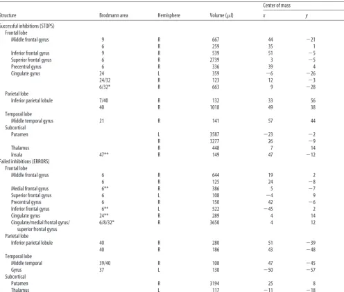
Activated areas were primarily in the right hemisphere and
in-cluded dorsolateral PFC, ACC, inferior parietal lobule, and
bilat-eral putamen (Table 1). The activation patterns of cocaine users
were similar to controls, but, in some cases, the volume of
acti-vation was smaller in users (Fig. 2). The fMRI data revealed
sig-nificantly less mean activation in cocaine users for STOPS in the
anterior cingulate and right insula when compared with
nonco-caine users (Fig. 3). Notably, other areas, including those
sug-gested previously to be important for inhibitory control (e.g.,
right inferior parietal lobule and right dorsolateral prefrontal
cor-Table 1. Regions activated for successful inhibitions (STOPS) and failed inhibitions (ERRORS)Structure Brodmann area Hemisphere Volume (l)
Center of mass
x y z
Successful inhibitions (STOPS) Frontal lobe
Middle frontal gyrus 9 R 667 44 ⫺21 28
6 R 259 35 1 50
Inferior frontal gyrus 9 R 539 51 ⫺5 20
Superior frontal gyrus 6 R 2739 3 ⫺5 52
Precentral gyrus 6 R 336 39 4 42
Cingulate gyrus 24 L 359 ⫺6 ⫺26 22
24/32 R 123 12 ⫺3 44
6/32* R 663 9 ⫺28 32
Parietal lobe
Inferior parietal lobule 7/40 R 132 33 56 40
40 R 1018 49 38 42
Temporal lobe
Middle temporal gyrus 21 R 141 57 44 ⫺2
Subcortical
Putamen L 3587 ⫺23 ⫺2 6
R 3277 26 ⫺9 4
Thalamus R 448 7 14 5
Insula 47** R 149 47 ⫺12 ⫺4
Failed inhibitions (ERRORS) Frontal lobe
Middle frontal gyrus 6 R 644 19 2 60
6 R 125 24 ⫺8 50
Medial frontal gyrus 6** R 386 5 ⫺7 60
Superior frontal gyrus 6 L 108 ⫺4 9 56
Precentral gyrus 6 R 150 42 ⫺6 39
Inferior frontal gyrus 6** L 522 ⫺45 2 31
Cingulate gyrus 24** R 289 4 14 31
Cingulate/medial frontal gyrus/ 6/8/32* R 3650 4 12 46
superior frontal gyrus Parietal lobe
Inferior parietal lobule 40 R 280 51 ⫺39 40
40 R 186 43 ⫺48 39
Temporal lobe
Middle temporal 39/40 R 108 47 ⫺45 27
Gyrus 37 L 130 ⫺50 ⫺57 5
Subcortical
Putamen R 3194 25 8 4
Thalamus L 117 ⫺11 ⫺18 11
Insula 13** L 1080 ⫺30 13 6
[image:3.603.46.543.73.495.2]tex) (Garavan et al., 1999, 2002; Konishi et
al., 1999; Fuster, 2001; Rubia et al., 2001), did
not differ in activity between the groups for
STOPS, underscoring the specificity of the
hypoactive areas. In addition, a number of
anterior cingulate clusters, as well as the right
medial frontal gyrus/pre-SMA, left insula,
and left inferior frontal gyrus were also
sig-nificantly hypoactive in users relative to
non-users for ERRORS (Fig. 3) (for a complete
list of structures activated by this task, see
Table 1).
The levels of difficulty performed by
the two groups were not equivalent. To
determine whether this may underlie the
observed effects, an analysis between
matched groups (i.e., the subset of the
present sample that can be chosen to
en-sure equal representation of each level of
task difficulty) was performed, and similar
patterns of hypoactivity were observed in
the cocaine users. Additionally,
multivari-ate ANOVA was run with all subjects
ex-amining the mean activation for each
re-gion of interest used in the analyses for
both successful inhibitions and errors.
Al-though no group
⫻
rate effect was seen, a
group effect was present for both incorrect
and correct (as expected per the overall
findings of this study). There was also no
effect for rate. This would suggest that
there is not an interaction based on rate of
presentation and that group differences
persist even when rate is factored in.
Discussion
These data demonstrate that certain
corti-cal areas, especially midline areas of the
anterior cingulate that are critical for
cog-nitive control, are less responsive in
chronic cocaine users. STOP-related
hy-poactivity was observed in the right insula
and a rostral region of the ACC, two regions that have been
identified with emotional processes (Whalen et al., 1998). The
ACC has also been implicated previously in inhibitory control
(Casey et al., 1996; Ponesse et al., 1998). It has been suggested to
play a critical role in urgent inhibitions (i.e., when time pressures
preclude the involvement of a “controlled” response inhibition
mediated by dorsolateral prefrontal areas) and has been shown to
be relied on more by highly absentminded subjects (Garavan et
al., 2002). Furthermore, we demonstrated that ACC function is
compromised not only for successful STOPS but also for
ER-RORS. Others (Carter et al., 1998; Kiehl et al., 2000) have
ob-served error-related activations in both the ACC and left
prefron-tal cortex, two areas observed to be hypoactive in cocaine users.
This internal replication of ACC hypoactivity in cocaine users in
the presence of comparable activation levels in many other
task-related cortical areas demonstrates that differences in cortical
function in users are anatomically specific and not ubiquitous.
These results, therefore, enable us to link existing evidence of
cognitive control impairment in addicts with the existing
evi-dence of ACC dysfunction in this group.
Alterations in blood oxygenation level-dependent response in
these midline action-monitoring regions have been
demon-strated in other pathological populations. For example, the ACC
is hyperactive in patients suffering from OCD and is hypoactive
in schizophrenic patients (Carter et al., 1998; Ursu et al., 2001).
These findings are consistent with the clinical profiles of these
groups, suggesting an overactive action monitoring system and a
heightened attentiveness to corrective behavior in OCD, and a
failure to properly monitor and integrate stimuli in the
environ-ment in schizophrenia. Similarly, the hypoactivity of cocaine
us-ers would appear consistent with their pathological drug use
pat-tern. Reduced inhibitory control, diminished action monitoring,
and diminished responsivity to one’s errors may represent an
executive function profile of cocaine users that may, at its least,
serve to prolong the maintenance of drug abuse. Recently, similar
findings of ACC hypoactivity have been observed in opiate
ad-dicts (S. D. Forman, personal communication).
[image:4.603.219.544.61.256.2]These dysexecutive sequelae of drug abuse suggest that
co-caine users may be compromised in the endogenous and
voli-tional control of their behavior. Consequently, their behavior
may be disproportionately determined by environmental
contin-gencies, environmental cues (e.g., drug craving cues), and
au-Figure 2. Sagittal sections show midline regions involved in the inhibitory control task. Examination of successful inhibitions (left column) and failed inhibitions (right column) between cocaine using subjects (upper row) and control subjects (bottom row) demonstrate consistent regions of activation for both groups. However, smaller volumes of activation survive thresholding for cocaine subjects for these regions, consistent with significant hypoactivity for cocaine users in midline structures. [image:4.603.217.547.319.476.2]tomatized or habitual behaviors. The effect of this would be to
compound the maintenance of drug abuse: if chronic cocaine
users are especially influenced by environmental contingencies
and cues, then an inhibitory dysfunction may reduce their
capac-ity to inhibit these external influences. Although these findings
are quite consistent with previous reports of metabolic and
neu-roanatomical dysfunctions in cocaine users, the current design
does not permit an examination of the time course of these changes.
In effect, it is not possible to determine whether these changes
pre-date the overt expression of a cocaine addiction or whether they are
the outcome of a history of cocaine abuse. Additionally, although the
current study has demonstrated a significant ACC dysfunction
relat-ing to one aspect of inhibitory control, namely the response
inhibi-tions required of a GO–NOGO task, it remains to be seen whether
other types of inhibitory control (Bechara et al., 2001; Bechara, 2003)
would show a similar deficit. These caveats notwithstanding, an
emerging knowledge of the cognitive profile of cocaine users and its
associated functional neuroanatomy may inform optimal
therapeu-tic interventions and help identify casual users most at risk for
be-coming drug dependent.
References
Aron AR, Fletcher PC, Bullmore ET, Sahakian BJ, Robbins TW (2003) Stop-signal inhibition disrupted by damage to right inferior frontal gyrus in humans. Nat Neurosci 6:115–116.
Bechara A (2003) Risky business: emotion, decision-making, and addiction. J Gambl Stud 19:23–51.
Bechara A, Dolan S, Denburg N, Hindes A, Anderson SW, Nathan PE (2001) Decision-making deficits, linked to a dysfunctional ventromedial pre-frontal cortex, revealed in alcohol and stimulant abusers. Neuropsycho-logia 39:376 –389.
Botvinick MM, Braver TS, Barch DM, Carter CS, Cohen JD (2001) Conflict monitoring and cognitive control. Psychol Rev 108:624 – 652.
Brass M, Zysset S, von Cramon DY (2001) The inhibition of imitative re-sponse tendencies. NeuroImage 14:1416 –1423.
Braver TS, Barch DM, Gray JR, Molfese DL, Snyder A (2001) Anterior cin-gulate cortex and response conflict: effects of frequency, inhibition and errors. Cereb Cortex 11:825– 836.
Carter CS, Braver TS, Barch DM, Botvinick MM, Noll D, Cohen JD (1998) Anterior cingulate cortex, error detection, and the online monitoring of performance. Science 280:747–749.
Casey BJ, Trainor R, Orendi J, Schubert A (1996) A functional Magnetic resonance imaging (fMRI) study of ventral prefrontal cortex mediation of response inhibition. Soc Neurosci Abstr 22:1107.
Cox RW (1996) AFNI: software for analysis and visualization of functional magnetic resonance neuroimages. Comput Biomed Res 29:162–173. de Zubicaray GI, Andrew C, Zelaya FO, Williams SCR, Dumanoir C (2000)
Motor response suppression and the prepotent tendency to respond: a parametric fMRI study. Neuropsychologia 38:1280 –1291.
Di Chiara G (1999) Drug addiction as dopamine-dependent associative learning disorder. Eur J Pharmacol 375:13–30.
Di Sclafani V, Truran DL, Bloomer C, Tolou-Shams M, Clark HW, Norman D, Hannauer D, Fein G (1998) Abstinent chronic crack-cocaine and crack-cocaine/alcohol abusers evidence normal hippocampal volumes on MRI despite persistent cognitive impairments. Addict Biol 3:261–270. Easton C, Bauer LO (1997) Neuropsychological differences between
alcohol-dependent and cocaine-dependent patients with or without problematic drinking. Psychiatry Res 71:97–103.
Fillmore MT, Rush CR (2002) Impaired inhibitory control of behavior in chronic cocaine users. Drug Alcohol Depend 66:265–273.
Fillmore MT, Rush CR, Hays L (2002) Acute effects of oral cocaine on in-hibitory control of behavior in humans. Drug Alcohol Depend 67:157–167.
Franklin TR, Acton PD, Maldjian JA, Gray JD, Croft JR, Dackis CA, O’Brian CP, Childress AR (2002) Decreased gray matter concentration in the insular, orbitofrontal, cingulate, and temporal cortices of cocaine pa-tients. Soc Biol Psychiatry 51:134 –142.
Fuster JM (2001) The prefrontal cortex—an update: time is of the essence. Neuron 30:319 –333.
Garavan H, Ross TJ, Stein EA (1999) Right hemispheric dominance of in-hibitory control: an event-related functional MRI study. Proc Natl Acad Sci USA 96:8301– 8306.
Garavan H, Ross TJ, Murphy K, Roche RAP, Stein EA (2002) Dissociable executive functions in the dynamic control of behavior: inhibition, error detection and correction. NeuroImage 17:1820 –1829.
Hollander E, Rosen J (2000) Impulsivity. J Psychopharmacol 14:S39 –S44. Johannes S, Wieringa BM, Mantey M, Nager W, Rada D, Muller-Vahl KR,
Emrich HM, Dengler R, Munte TF, Dietrich D (2001) Altered inhibition of motor responses in Tourette Syndrome and Obsessive-Compulsive Disorder. Acta Neurol Scand 104:36 – 43.
Kiehl KA, Liddle PF, Hopfinger JB (2000) Error processing and the rostral anterior cingulate: an event-related fMRI study. Psychophysiology 37:216 –223.
Konishi S, Nakajima K, Uchida I, Kikyo H, Kameyama M, Miyashita Y (1999) Common inhibitory mechanism in human inferior prefrontal cortex revealed by event-related functional MRI. Brain 122:981–991. Koob GF, Bloom FE (1988) Cellular and molecular mechanisms of drug
dependence. Science 242:715–723.
Liddle PF, Kiehl KA, Smith AM (2001) Event-Related fMRI study of re-sponse inhibition. Hum Brain Mapp 12:100 –109.
Liu X, Matochik JA, Cadet JL, London ED (1998) Smaller volume of pre-frontal lobe in polysubstance abusers: a magnetic resonance imaging study. Neuropsychopharmacology 18:243–252.
Lyvers M (2000) “Loss of control” in alcoholism and drug addiction: a neu-roscientific interpretation. Exp Clin Psychopharmacol 8:225–249. Menon V, Adleman NE, White CD, Glover GH, Reiss AL (2001)
Error-related brain activation during a Go/NoGo response inhibition task. Hum Brain Mapp 12:131–143.
O’Malley S, Adamse M, Heaton RK, Gawin FH (1992) Neuropsychological impairment in chronic cocaine abusers. Am J Drug Alcohol Abuse 18:131–144.
Pliszka SR, Liotti M, Woldorff MG (2000) Inhibitory control in children with attention deficit/hyperactivity disorder: event-related potentials identify the processing component and timing of an impaired right-frontal response-inhibition mechanism. Biol Psychiatry 48:238 –246. Ponesse JS, Logan WJ, Schacher RS, Tannock R, Crawley AP, Mikulis DJ
(1998) Functional neuroimaging of the inhibition of a motor response. NeuroImage 7:S972.
Rabbitt PM (1966) Errors and error correction in choice-response tasks. J Exp Psychol 71:264 –272.
Rubia K, Russell T, Overmeyer S, Brammer MJ, Bullmore ET, Sharma T, Simmons A, Williams SC, Giampietro V, Andrew CM, Taylor E (2001) Mapping motor inhibition: conjunctive brain activations across different versions of go/no-go and stop tasks. NeuroImage 13:250 –261. Stewart JA, Tannock R (1999) Inhibitory control differences following mild
head injury. Brain Cogn 41:411– 416.
Talairach J, Tournoux P (1988) Co-planar stereotaxic atlas of the human brain. New York: Thieme Medical.
Ullsperger M, von Cramon DY (2001) Subprocesses of performance moni-toring: a dissociation of error processing and response competition re-vealed by event-related fMRI and ERPs. NeuroImage 14:1387–1401. Ursu S, van Veen V, Siegle G, MacDonald A, Stenger A, Carter C (2001)
Executive control and self-evaluation in obsessive-compulsive disorder: an event related fMRI study. Paper presented at the Cognitive Neuro-science Society Meeting, New York, NY, March.
Volkow ND, Fowler JS, Wang GJ, Hitzemann R, Logan J, Schlyer DJ, Dewey SL, Wolf AP (1993) Decreased dopamine D2 receptor availability is associated with reduced frontal metabolism in cocaine abusers. Synapse 14:169 –177. Watanabe J, Sugiura M, Sato K, Sato Y, Maeda Y, Matsue Y, Fukuda H,
Kawashima R (2002) The human prefrontal and parietal association cortices are involved in NO-GO performances: an event-related fMRI study. NeuroImage 17:1207–1216.
Whalen PJ, Bush G, McNally RJ, Wilhelm S, McInerney SC, Jenike MA, Rauch SL (1998) The emotional counting Stroop paradigm: a functional magnetic resonance imaging probe of the anterior cingulate affective di-vision. Biol Psychiatry 44:1219 –1228.