Original Article
Electroacupucture accelerates peripheral nerve
regeneration via enhancement of BDNF,
NGF, and GAP43 in rats
Li Li1, Guoying Li1, Yanyan Dong2, Yilin Ye3
1Department of Pharmacology, Anhui University of Chinese Medicine, China; 2Department of Pharmacy, The Third People’s Hospital of Hefei, China; 3College of Nursing, Anhui University of Chinese Medicine, China
Received September 29, 2018; Accepted February 11, 2019; Epub May 15, 2019; Published May 30, 2019
Abstract: Electroacupucture (EA) is a modified Traditional Chinese Medicine approach. Acupuncture has been used for treatment of sciatic nerve injuries, showing excellent clinical effects. This study aimed to investigate the effects of electroacupucture (EA) on peripheral nerve regeneration. A total of 104 Sprague-Dawley rats were randomly divided into four groups, the sham group, sciatic nerve crush model group, EA treatment group, and EA + K252α treatment group. The rats received sham/sciatic nerve crush injury surgery, in respective groups, and adopted different methods of treatment for 8 days. Present results indicate that EA significantly improved axonal regenera -tion and recovery, both structurally and func-tionally. Brain-derived neurotrophic factor (BDNF), nerve growth factor (NGF), and growth associated protein 43 (GAP-43) were significantly increased after EA administration. Interestingly, blockage of BDNF downstream signaling abolished EA regeneration promoting actions. In summary, EA is beneficial for axonal regeneration after sciatic nerve injuries, potentially through elevation of levels of BDNF, NGF, and GAP-43.
Keywords: Electroacupucture, sciatic nerve crush, BDNF, NGF, sciatic function index
Introduction
Peripheral nerve injuries are devastating to humans, leading to high rates of morbidity and extreme healthcare expenses. Although the peripheral nervous system obtains a certain degree of capacity for axonal regrowth and regeneration, recovery of injured peripheral nerves has been unsatisfactory [1]. As a result, development of reagents or methods that can foster and promote axonal regeneration after peripheral nerve injuries is demanding.
Neurotrophins, including BDNF and NGF, sup-port and nurture growth and regeneration of axons during development and after injures [2, 3]. BDNF and NGF are members of a small fam-ily of secreted proteins. Expression, secretion, and actions of BDNF and NGF have been
stud-ied extensively. They regulate neural activity in
the mammalian nervous system [2]. BDNF and NGF are secreted by Schwann cells following peripheral nerve injuries, promoting axonal regeneration capacity and enhancing myelina-tion [3, 4].
Growth-associated protein 43 (GAP-43) is a presynaptic protein. Located at the inner sur-face of axon terminals, it is actively transported at nerve terminals between neurons to regulate neurite outgrowth and branching [5]. GAP-43 is highly upregulated during the regeneration
pro-cesses when neurons are damaged [6].
The-refore, increased GAP-43 during neural
regen-eration is beneficial for repair.
EA is a modified Traditional Chinese Medicine
approach. EA may improve clinical outcomes in multiple conditions. EA has been recently reported to improve locomotive and sensory functions after spinal cord transaction [7]. EA effects on neuroplasticity have been associat-ed with increasassociat-ed neurotrophins (NGF, BDNF) and GAP-43 [8, 9]. However, underlying
mecha-nisms of EA after sciatic nerve injuries and fine
protocols have not been fully elucidated.
explored the recovery of sensory and motor function of the sciatic nerve in vivo.
Materials and methods
Experimental animals
Sprague-Dawley rats (180-220 g) were pur-chased from the Animal Center of Anhui Pro- vince. All animal protocols were approved by Anhui Chinese University Animal Care and Use Committee. All procedures were designed to minimize discomfort for the animals.
A total of 104 animals were randomly divided into four groups (n = 26). The sham group
re-ceived sham sciatic nerve crush injury surgery.
The injury group received sciatic nerve crush injury surgery. The injury + EA group received
sciatic nerve crush injury surgery and EA
treat-ment on day 1 to day 8 after surgery. The in-jury + EA + K252α group received sciatic nerve
crush injury surgery, injections of BDNF
antago-nist K252α, and EA treatment on day 1 to day 8
after surgery. As previously reported, stock
solutions (10 mM) of K252α were prepared in DMSO and final concentrations of 100 nM were
supplemented in the cell culture medium for 1 hour [10].
Sciatic nerve crush model
Rats were anesthetized with 2% pentobarbital
sodium (35 mg/kg, ip). The sciatic nerve of
right hind leg was exposed by blunt dissection
through the biceps femoris. The sciatic nerve
was crushed at 6 mm distal to ischial
tuberosi-ty. The crush was achieved by applying consis -tent pressure with a pair of needles held for 10
seconds. This was repeated three times with
intervals of 10 seconds. About 2 mm of the nerve was injured. All crushing was done by one person. Finally, the muscles and skin were sutured separately.
To build a sciatic nerve regeneration chamber
model, 3 mm of the sciatic nerve was removed from the middle and bridged by a silicone tube. Sham surgery (exposure of the sciatic nerves without ligation) was used for the Sham control group.
Electroacupuncture treatment
Two acupuncture needles, 0.25 mm in diame -ter, were inserted into right hind leg, one in the
Zusanli point and the other in the Huantiao point. Electrical stimulation was applied using a PCE-A acupuncture stimulation machine (Anhui
Tianheng Technology, China) for 20 minutes,
with 1.5 mA intensity and 2 Hz frequency from day 1 to day 7 after surgery, once a day. Rats in the Sham group and Injury group did not receive electroaupuncture (EA) treatment. After EA treatment, 16 rats in each group were random-ly selected for sciatic functional index (SFI) determination, electrophysiological examina-tion, histological examinaexamina-tion, immunohisto-chemical staining, real-time PCR, and Western blotting. For the remaining rats, a sciatic nerve regeneration chamber model was produced. After a week of regular feeding, BDNF
concen-trations in the regeneration fluid were tested by
ELISA.
After SFI determination and electrophysiologi-cal testing, the right sciatic nerve was removed
and fixed for histological examination and immunohistochemical staining. The ventral
horn of L4-L6 spinal cord was also removed and frozen for real-time PCR and Western blotting.
Sciatic functional index (SFI) determination
Functional evaluation of sciatic nerve regenera-tion was expressed by the sciatic funcregenera-tional
index (SFI). The hind paws were dipped in blue
ink and the rats walked through a 50 cm × 8 cm
tunnel. Thus, the footprints could be recorded
on paper loaded onto the bottom of the tunnel, with three or four footprints for each hind paw.
The distance between the third toe ant heel (PL), first and fifth toe (TS), and second and fourth toe (ITS) was measured on the experi
-mental side (EPL, ETS, and EITS, respectively) and the contra lateral normal side (NPL, NTS, and NITS, respectively). SFI was calculated
as follows: SFI = -38.3 × (EPLNPL)/NPL + 109.5
× (ETS - NTS)/NTS + 13.3 × (EITSNITS)/NITS -
8.8. In general, the SFI oscillates around 0 for normal nerve function. Approximately -100 SFI represents total dysfunction.
Electrophysiological study
Stimulation parameters were set, including square wave 0.2 ms, frequency 2-3 Hz, and su- percurrent 2.6 mA. Recording parameters in- cluded sensitivity 2 mV/D and scanning speed 2 ms/D. Stimulation and recording were per-formed twice per time group.
Latency and maximum evoked potential of CMAP, on both sides, were recorded. Nerve conduction velocity (NCV) of the nerve trunk potential was also calculated. During the exper-iment, the muscles were moistened with warm saline to avoid contact of the stimulating elec-trode with surrounding tissue. Room
tempera-ture was maintained at 25°C. The recovery rate
was calculated by dividing the delay rate and maximum evoked potential of sciatic nerve latency on the surgical side by the correspond-ing measurements on the contralateral side.
Immunohistochemistry
Nerve samples were taken at 0.5 cm from the distal and proximal ends of the crushing injury
point. Samples were then fixed in 10% formal
-dehyde (formalin), embedded in paraffin, and
cut into 5 µm sections. Hematoxylin-eosin (HE) staining and luxol fast blue staining were per-formed. After luxol fast blue staining,
myelinat-ed fibers (including phospholipids) were
blue-green and the background was colorless. Samples were observed using an Olympus biomicroscope.
The number of all axons on the proximal and
distal cross-sections of the sciatic nerve injury
segment was then counted under a magnifica -tion of 400 times using an Olympus
biomicro-scope. The recovery rate was calculated by
dividing the distal measurement by the proxi-mal measurement. Using the IMS/color image
analysis system, (the magnification of the eye -piece is 40 times and the calibration value is
0.38 μm/pixel), the cross-sectional area of
axons in the proximal and distal cross sections of the lesion was determined. A straight line passing through the center was randomly
spec-ified and the cross-sectional area of all the
nerve axons on the straight line was measured.
The average value was taken as the sectional
area of the axon of the nerve cross section. For BDNF staining,tissues were embedded in
paraffin, and cut into 5 µm consecutive sec -tions. Antigen retrieval was performed. After exposure to 0.3% hydrogen peroxide and
block-ing, the sections were incubated sequentially with BDNF antibody (1:1000) (ab108319, Abcam, UK) overnight at 4°C and HRP conju-gated secondary antibodies for 30 minutes at room temperature. Bound peroxidase was visu-alized after incubation with 0.05% diaminoben-zidine. Once the color change was observed,
the sections were rinsed with water. They were
then stained with hematoxylin. After gradient alcohol dehydration and xylene transparent, the sections were mounted with neutral gum and examined under the optical microscopy.
ELISA
Sciatic nerve regeneration fluid was extracted
and BDNF content was detected using a rat brain BDNF ELISA kit (RA20017, Bioswamp).
Western blotting
Extracted tissues were homogenized in ice-cold RIPA lysis buffer (BYL40825, Jrdun bio-technology, Shanghai, China) with 1 mM
phenyl-methylsulfonyl fluoride, 1 mM phosphatase, and proteinase inhibitors (EDTA-free protease
inhibitor cocktail, Roche, USA). Homogenates were centrifuged at 12,000 g for 15 minutes at 4°C and the supernatant was collected. Protein concentrations were measured using a BCA assay kit (Pierce Biotechnology).
A total of 50 µg in each sample was subjected to SDS-PAGE, using 12% running gels, and transferred onto PVDF membranes. Membranes
were blocked with 5% BSA in TBST (50 mM tris-HCl at pH 7.5, 150 mM NaCl, and 0.1% Tween
20) for 1 hour at room temperature, then incu-bated overnight at 4°C with the following pri-mary antibodies: BDNF (1:1000, Ab108319, Abcam), NGF (1:1000, Ab52918, Abcam), GAP-43 (1:20000, Ab75810, Abcam), and GAPDH
(1:2000, 5174, CST). HRP-conjugated antibody
(1:1000, A0208, A0181, and A0216, Beyo- time Biotechnology, China) was used as the secondary antibody. Blots were examined with the lightening chemiluminescence kit (A0216WBKLS0100, Millipore). For analysis,
films were scanned using a Tanon-5200 chemi
-luminescent imaging system (Tanon Science & Technology, Shanghai, China).
RT-PCR and real-time PCR
Carlsbad, CA, USA). D260 values and D260/280 ratios were determined using an ultraviolet
spectrophotometer. Total RNA concentrations
and purities were also calculated. Reverse
tran-scription was performed using a RT-PCR kit (Tiangen, Beijing, China).
Primers (Sangon Biotech, Shanghai, China) for real-time PCR were as follows: GAP-43,
forward 5’-ACGATCA TGCTGTGCTGTATGAGAAG-3’, and reverse 5’-CTGGCTGCTCTGCCCGGTG-3’; BDNF, forward 5’-TAGGCAGAATGAGCAATG-TC-3’, and reverse 5’-CCCAAGAGGTAAAGTGTA-GAAG-3’; NGF, forward 5’-GACATCAAGGGCAA-GGAG-3’, and reverse 5’-GTCGTGGTGCAGTA-TGAG-3’; GAPDH (as a loading control), for-ward 5’-GTCAACGGATTTGGTCGTATT-3’, back
-ward 5’-AGTCTTCTGGGTGGCAGTCAT-3’.
Thermocycling conditions for real-time PCR:
Initial denaturation at 95°C for 2 minutes. Denaturation at 94°C for 40 seconds, anneal-ing at 58°C for 40 seconds, and Extension at 72°C for 40 seconds, repeated for 35 cycles in total. Final extension was at 72°C for 5 min-utes, held at 4°C.
Statistical analysis
Values are presented as mean ± SEM. GraphPad Prism was used for statistical analy-sis. One-way ANOVA, followed by Newman-Keuls post hoc tests, was used for immunohis-tochemistry, real-time PCR, Western blotting, and ELISA data. P < 0.05 indicates statistical
significance.
Results
Elevated BDNF, NGF, and GAP-43 mRNA ex-pression in right L4-L6 spinal ventral horn
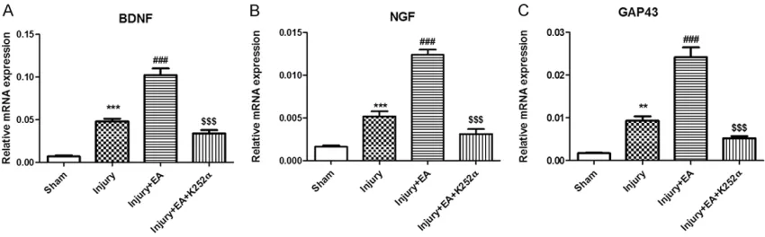
The current study examined changes in BDNF,
NGF, and GAP-43 mRNA expression levels after EA in the right L4-L6 spinal ventral horn. Effects
of K252α, a pharmacological blocker of Trk sig -naling, were evaluated by real-time PCR. BDNF, NGF, and GAP-43 mRNA expression levels were
significantly increased in the Injury group, com -pared to the Sham group (Figure 1). An increase in BDNF, NGF, and GAP-43 mRNA expression levels was observed in the Injury + EA group (compared with the Injury group), suggesting that EA treatment upregulated mRNA expres-sion. Moreover, a reduction in BDNF, NGF, and GAP-43 mRNA expression levels was observed
in the Injury + EA + K252α group (compared
with the Injury + EA group), suggesting that
K252α reversed the effects of EA on expres -sion of BDNF, NGF, and GAP-43 mRNA.
Elevated BDNF, NGF, and GAP-43 protein ex-pression in the right L4-L6 spinal ventral horn
BDNF, NGF, and GAP-43 protein expression lev-els in the ipsilateral (right side) L4-L6 spinal ventral horn were analyzed by Western blotting.
BDNF, NGF, and GAP-43 were significantly
upregulated in the Injury group, compared with the Sham group, suggesting that peripheral nerve injuries induced BDNF, NGF, and GAP-43
[image:4.612.93.518.73.205.2]expression in the spinal ventral horn. The Injury + EA group showed significant increases in
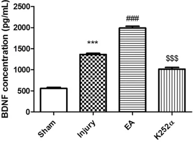
K252α reversed the effects of EA on BDNF
release.
Effects of EA and K252 on structures of in-jured sciatic nerves
H&E staining was performed to observe the
structures of ipsilateral sciatic nerves. No
inflammatory cell infiltration was observed in
the Sham control sample (Figure 4). After nerve
injury, inflammatory cells, including neutrophils, T lymphocytes, and macrophages, were noted
in sciatic nerves from animals in the Injury group. Notable Schwann cell proliferation was
found in the Injury group. There was less inflam
-matory cell infiltration in the Injury + EA group,
compared with the Injury group, suggesting
[image:5.612.91.369.74.336.2]that EA relieved local inflammatory response. There was more inflammatory cell infiltration in the Injury + EA + K252α group, compared with the Injury + EA group, suggesting that K252α reversed the effects of EA on local inflammato -ry response (Figure 4).
Figure 2. Changes in GAP-43, NGF, and BDNF protein expression in the L4-L6 spinal ventral horn after different treatments on day 8 after surgery. (A) Representative Western blots of GAP-43, NGF, and BDNF in L4-L6 spinal ventral horn on day 8 after different treatments. GAPDH was used as a load-ing control. Quantification analysis of the optical density of these bands, (B) GAP-43, (C) NGF, and (D) BDNF. Columns represent means ± SEM from two separate experiments. (*** P < 0.001, compared with the Sham group; ### P < 0.001, compared with the Injury group; $$$ P < 0.001, compared with the Injury + EA group).
Figure 3. Changes in BDNF concentrations in sciatic nerve regeneration chambers after different treat-ments on day 14 after surgery. BDNF was quantified by standard ELISA (*** P < 0.001, compared with the Sham group; ### P < 0.001, compared with the Injury group; $$$ P < 0.001, compared with the In-jury + EA group).
BDNF, NGF, and GAP-43 expression, compared to the Injury group, suggesting that EA further
stimulated BDNF expression in the spinal ventral horn. Ex- pression levels of these
pro-teins were significantly decrea-sed in the Injury + EA + K252α
group, compared to the Injury + EA group (Figure 2), indicating
that K252α reversed the
ef-fects of EA on BDNF, NGF, and GAP-43 protein expression.
Elevated BDNF concentrations in sciatic nerve regeneration fluid
BDNF concentrations in sciatic
nerve regeneration fluid were
examined by ELISA. BDNF
con-centrations were significantly
upregulated in the Injury group, compared to the Sham group.
The Injury + EA group showed a significant increase in BDNF
concentrations, compared to the Injury group, suggesting that EA stimulated BDNF re- lease after sciatic nerve injury. BDNF concentrations were
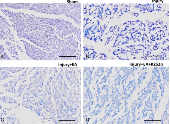
[image:5.612.92.286.464.607.2]Cross-sections of sciatic nerves were stained with luxol fast blue, a stain commonly used to observe myelin, revealing the structure of the
myelin sheath. The sciatic nerve myelin sheath
was found to be regular and uniform in the Sham control group (Figure 5). In contrast, the sciatic nerve myelin sheath was irregular, sug-gesting that myelins were degenerated and
demyelination occurred after injury. The struc -ture of sciatic nerve myelin sheath was partially
serve as an efficient approach to promote
recovery from sciatic nerve injuries in future clinical settings.
[image:6.612.91.373.76.278.2]Currently, methods used for spinal cord injuries are limited due to severe limb movement dys-function and locomotor impairment found in patients. In recent years, the recovery of motor function after spinal cord injuries has been studied extensively [11]. Electrical stimulation
Figure 4. Representative images showing the structures of ipsilateral sci-atic nerves for (A) Sham, (B) Injury, (C) Injury + EA, and (D) Injury + EA + K252α group after H&E staining. Scale bar: 100 μm.
Figure 5. Representative images showing the myelin sheath for (A) Sham, (B) Injury, (C) Injury + EA, and (D) Injury + EA + K252α group after luxol blue staining. Scale bar: 100 μm.
recovered after EA treatment, suggesting that EA promoted sciatic nerve myelin sheath
regeneration and that K252α
blocked this effect of EA (Figure 5).
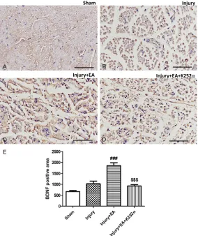
Elevated BDNF expression in injured sciatic nerves
Expression of BDNF in injured sciatic nerves was detected by immunohistochemical stain-ing. Results showed that, in the Sham control group, BDNF was mildly expressed in the cytoplasm (Figure 6). In the In- jury group, sciatic nerves show- ed degeneration and BDNF ex- pression was slightly increa- sed. BDNF staining was stron-ger in the Injury + EA group, suggesting that EA treatment increased BDNF expression. A reduction in BDNF expression was observed in the Injury +
EA + K252α group (compared
with Injury + EA group),
sug-gesting that K252α reversed
the effects of EA on expression of BDNF (Figure 6).
Discussion
Current study observations provide compelling evidence
that BDNF, TrkB, and GAP-43
contribute to sciatic nerve inju-ries and that EA exaggerates
the upregulation of BDNF, TrkB,
and GAP-43. In addition, EA improves the functional and electrical recovery of nerves
after damage. The current
[image:6.612.92.374.343.549.2]increases nerve regeneration with no effects
on neuropathic pain [12]. Therefore, electrical
stimulation plays an important role in the treat-ment of nerve injury conditions, depending on the parameters and protocols of the adminis-tration. EA has been utilized to alleviate chronic neuropathic pain after spinal cord injuries [13, 14]. Consistent with present results, EA im-
proved gait locomotion, reflex, and ventral root
potential in a spinal compression injured model [15], although different EA protocols and sites were administered.
During peripheral nerve injuries, distal axons are derived from nutrition support from soma.
Therefore, they undergo collapse [16].
Sur-levels decrease after regeneration. Therefore,
it has been considered the internal mediator for axonal growth and branching. Recent research has suggested that BDNF affects GAP-43 and promotes neuronal regeneration after injuries. Present results demonstrate that GAP-43 is highly regulated after sciatic nerve injuries. In addition, EA (14 days after treat-ment) promotes GAP-43 expression, suggest-ing that EA mediated GAP-43 is BDNF-related. Acknowledgements
This study was supported by the Natural
[image:7.612.90.374.72.410.2]Sci-ence Foundation of Ministry of Education of Anhui Province (KJ2015A116).
Figure 6. Changes in BDNF immunoreactivity in ipsilateral sciatic nerves af-ter different treatments on day 8 afaf-ter surgery. Representative immunohis-tochemistry image showing expression of BDNF in ipsilateral sciatic nerves for (A) Sham, (B) Injury, (C) Injury + EA, (D) Injury + EA + K252α group; (E) Average BDNF positive area in ipsilateral sciatic nerves after different treatment. Data are presented as mean ± SEM. (### P < 0.001, compared with the Injury group; $$$ P < 0.001, compared with the Injury + EA group). Scale bar: 100 μm.
rounding glia cells (Schwann cells) become activated and proliferate to engulf disorga-nized distal axons with macro-phages. Activated Schwann cells recreate growth factors, including NGF, BDNF, and Laminin [3].
NGF and BDNF promote axonal regrowth after sciatic nerve
injuries. The use of induced
pluripotent stem cells to differ-entiate into Schwann cells to boost the elevation of NGF and BDNF is an ongoing novel approach towards the treat-ment of spinal cord regenera-tion [17]. EA has been
demon-strated to enhance
Wnt/β-catenin signaling, which pro-motes the proliferation and differentiation of neural stem
cells [18]. These results par -tially explain the elevation of NGF and BDNF after EA admin-istration. However, further stu- dies are warranted to elucidate the detailed signaling trans- duction.
GAP-43 is a rapidly
transport-ed axonal protein, significantly
Disclosure of conflict of interest
None.
Address correspondence to: Yilin Ye, College of Nur- sing, Anhui University of Chinese Medicine, B7-459, No. 1 Qianjiang Road, Xinzhan District, Hefei 230012, Anhui Province, China. Tel: +86 0551-68129263; Fax: +86 0551-68129165; E-mail: [email protected]
References
[1] Fu SY and Gordon T. The cellular and molecular basis of peripheral nerve regeneration. Mol Neurobiol 1997; 14: 67-116.
[2] Park H and Poo MM. Neurotrophin regulation of neural circuit development and function. Nat Rev Neurosci 2013; 14: 7-23.
[3] Richner M, Ulrichsen M, Elmegaard SL, Dieu R, Pallesen LT and Vaegter CB. Peripheral nerve injury modulates neurotrophin signaling in the peripheral and central nervous system. Mol Neurobiol 2014; 50: 945-970.
[4] Thompson DM and Buettner HM. Neurite out -growth is directed by schwann cell alignment in the absence of other guidance cues. Ann Biomed Eng 2006; 34: 161-168.
[5] Zakharov VV, Bogdanova MN and Mosevitsky MI. Specific proteolysis of neuronal protein GAP-43 by calpain: characterization, regula-tion, and physiological role. Biochemistry (Mosc) 2005; 70: 897-907.
[6] Aigner L, Arber S, Kapfhammer JP, Laux T, Schneider C, Botteri F, Brenner HR and Caroni P. Overexpression of the neural growth-associ-ated protein GAP-43 induces nerve sprouting in the adult nervous system of transgenic mice. Cell 1995; 83: 269-278.
[7] Hu LN, Tian JX, Gao W, Zhu J, Mou FF, Ye XC, Liu YP, Lu PP, Shao SJ and Guo HD. Electroacu-puncture and moxibustion promote regenera-tion of injured sciatic nerve through Schwann cell proliferation and nerve growth factor se-cretion. Neural Regen Res 2018; 13: 477-483. [8] Wang X, Ju S, Chen S, Gao W, Ding J, Wang G,
Cao H, Tian H and Li X. Effect of electro-acu -puncture on neuroplasticity of spinal cord-tran-sected rats. Med Sci Monit 2017; 23: 4241-4251.
[9] Chen J, Qi JG, Zhang W, Zhou X, Meng QS, Zhang WM, Wang XY and Wang TH. Electro-acupuncture induced NGF, BDNF and NT-3 ex -pression in spared L6 dorsal root ganglion in cats subjected to removal of adjacent ganglia. Neurosci Res 2007; 59: 399-405.
[10] Seow SLS, Hong SL, Lee GS, Malek SNA and Sabaratnam V. 6-shogaol, a neuroactive com-pound of ginger (jahe gajah) induced neurito-genic activity via NGF responsive pathways in PC-12 cells. BMC Complementary and Alterna-tive Medicine 2017; 17: 334.
[11] Sharp KG, Dickson AR, Marchenko SA, Yee KM, Emery PN, Laidmae I, Uibo R, Sawyer ES, Stew-ard O and Flanagan LA. Salmon fibrin treat -ment of spinal cord injury promotes functional recovery and density of serotonergic innerva-tion. Exp Neurol 2012; 235: 345-356. [12] Su HL, Chiang CY, Lu ZH, Cheng FC, Chen CJ,
Sheu ML, Sheehan J and Pan HC. Late admin-istration of high-frequency electrical stimula-tion increases nerve regenerastimula-tion without ag-gravating neuropathic pain in a nerve crush injury. BMC Neurosci 2018; 19: 37.
[13] Wang JY, Gao YH, Qiao LN, Zhang JL, Duan-Mu CL, Yan YX, Chen SP and Liu JL. Repeated elec-troacupuncture treatment attenuated hyperal-gesia through suppression of spinal glial acti-vation in chronic neuropathic pain rats. BMC Complement Altern Med 2018; 18: 74.
[14] Xu J, Chen XM, Zheng BJ and Wang XR. Electroacupuncture relieves nerve injury-in-duced pain hypersensitivity via the inhibition of spinal P2X7 receptor-positive microglia. Anesth Analg 2016; 122: 882-892.
[15] Escobar-Corona C, Torres-Castillo S, Rodriguez-Torres EE, Segura-Alegria B, Jimenez-Estrada I and Quiroz-Gonzalez S. Electroacupuncture improves gait locomotion, H-reflex and ventral root potentials of spinal compression injured rats. Brain Res Bull 2017; 131: 7-17.
[16] Chen X, Chen C, Hao J, Qin R, Qian B, Yang K, Zhang J and Zhang F. AKR1B1 Upregulation contributes to neuroinflammation and astro -cytes proliferation by regulating the energy me-tabolism in rat spinal cord injury. Neurochem Res 2018; 43: 1491-1499.
[17] Huang CW, Huang WC, Qiu X, Fernandes Ferreira da Silva F, Wang A, Patel S, Nesti LJ, Poo MM and Li S. The differentiation stage of transplanted stem cells modulates nerve re-generation. Sci Rep 2017; 7: 17401.