DESIGN OF AUTOMATED BIOMEDICAL WASTE SEGREGATION UNIT USING IMAGE PROCESSING
1
Sandesh, S.,
2Balaji,
Department of Biomedical Engineering, Sri Ramakrishna Engineering College, Anna University, Coimbatore
ARTICLE INFO ABSTRACT
The management of health care waste is a subject of considerable concern to public health and infections
transmission of communicabl
blood and infections of body fluids. Diseases are spread by improper treatment and disposal of wastes while handling items like needles, surgical
medica
be memorised by any one and interpreted correctly. There is also a possibility of infections when hospital staff try to dispose the waste themselves without the kn
prevent the disease spreading and color
an automated waste segregation unit without human contact.
Copyright ©2015 Sandesh et al. This is an open access
distribution, and reproduction in any medium, provided the original work is properly cited.
INTRODUCTION
The aim of this project is to prevent infection that spreads to hospital staffs due to manual and sometimes improper disposal of wastes. Mixing the two color codes wastes to be avoided and also prevent the air pollution and sand pollution of improper segregation of wastes.
HARDWARE COMPONENTS
The hardware used is as follows:
Arduino Mega
DC motor
Relay Circuit
Transformer
Voltage regulator
Interface Max232
Conveyor belt
2x8 Liquid Crystal Display
The Arduino mega is used here for control the dc
pusher gun for waste segregation. Initially Biomedical or biological Waste will be placed onto a conveyor belt
then captured by the camera further there will be image processingprocess involved onto the captured image. Then the captured image is analyzed within the database which then
*Corresponding author: G. Duraivarun,
Department of Biomedical Engineering, Sri Ramakrishna
Engineering College, Anna University, Coimbatore
ISSN: 0975-833X
Article History:
Received 16th December, 2014
Received in revised form
05th January, 2015
Accepted 23rd February, 2015
Published online 31st March,2015
Key words:
Color codes, Medical waste, Communicable diseases
RESEARCH ARTICLE
OF AUTOMATED BIOMEDICAL WASTE SEGREGATION UNIT USING IMAGE PROCESSING
Balaji, T.,
3,*Duraivarun, G. and
4Anandhakumar,
Department of Biomedical Engineering, Sri Ramakrishna Engineering College, Anna University, Coimbatore
ABSTRACT
The management of health care waste is a subject of considerable concern to public health and infections-control specialists, as well as the general public. Careless disposal of wastes may lead to transmission of communicable disease spreading through air, water and direct human contact with blood and infections of body fluids. Diseases are spread by improper treatment and disposal of wastes while handling items like needles, surgical gloves, blood bags. This paper
medical waste automatically instead of the conventional colour coding method. Colour codes cannot be memorised by any one and interpreted correctly. There is also a possibility of infections when hospital staff try to dispose the waste themselves without the knowledge of becoming infected. So to prevent the disease spreading and color-codes identifications, we have proposed the idea to develop an automated waste segregation unit without human contact.
is an open access article distributed under the Creative Commons Attribution License, which distribution, and reproduction in any medium, provided the original work is properly cited.
The aim of this project is to prevent infection that spreads to hospital staffs due to manual and sometimes improper disposal of wastes. Mixing the two color codes wastes to be avoided and also prevent the air pollution and sand pollution
The Arduino mega is used here for control the dc motor and Initially Biomedical or conveyor belt which is then captured by the camera further there will be image processingprocess involved onto the captured image. Then the captured image is analyzed within the database which then
eering, Sri Ramakrishna
Engineering College, Anna University, Coimbatore-22
activates the pusher gun. The input w the gun and are dragged into the indication of this process is display
SOFTWARE COMPONENTS
MATLAB software is used in the
integration for the project. The source code acquisition, pusher gun control is developed o mega microcontroller board.
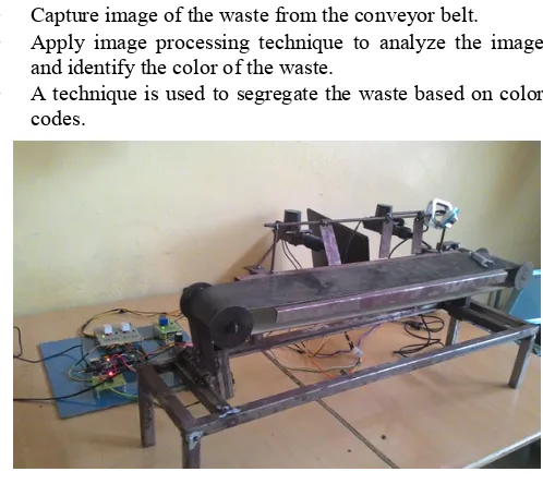
HARDWARE SETUP
Camera is mounted in front of the conveyor belt and connected to the computer.The image processing is widely used in automation of process
consists of a conveyor belt on which the wastes are moving with a constant speed. The conveyor
of 1m/s, making a rate of 3 different color wastes per second. The system used in this paper is that
controlled by the user from the fro
Arduino software. The wastes before being disp be checked and segregated based on the type of
code.TheDisposal of wastes is carried out by the conveyor belt running at a high speed making it essential for the proposed process of segregation through automation
mixing of different color coded wastes.Once the wastes are segregated it is separatedin individual bags. Automation in the waste segregation is a very essential activity of a hospital waste management.
International Journal of Current Research
Vol. 7, Issue, 03, pp.13744-13748, March, 2015
OF AUTOMATED BIOMEDICAL WASTE SEGREGATION UNIT USING IMAGE PROCESSING
Anandhakumar, R.
Department of Biomedical Engineering, Sri Ramakrishna Engineering College, Anna University, Coimbatore-22
The management of health care waste is a subject of considerable concern to public health and control specialists, as well as the general public. Careless disposal of wastes may lead to , water and direct human contact with blood and infections of body fluids. Diseases are spread by improper treatment and disposal of wastes gloves, blood bags. This paper is about segregation of l waste automatically instead of the conventional colour coding method. Colour codes cannot be memorised by any one and interpreted correctly. There is also a possibility of infections when owledge of becoming infected. So to codes identifications, we have proposed the idea to develop
ribution License, which permits unrestricted use,
. The input wastes is pushed through dragged into the color coded waste bag the
displayed on the LCD.
used in the process of development and integration for the project. The source code for the image usher gun control is developed on the Arduino
in front of the conveyor belt and is connected to the computer.The image processing is widely of processindustries. The process setup consists of a conveyor belt on which the wastes are moving with a constant speed. The conveyor belt is operated at a speed 1m/s, making a rate of 3 different color wastes per second. system used in this paper is that speed of the motor is controlled by the user from the front panel window of the stes before being disposed needs to be checked and segregated based on the type of the color is carried out by the conveyor belt at a high speed making it essential for the proposed through automation which avoids the different color coded wastes.Once the wastes are segregated it is separatedin individual bags. Automation in the waste segregation is a very essential activity of a hospital waste
PROBLEM STATEMENT
To achieve the objectives of the proposed work it is required to choose a non-contact method for segregation of biomedical waste. To achieve this objective following steps are involved.
Capture image of the waste from the conveyor belt.
Apply image processing technique to analyze the image and identify the color of the waste.
A technique is used to segregate the waste based on color
[image:2.595.40.289.110.332.2]codes.
Fig. 1. Biomedical Waste Segregation Proposed System
Fig. 2. Different Types of Color Codes for Biomedical Wastes
PROPOSED SOLUTION
Having obtained the image of conveyor belt on which the biomedical wastes are moving it is processed to identify the color coded wastes.Once the segregation of wastes is made based on the color coded wastes, it is subjected to avoid the mixing of two different color code wastes.The whole process is divided into two stages a. color coded waste identification b. Segregation.
COLOR CODED WASTE IDENTIFICATION
The color coded wastes which are moving on the conveyor belt needs to be first identified. For identification of color coded wastes the following process are incorporated
Selection of region of interest
Edge detection
Identification of color coded waste
REGION OF INTEREST
[image:2.595.313.557.212.386.2]Rectangular type of ROI with coordinate values for left, top, right, bottom is created from the entire image. Also we can use different types of ROI like point, line, oval, annulus etc. The inspection will automatically pass if measurements necessary to determine the ROI are available and the coordinates of the ROI are valid. ROI is drawn on top of an image using the operation ImageGenerateROIMask.
Fig.3. Region of Interest in the Input Image
EDGE DETECTION
Edge detection refers to the process of identifying and locating sharp discontinuities in an image. The discontinuities are abrupt changes in pixel intensity which characterize boundaries of objects in a scene. Classical methods of edge detection involve convolving the image with an operator (a 2D filter), which is constructed to be sensitive to large gradients in the image while returning values of zero in uniform regions.
[image:2.595.40.291.362.544.2] [image:2.595.318.551.534.714.2]IDENTIFICATION OF COLOR CODED WASTE
Having obtained the region where the color detection should be carried out the edge detection algorithm is used to identify the borders of images. The acquired biomedical waste image is subjected to various processes like image segmentation, feature extraction, threshold image for saturation plane, histogram image, median filtering, Gaussian image, homogenized image, HSV image, hue image, saturation image. These image processing steps used to identify the type of color coded waste.
SEGREGATION OF BIOMEDICAL WASTE
[image:3.595.318.553.47.598.2]Once the color coded biomedical waste image is identified, segregation of wastes is to be done. For segregation in the proposed work three solenoid trigger guns are used.
Fig. 5. Histogram Equalized Image
Fig. 6. HSV Image
Fig. 7. Hue Image
Fig. 8. Saturation Image
[image:3.595.326.547.58.265.2]Fig. 9. Theshold Image
Fig.10. Brightness of the Image
[image:3.595.60.273.225.781.2] [image:3.595.323.549.589.742.2]Fig.12. Segmentation Image
Fig. 13. Result displayed in the message box
Based on a particular biomedical waste color codes, respective trigger is initialized to trigger the biomedical wastes out of the conveyor. The conveyor belt consist of three trigger guns of different tension levels so as to shoot the biomedical wastes to different conveyors running in parallel, which carry 3 different colors yellow, green and blue respectively. (But in the proposed setup we have placed three baskets. Since, it was not economical to fabricate three conveyors in the facility. The distance between the camera and the trigger guns are fixed.
Since the speed of the conveyor is controlled by the program based on user inputs, the time required for a biomedical waste to travel from camera to trigger gun can be computed.
Once the program identifies the color coded of biomedical wastes, respective trigger gun is initialized to trigger so as to move the biomedical waste to its respective conveyor. In the proposed model, only three color coded biomedical wastes are considered so we use three trigger guns. This can be increased or decreased based on the application.
PROPOSED SOLUTION IMPLEMENTATION
Implementation in the proposed work, MATLAB platform is used for programming. The MATLAB window shows the image on which segregation and its analysis are done. Arduino mega is used to control the trigger guns and operating the whole setup. Control board consists of transformer, regulator circuit, three relay circuits, Arduino mega board, interface circuit (max232 IC, capacitor). Once the program is executed the camera capture the images and the MATLAB used for image processing works such that the time reference image and captured images are compared then pusher guns used to move the wastes out of the conveyor belt. The corresponding waste is segregated by the appropriate waste box.
ANALYSIS
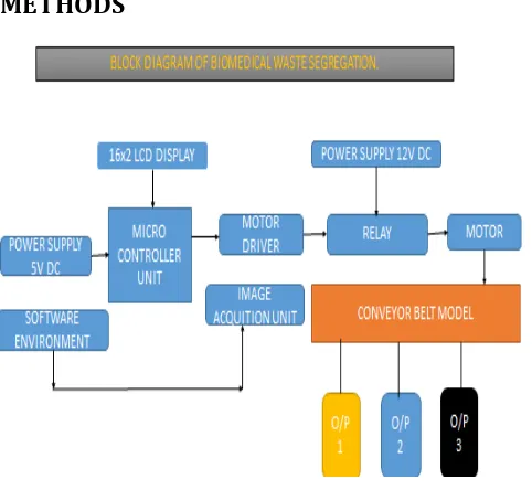
The proposed technique for segregation of biomedical wastes using image processing is programmed by using the block diagram of MATLAB. Three different colors coded biomedical wastes are classified by this proposed technique. If a scenario wherein unwanted waste are placed on the conveyor belt then the three pusher guns will not work and finally the waste drops straight into the non-color coded waste bag.
DISCUSSION
[image:4.595.44.285.468.690.2]An automated technique for classification and segregation of biomedical wastes based on color codes is reported in this paper using image processing techniques. The images once acquired are subjected to edge detection, histogram normalization for the classification of color coded wastes using support vector machine algorithms.
Fig. 15. Result is the Yellow Color Coded Biomedical Waste to be Identified
METHODS
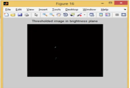
[image:4.595.304.579.526.730.2]Fig. 16. Result is the Red Color Coded Biomedical Waste to be Identified
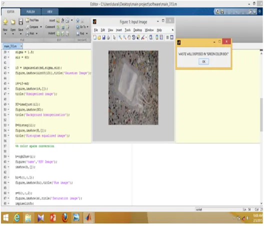
Fig. 17. Result is the Green Color Coded Biomedical Waste to be Identified
Finally, on identifying the color coded wastes, segregation of wastes is done using three trigger guns placed near the conveyor. Result of the proposed technique for test cases shows the successful implementation with an accuracy of 95%. Efficiency of the technique can be improved in future.
RESULTS
An image processing based automated classification system in biomedical waste management for segregation of biomedical wastes based on color codes is implemented using MATLAB.The proposed technique was subjected to test with three different color codes like red, yellow, blue color wastes. By this process many infectious disease can be prevented by spreading in the hospital facility.
REFERENCES
Ballard, D. H. 1981. Generalizing the Hough Transform to Detect Arbitrary Shapes. Pattern Recognition, Vol. 13, (1981), 111-122.
Cosgriff, R. L. 1960. Identification of Shape. Ohio State University Research Foundation, Columbus, Rep. 82011. Fast discriminationand counting of filled/unfilled rice
spikelets based on bi-modal imaging. Computers and
Electronics in Agriculture, Vol. 75, (2011), 196–203. International Conference on Wavelet Analysis and Pattern
Recognition, Hong Kong.
LingfengDuan, Wanneng Yang, Kun Bi, Shangbin Chen, and QingmingLuo, Qian Liu. 2011.
Monika Bhatnagar and Prashant Kumar Singh. 2014. An Efficient Method of Image Segmentation for Harvest Time Identification, International Journal of Computer Applications, Volume 87, No.7, (February 2014), 31-34. Pornpanomchai, C. Liamsanguan, and Vannakosit, V. 2008.
Vehicle Detection and Counting From A Video Frame. In Proceedings of the 2008
Pushpendra Kumar, RekhaPandit, and VineetRichhariya. 2014. Retinal Image Segmentation by using Gradient Descent. International Journal of Computer Applications, Vol. 86, No 10, (January 2014), 1-7.
Santhosh, K. V. and Bhagya, R Navada.2014. Online Classification and Measurement of Pencils using Image
Processing Techniques, International Journal of
Computer Applications, Volume 96, No.4, (June 2014), 25-30.
SugataBanerji, N. AtreyeeSinha, and Chengjun Liu. 2013. New image descriptors based on color, texture, shape, and wavelets for object and scene image classification.
Neurocomputing, Vol. 117, (2013), 173– 185.
Weyricha, M., Wanga, Y., Winkela, J. and Laurowskib, M. 2012. High Speed Vision Based Automatic Inspection and Path Planning for Processing Conveyed Objects”, Procedia CIRP, Vol. 3, Elsevier, (2012), 442 – 447.
[image:5.595.35.304.291.517.2]