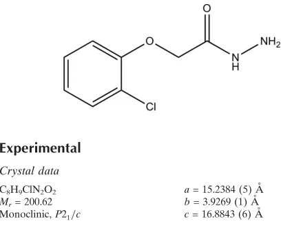
2-(2-Chlorophenoxy)acetohydrazide
Hoong-Kun Fun,a*‡ Ching Kheng Quah,a§ Arun M. Isloor,bDhanya Sunilcand Prakash Shettyd
a
X-ray Crystallography Unit, School of Physics, Universiti Sains Malaysia, 11800 USM, Penang, Malaysia,bDepartment of Chemistry, National Institute of Technology-Karnataka, Surathkal, Mangalore 575 025, India,cDepartment of
Chemistry, Manipal Institute of Technology, Manipal University, 576 104, India, and
dDepartment of Printing and Media Engineering, Manipal Institute of Technology,
Manipal University, 576 104, India Correspondence e-mail: [email protected]
Received 26 November 2009; accepted 28 November 2009
Key indicators: single-crystal X-ray study;T= 100 K; mean(C–C) = 0.003 A˚; Rfactor = 0.050;wRfactor = 0.121; data-to-parameter ratio = 20.5.
In the title compound, C8H9ClN2O2, the acetohydrazide group is approximately planar, with the maximum deviation of 0.031 (2) A˚ . In the crystal, the molecules are linked by N— H N, N—H O and C—H O hydrogen bonds, with the acetohydrazide O atom accepting two C—H O links and one N—H O link. This results in infinite sheets lying parallel to (100).
Related literature
For general background to and biological properties of hydrazine derivatives, see: Rando et al.(2008); Kumar et al. (2009); Kamalet al.(2007); Masunari & Tavares (2007); Rando et al.(2002). For related structures, see: Funet al.(2009, 2010). For the preparation, see: Holla & Udupa (1992). For the stability of the temperature controller used for the data collection, see: Cosier & Glazer (1986).
Experimental
Crystal data
C8H9ClN2O2
Mr= 200.62 Monoclinic,P21=c
a= 15.2384 (5) A˚ b= 3.9269 (1) A˚ c= 16.8843 (6) A˚
= 117.269 (2)
V= 898.07 (5) A˚3
Z= 4
MoKradiation
= 0.39 mm1
T= 100 K
0.280.100.09 mm
Data collection
Bruker SMART APEXII CCD diffractometer
Absorption correction: multi-scan SADABS(Bruker, 2005) Tmin= 0.897,Tmax= 0.965
11351 measured reflections 2662 independent reflections 2029 reflections withI> 2(I) Rint= 0.050
Refinement
R[F2> 2(F2)] = 0.050
wR(F2) = 0.121
S= 1.05 2662 reflections 130 parameters
H atoms treated by a mixture of independent and constrained refinement
max= 0.44 e A˚ 3
[image:1.610.46.258.554.718.2]min=0.30 e A˚ 3
Table 1
Hydrogen-bond geometry (A˚ ,).
D—H A D—H H A D A D—H A
N1—H1N1 N2i
0.83 (3) 2.20 (2) 2.930 (3) 148 (2) N2—H1N2 O2ii
0.91 (3) 2.36 (2) 3.070 (2) 134 (2)
C1—H1A O2iii 0.93 2.54 3.443 (3) 164
C7—H7A O2iv
0.97 2.37 3.317 (2) 165
Symmetry codes: (i)xþ2;yþ1;zþ2; (ii)x;yþ1;z; (iii)xþ2;y1 2;zþ
3 2;
(iv)xþ2;yþ1 2;zþ
3 2.
Data collection:APEX2(Bruker, 2005); cell refinement:SAINT
(Bruker, 2005); data reduction:SAINT; program(s) used to solve structure: SHELXTL (Sheldrick, 2008); program(s) used to refine structure:SHELXTL; molecular graphics:SHELXTL; software used to prepare material for publication:SHELXTLandPLATON(Spek, 2009).
HKF and CKQ thank Universiti Sains Malaysia (USM) for the Research University Golden Goose Grant (1001/PFIZIK/ 811012). CKQ thanks USM for a Research Fellowship. AMI is grateful to the Director, NITK-Surathkal, India, for providing research facilities and the Head of the Department of Chemistry & Dean R&D, NITK Surathkal, for their encour-agement.
Supplementary data and figures for this paper are available from the IUCr electronic archives (Reference: HB5257).
References
Bruker (2005).APEX2,SAINTandSADABS. Bruker AXS Inc., Madison, Wisconsin, USA.
Fun, H.-K., Quah, C. K., Isloor, A. M., Sunil, D. & Shetty, P. (2010).Acta Cryst. E66, o53–o54.
Fun, H.-K., Quah, C. K., Sujith, K. V. & Kalluraya, B. (2009).Acta Cryst.E65, o1184–o1185.
Cosier, J. & Glazer, A. M. (1986).J. Appl. Cryst.19, 105–107. Holla, B. S. & Udupa, K. V. (1992). Farmaco,47, 305–318
Kamal, A., Khan, N. A., Reddy, K. S. & Rohini, K. (2007).Bioorg. Med. Chem. 15, 1004–1013.
Kumar, P., Narasimhan, B., Sharma, D., Judge, V. & Narang, R. (2009).Eur. J. Med. Chem.44, 1853–1863.
Masunari, A. & Tavares, L. C. (2007).Bioorg. Med. Chem.15, 4229–4236. Rando, D. G., Avery, M. A., Tekwani, B. L., Khan, S. I. & Ferreira, E. I. (2008).
Bioorg. Med. Chem.16, 6724–6731.
organic compounds
Acta Cryst.(2010). E66, o31–o32 doi:10.1107/S1600536809051356 Funet al.
o31
Acta Crystallographica Section E
Structure Reports Online
ISSN 1600-5368
Rando, D. G., Sato, D. N., Siqueira, L., Malvezzi, A., Leite, C. Q. F., do Amaral, A. T., Ferreira, E. I. & Tavares, L. C. (2002).Bioorg. Med. Chem.10, 557– 560.
supporting information
sup-1
Acta Cryst. (2010). E66, o31–o32
supporting information
Acta Cryst. (2010). E66, o31–o32 [doi:10.1107/S1600536809051356]
2-(2-Chlorophenoxy)acetohydrazide
Hoong-Kun Fun, Ching Kheng Quah, Arun M. Isloor, Dhanya Sunil and Prakash Shetty
S1. Comment
Hydrazine derivatives have been reported to possess several biological properties. 5-nitro-2-heterocyclic benzylidine
hydrazides were found to possess antileishmanial activities (Rando et al., 2008). Many substituted benzoic acid
furan-2-yl-methylene hydrazides showed potent antimicrobial properties(Kumar et al., 2009). Hydrazine derivatives were also
associated with remarkable anticancer (Kamal et al., 2007), antibacterial (Masunari & Tavares, 2007) and tuberculostatic
(Rando et al., 2002) activities.
The molecular structure is shown in Fig. 1. The acetohydrazide group (C7/C8/N1/N2/O2) is approximately planar, with
the maximum deviation of 0.031 (2) Å for atom N1. Bond lengths and angles are within normal ranges, and comparable
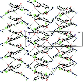
to closely related structures (Fun et al., 2009, 2010). In the solid state (Fig. 2), the molecules are linked via
intermolecular N2—H1N2···O2, C1—H1A···O2 and C7—H7A···O2 trifurcated acceptor bonds, together with N1—
H1N1···N2 hydrogen bonds, into infinite two-dimensional networks parallel to plane (1 0 0).
S2. Experimental
O-chloro phenol (11 ml, 1.00 mmol), ethyl chloroacetate (10.7 ml, 1.00 mmol) and potassium carbonate (20.75 g, 1.50
mmol) were refluxed in acetone (100 ml) at 80 °c for 18 h. The reaction mixture is then filtered, distilled to remove the
acetone and poured into ice cold water with vigorous stirring. The ester, phenoxy ethyl acetate was extracted using ether.
The solution was distilled to remove ether. Phenoxy ethyl acetate (8.2 ml, 0.50 mmol) was heated at 100 °C for 10h in an
absolute alcohol medium (40 ml) with hydrazine hydrate (2.5 ml, 0.50 mmol). The reaction mixture was allowed to cool,
the solid separated was filtered, dried and recrystallized from ethanol. The yield was found to be 7.1 g (71 %). M. p.
384-385 K (Holla & Udupa, 1992).
S3. Refinement
Atoms H1N1, H1N2 and H2N2 were located from the difference Fourier map and refined freely. The remaining H atoms
Figure 1
The molecular structure of (I), showing 50% probability displacement ellipsoids.
Figure 2
The crystal structure of (I) viewed along the a axis. H atoms not involved in intermolecular interactions (dashed lines)
[image:4.610.127.483.306.657.2]supporting information
sup-3
Acta Cryst. (2010). E66, o31–o32
2-(2-Chlorophenoxy)acetohydrazide
Crystal data
C8H9ClN2O2 Mr = 200.62 Monoclinic, P21/c
Hall symbol: -P 2ybc
a = 15.2384 (5) Å
b = 3.9269 (1) Å
c = 16.8843 (6) Å
β = 117.269 (2)°
V = 898.07 (5) Å3 Z = 4
F(000) = 416
Dx = 1.484 Mg m−3
Mo Kα radiation, λ = 0.71073 Å Cell parameters from 3388 reflections
θ = 2.4–30.1°
µ = 0.39 mm−1 T = 100 K Block, colourless 0.28 × 0.10 × 0.09 mm
Data collection
Bruker SMART APEXII CCD diffractometer
Radiation source: fine-focus sealed tube Graphite monochromator
φ and ω scans
Absorption correction: multi-scan
SADABS (Bruker, 2005)
Tmin = 0.897, Tmax = 0.965
11351 measured reflections 2662 independent reflections 2029 reflections with I > 2σ(I)
Rint = 0.050
θmax = 30.2°, θmin = 1.5°
h = −21→20
k = −5→4
l = −22→23
Refinement
Refinement on F2
Least-squares matrix: full
R[F2 > 2σ(F2)] = 0.050 wR(F2) = 0.121 S = 1.05 2662 reflections 130 parameters 0 restraints
Primary atom site location: structure-invariant direct methods
Secondary atom site location: difference Fourier map
Hydrogen site location: inferred from neighbouring sites
H atoms treated by a mixture of independent and constrained refinement
w = 1/[σ2(F
o2) + (0.0585P)2 + 0.3771P]
where P = (Fo2 + 2Fc2)/3
(Δ/σ)max < 0.001
Δρmax = 0.44 e Å−3
Δρmin = −0.30 e Å−3
Special details
Experimental. The crystal was placed in the cold stream of an Oxford Cyrosystems Cobra open-flow nitrogen cryostat (Cosier & Glazer, 1986) operating at 100.0 (1) K.
Geometry. All esds (except the esd in the dihedral angle between two l.s. planes) are estimated using the full covariance matrix. The cell esds are taken into account individually in the estimation of esds in distances, angles and torsion angles; correlations between esds in cell parameters are only used when they are defined by crystal symmetry. An approximate (isotropic) treatment of cell esds is used for estimating esds involving l.s. planes.
Refinement. Refinement of F2 against ALL reflections. The weighted R-factor wR and goodness of fit S are based on F2,
conventional R-factors R are based on F, with F set to zero for negative F2. The threshold expression of F2 > 2sigma(F2) is
used only for calculating R-factors(gt) etc. and is not relevant to the choice of reflections for refinement. R-factors based on F2 are statistically about twice as large as those based on F, and R- factors based on ALL data will be even larger.
Fractional atomic coordinates and isotropic or equivalent isotropic displacement parameters (Å2)
x y z Uiso*/Ueq
Cl1 0.69094 (3) 0.22956 (13) 0.86423 (3) 0.02146 (15)
O2 1.09342 (9) −0.0760 (4) 0.87110 (8) 0.0183 (3)
N1 1.01406 (11) 0.2382 (4) 0.93081 (10) 0.0158 (3)
N2 1.10162 (11) 0.3687 (5) 1.00234 (10) 0.0175 (3)
C1 0.72527 (14) −0.1775 (5) 0.66271 (12) 0.0192 (4)
H1A 0.7768 −0.2398 0.6506 0.023*
C2 0.62775 (14) −0.2331 (5) 0.59963 (12) 0.0232 (4)
H2A 0.6144 −0.3314 0.5452 0.028*
C3 0.55039 (14) −0.1436 (6) 0.61715 (13) 0.0236 (4)
H3A 0.4855 −0.1815 0.5746 0.028*
C4 0.57002 (12) 0.0024 (5) 0.69829 (12) 0.0197 (4)
H4A 0.5185 0.0633 0.7105 0.024*
C5 0.66699 (13) 0.0571 (5) 0.76107 (12) 0.0177 (4)
C6 0.74518 (12) −0.0283 (5) 0.74390 (11) 0.0161 (4)
C7 0.91785 (12) −0.0685 (5) 0.79289 (11) 0.0154 (4)
H7A 0.9113 0.0356 0.7383 0.018*
H7B 0.9149 −0.3137 0.7851 0.018*
C8 1.01638 (12) 0.0305 (5) 0.86985 (11) 0.0147 (4)
H1N1 0.9633 (17) 0.307 (5) 0.9322 (14) 0.014 (5)*
H1N2 1.1345 (16) 0.478 (6) 0.9761 (14) 0.018 (6)*
H2N2 1.1333 (17) 0.195 (6) 1.0302 (16) 0.022 (6)*
Atomic displacement parameters (Å2)
U11 U22 U33 U12 U13 U23
Cl1 0.0139 (2) 0.0305 (3) 0.0168 (2) 0.00075 (17) 0.00427 (15) −0.00393 (19)
O1 0.0092 (5) 0.0272 (8) 0.0117 (6) −0.0005 (5) 0.0012 (4) −0.0029 (5)
O2 0.0137 (6) 0.0242 (8) 0.0163 (6) 0.0013 (5) 0.0063 (5) −0.0006 (5)
N1 0.0092 (6) 0.0219 (8) 0.0123 (7) 0.0002 (6) 0.0016 (5) −0.0024 (6)
N2 0.0113 (6) 0.0238 (9) 0.0114 (7) −0.0012 (6) −0.0002 (6) −0.0012 (6)
C1 0.0177 (8) 0.0214 (10) 0.0142 (8) −0.0010 (7) 0.0035 (7) −0.0001 (7)
C2 0.0226 (9) 0.0242 (10) 0.0136 (8) −0.0030 (8) 0.0003 (7) −0.0023 (8)
C3 0.0145 (8) 0.0259 (11) 0.0201 (9) −0.0035 (7) −0.0011 (7) −0.0010 (8)
C4 0.0113 (7) 0.0224 (10) 0.0205 (9) −0.0013 (7) 0.0029 (7) 0.0006 (8)
C5 0.0154 (8) 0.0184 (10) 0.0140 (8) −0.0007 (7) 0.0023 (6) 0.0002 (7)
C6 0.0115 (7) 0.0175 (9) 0.0140 (8) −0.0011 (7) 0.0012 (6) 0.0019 (7)
C7 0.0124 (7) 0.0195 (10) 0.0116 (7) −0.0005 (7) 0.0031 (6) −0.0007 (7)
C8 0.0142 (7) 0.0162 (9) 0.0115 (7) 0.0002 (7) 0.0041 (6) 0.0032 (6)
Geometric parameters (Å, º)
Cl1—C5 1.7443 (19) C1—H1A 0.9300
O1—C6 1.3756 (19) C2—C3 1.386 (3)
O1—C7 1.430 (2) C2—H2A 0.9300
O2—C8 1.237 (2) C3—C4 1.385 (3)
N1—C8 1.326 (2) C3—H3A 0.9300
N1—N2 1.4231 (19) C4—C5 1.385 (2)
N1—H1N1 0.83 (2) C4—H4A 0.9300
supporting information
sup-5
Acta Cryst. (2010). E66, o31–o32
N2—H2N2 0.84 (2) C7—C8 1.519 (2)
C1—C6 1.391 (3) C7—H7A 0.9700
C1—C2 1.393 (2) C7—H7B 0.9700
C6—O1—C7 115.63 (14) C5—C4—H4A 120.3
C8—N1—N2 122.15 (15) C3—C4—H4A 120.3
C8—N1—H1N1 125.4 (15) C4—C5—C6 121.25 (17)
N2—N1—H1N1 112.5 (15) C4—C5—Cl1 119.09 (15)
N1—N2—H1N2 105.5 (13) C6—C5—Cl1 119.65 (13)
N1—N2—H2N2 104.8 (16) O1—C6—C5 116.80 (16)
H1N2—N2—H2N2 110 (2) O1—C6—C1 124.09 (17)
C6—C1—C2 119.63 (18) C5—C6—C1 119.11 (16)
C6—C1—H1A 120.2 O1—C7—C8 110.19 (14)
C2—C1—H1A 120.2 O1—C7—H7A 109.6
C3—C2—C1 120.73 (18) C8—C7—H7A 109.6
C3—C2—H2A 119.6 O1—C7—H7B 109.6
C1—C2—H2A 119.6 C8—C7—H7B 109.6
C4—C3—C2 119.78 (17) H7A—C7—H7B 108.1
C4—C3—H3A 120.1 O2—C8—N1 123.86 (16)
C2—C3—H3A 120.1 O2—C8—C7 119.11 (16)
C5—C4—C3 119.50 (18) N1—C8—C7 117.00 (15)
C6—C1—C2—C3 −0.5 (3) C4—C5—C6—C1 −1.3 (3)
C1—C2—C3—C4 −0.1 (3) Cl1—C5—C6—C1 177.58 (15)
C2—C3—C4—C5 0.0 (3) C2—C1—C6—O1 −179.16 (18)
C3—C4—C5—C6 0.7 (3) C2—C1—C6—C5 1.1 (3)
C3—C4—C5—Cl1 −178.13 (16) C6—O1—C7—C8 178.73 (15)
C7—O1—C6—C5 176.26 (16) N2—N1—C8—O2 2.4 (3)
C7—O1—C6—C1 −3.5 (3) N2—N1—C8—C7 −175.28 (16)
C4—C5—C6—O1 179.00 (18) O1—C7—C8—O2 172.20 (16)
Cl1—C5—C6—O1 −2.1 (2) O1—C7—C8—N1 −10.0 (2)
Hydrogen-bond geometry (Å, º)
D—H···A D—H H···A D···A D—H···A
N1—H1N1···N2i 0.83 (3) 2.20 (2) 2.930 (3) 148 (2)
N2—H1N2···O2ii 0.91 (3) 2.36 (2) 3.070 (2) 134 (2)
C1—H1A···O2iii 0.93 2.54 3.443 (3) 164
C7—H7A···O2iv 0.97 2.37 3.317 (2) 165