Jamshid Ahmadi1 Charles M. North1
Hervey D. Segall1 Chi-Shing Zee 1 Martin H. Weiss2
This article appears in the November/Decem-ber 1985 issue of AJNR and the February 1985 issue of AJR.
Received December 12, 1984; accepted after revision May 14, 1985.
1 Department of Radiology, Section of Neurora-diology, University of Southern California School of Medicine, Los Angeles, CA 90033. Address reprint requests to J. Ahmadi, LAC/USC Medical Center, Box 2,1200 N. State St., Los Angeles, CA 90033. 2 Department of Neurological Surgery, University of Southern California School of Medicine, Los An-geles, CA 90033.
AJNR 6:893-898, November/December 1985 0195-6108/85/0606-0893
© American Roentgen Ray Society
893
Cavernous Sinus Invasion by
Pituitary Adenomas
One hundred ninety-eight surgically explored pituitary adenomas were evaluated preoperatively by high-resolution computed tomography (CT). At surgery, evidence of direct cavernous sinus invasion was demonstrated in 19. CT findings in these cases included cavernous sinus expansion (17 patients) and visible encasement of the internal carotid artery (14 patients). The invasive tumor often enhanced to a lesser degree than the cavernous sinuses and ipsilateral internal carotid artery. Intracavernous cranial nerve compression, obliteration, or displacement (14 patients), invasion of the lateral wall of the cavernous sinus (seven patients), and diffuse bone destruction (seven cases) were other findings. MagnetiC resonance imaging in three patients provided excellent demonstration of intracavernous internal carotid artery encasement, but displacement and obliteration of intracavernous cranial nerves was not shown as well as it was with CT. Histologically, only three patients showed anaplastic features and only one of them had distant metastases. There was no correlation between histologic features, hormone assays, and invasiveness. This experience indicates any type of pituitary adenorha, regardless of its endocrinologic activity, can invade the cavernous sinus. Cavernous sinus involvement makes complete surgical removal difficult. Preoperative recognition of invasive behavior of these tumors has prognostic value and aids in designing appropriate management. CT is the most useful technique generally available for evaluation and follow-up.
Although invasive pituitary adenomas are known to exist, only rarely has the diagnosis of cavernous sinus invasion been made preoperatively [1-4). Generally adenomas that penetrate into the cavernous sinus are difficult to resect completely, and they are associated with a greater risk of morbidity and mortality [4, 5).
Therefore, preoperative demonstration of cavernous sinus invasion may aid the
neurosurgeon and endocrinologist in planning appropriate medical and surgical
management. The purpose of this study was to determine the computed tomo -graphic (CT) (and, more recently, magnetic resonance [MR) imaging) signs of
cavernous sinus invasion. Such invasion was further correlated with tumor
histo-pathology and hormone assays. We performed a retrospective review of high-resolution CT studies in 19 surgically documented cases of cavernous sinus
invasion by pituitary tumor. Patients having bone invasion without cavernous sinus involvement were not included in this study.
Materials and Methods
894
AHMADI ET AL. AJNR:6, Nov/Dec 1985Fig. 1. -Pituitary adenocarcinoma extending into right cavernous sinus in 84-year-old patient. The In
-tracavernous part of partly calcified internal carotid
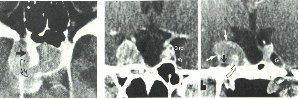
artery is engulfed and slightly displaced medially (solid arrow). Tumor had penetrated dural barrier a second time and extended into posterior fossa (open arrow).
Fig. 2.-Null-cell adenoma. Intra- and suprasellar part of tumor was resected 2 months earlier. Obvious compression and partial obliteration of right gasserian ganglion (black arrow) and nerves V, and III (cf. normal appearances of these nerves on opposite side). Engulfment of intracavernous right internal carotid artery is also seen as it exits from carotid canal (open arrow). White arrows show where enhancing dura mater, which forms lateral margins of cavernous sinus, is interrupted with tumor bulging locally through and beyond this area. G = gasserian ganglion.
A
B
c
Fig. 3.-Prolactin-cell adenoma. Displacement and compression of right gasserian ganglion/V,-V2 complex (solid arrows) on both coronal (A and B) and axial (C) views. (el. opposite side [open arrows).)
thickness and 2 mm spacing. The other two patients were examined with a Philips Tomoscan 310 scanner using 3 rnm collimation and 1.5 mrn spacing. Nine patients were scanned in the axial plane only (with coronal and sagittal reformation), three in the direct coronal plane only, and the other seven in both coronal and axial planes. Most CT scans were obtained after rapid drip infusion of iodinated contrast material (Reno-M-DIP, 42.3 g 1/300 ml solution). Four patients had an intravenous bolus plus a drip infusion of contrast material (71.3 g I). Three patients also had MR scanning. Medical records, including operative notes, of all these patients were reviewed. The results of pituitary hormone assays, histologic features of the surgical speci-mens, and CT were correlated. Surgical specimens in nine patients were also examined by immunohistologic staining techniques and electron microscopy.
Results
CT revealed unilateral involvement of the cavernous sinus
in 14 patients, with bilateral invasion in the other five cases.
Tumor size was variable. There was a suprasellar component (ranging from 1 to 6 em above the diaphragma sellae) in 16 patients; three patients had no suprasellar extension. In 17 patients, invasion of the cavernous sinus through the lateral dural envelope of the sella resulted in varying degrees of cavernous sinus expansion with bulging of the lateral margin of the sinus. In seven of these patients, the tumors had grown outside of the cavernous sinus (having penetrated the dural barrier a second time) into the middle or posterior cranial fossa (figs. 1 and 2). Invasive tumor often enhanced to a lesser degree than did the cavernous sinuses and internal carotid artery. The intracavernous internal carotid artery was clearly demonstrated in 14 patients because it enhanced to a greater degree than did the surrounding tumor. In the other
five patients, however, carotid artery engulfment was difficult
to distinguish from adjacent tumor because of an almost
equal degree of enhancement. A larger bolus of contrast
[image:2.612.58.559.79.246.2] [image:2.612.58.558.335.478.2]demonstra-AJNR:6, Nov/Dec 1985 PITUITARY ADENOMA IN CAVERNOUS SINUS 895
Fig. 4.-Corticotrophic cell adenoma. Rapid invasion of right cavernous sinus developed after bilateral adrenalectomy for control of hypercortisolism (Nelson syndrome). Displacement and engulfment of intracavernous right inter-nal carotid artery (arrow) is readily visible because it enhances to a greater degree than surrounding tumor.
Fig. 5.-Corticotrophic cell microadenoma with recent pituitary apoplexy and infarction. Minimal extension into right cavernous sinus (arrow) without clearly evident increase in width of cavernous sinus. At this stage, it would be
Fig. 7.-Null-cell adenoma. MR images. Coronal (A) and axial (8) spin-echo scans, 2 sec TR, 40 msec TE. Bilateral extension of pituitary tumor into cavernous sinuses, left greater than right (solid arrows), and engulfment of intracavernous internal ca-rotid arteries (open arrows).
A
tion of the internal carotid artery in more of these cases had
the study been performed prospectively.
Extension of the tumor into the cavernous sinus resulted in displacement, compression, or obliteration of the intracav-ernous cranial nerves in 14 patients (figs. 2 and 3). In 10
patients, both carotid artery engulfment and cranial nerve
compression were present (fig. 2). In four patients, carotid
artery engulfment could not be seen. However, compression
of the intracavernous cranial nerves was present (fig. 3). In four patients, the carotid artery was visibly engulfed, but we were unable to detect compression of the cranial nerves (fig.
4). In only one patient was there neither expansion of the
cavernous sinus nor engulfment or compression. In this case,
invasion was exhibited by very subtle bulging into the
cavern-ous sinus (fig. 5). This case presented the greatest difficulty in distinguishing extrinsic compression from invasion of the
cavernous sinus. Direct coronal scans might have been more
helpful in demonstrating subtle invasion. Nevertheless,
inva-sion into the cavernous sinus was proven at surgery.
In six patients, the significance of osseous abnormalities
could not be assessed easily because of previous
transsphe-noidal surgery. In seven of the other 13 patients, there were
somewhat difficult to distinguish invasion from focal displacement of cavernous sinus.
Fig. 6.-Null-cell adenoma. Axial CT scan of base of skull reveals extensive destruction of sphenoid body, greater sphenoid wings, pterygoid plates, vomer, petrous apices, clivus, and basiocciput (black arrows). Tumor extends into sphenoid and posterior ethmoid sinuses (white arrows). Histologically, it had benign microscopic appearance.
B
varying degrees of destruction of the sella and adjacent bones
(fig. 6). The CT findings in these 19 patients were cavernous
sinus expansion (17 patients), compression of the gasserian ganglion V, complex (14), visibly engulfed internal carotid
artery (14), invasion of the lateral wall of the cavernous sinus (seven), and diffuse bone destruction (seven). (Diffuse bone
destruction was evaluated in only 13 of the 19 patients
because of difficulties involved in differentiating surgical changes from tumor invasion.) MR imaging in three patients
clearly showed engulfment of the internal carotid artery by an invasive pituitary tumor. The low signal intenSity of flowing
blood in the carotid artery provided excellent contrast to the
high signal intensity of the surrounding tumor (fig. 7).
The serum prolactin level was substantially elevated
(rang-ing from 105 to 50,000 ngjml) in only eight patients (table 1). Hypopituitarism was present before surgery in four patients. Histologic examination of surgical specimens showed benign
pituitary adenoma in 16 patients. Histologically, marked cel-lular pleomorphism, multinuclear cells, and mitotic features were present in three patients, and only one of them had
896 AHMADI ET AL. AJNR:6, Nov/Dec 1985
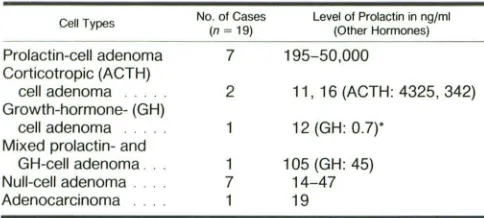
TABLE 1: Functional Classification of Invasive Pituitary Tumors
Cell Types No. of Cases Level of Prolactin in ng/ml
(n = 19) (Other Hormones)
Prolactin-cell adenoma 7 195-50,000 Corticotropic (ACTH)
cell adenoma 2 11, 16 (ACTH: 4325, 342) Growth-hormone-(GH)
cell adenoma 12 (GH: 0.7l' Mixed prolactin- and
GH-cell adenoma .. 1 105 (GH: 45) Null-cell adenoma . 7 14-47 Adenocarcinoma 1 19
Note.-Normal values: prolactin, <20 ng/ml: ACTH. 10-100 ng/ml: GH, <1 ng/ml.
• In this patient, serum GH hormone was normal. However, GH granules were identified in the tumor by electron microscopy and immunocytologic staining. Similar cases have been
well documented 161.
These patients have been followed for periods of 2 months to 5 years. Two patients with a small amount of cavernous sinus invasion had a total resection of their tumors with no recurrence (they have been followed 1112 and 3 years, respec-tively). Five patients were treated with bromocriptine and partial resection (the size of the residual tumor was reduced in three patients and remained unchanged in two other pa-tients). Nine patients had partial resection followed by radia -tion therapy. There was a dramatic response to radiation therapy in two of the nine patients. Both of these patients had anaplastic features histopathologically. There was a min-imal response to radiation therapy in another two. The residual tumor was unchanged in three patients and was actually larger in the other two. Two patients had partial resection and bromocriptine and radiation therapy. One of these had a mild response and the other had no response. One patient with cerebrospinal fluid and liver metastases died 2 months after
the diagnosis of pituitary adenocarcinoma was documented.
Discussion
On the basis of their biologic behavior, tumors that originate in the adenohypophysis may be divided into expanding ade-nomas, invasive adenomas, and carcinomas [1-5, 7]. Most pituitary adenomas are slow-growing and tend to grow in an upward direction into the suprasellar cistern. Intrasellar ex -pansion of these adenomas may erode the sella turcica and compress the cavernous sinuses laterally. The dura matter forming the medial walls of the cavernous sinus remains intact but is displaced somewhat because of pressure from the expanding pituitary adenoma. Most pituitary adenomas dem-onstrate a globular configuration regardless of their size. The diaphragma sellae, however, may impose a waistline inden-tation between the intrasellar and suprasellar components of large adenomas [5]. Occasionally, more rapid growth of one part of an adonoma results in an asymmetric configuration. Nodular outgrowths may burst (focally) through the thin mes -odermal envelope (pituitary adenomas do not have a capsule) encasing the adenoma. This has been likened to a bud perforating the skin of a potato [5]. In some cases this nodular outgrowth may destroy adjacent structures by invading them. Those tumors that invade directly into adjacent soft tissue
and osseous structures are often referred to as "invasive pituitary adenomas" [1-5, 7]. Most of the invasive pituitary tumors in our series had a benign microscopic appearance,
which demonstrates the poor correlation between biologically aggressive behavior and histopathologic features. A review of the literature revealed a diversity of opinion with regard to nomenclature in connection with pituitary adenomas. Several authors have indicated that features such as increased cel -lularity, cellular pleomorphism, and the presence of mitotic features are not necessarily proof of malignancy [2, 7-12]. They believe that the diagnosis of pituitary carcinoma can be
considered conclusively only when distant metastases occur.
Others consider anaplastic histopathologic features to be sufficient for the diagnosis of carcinoma [13-15].
Hardy [16] proposed a classification of pituitary tumors based on biologic behavior. Grade I is localized to the sella, grade II extends into the suprasellar cisterns, grade III refers to localized invasion of the floor of the sella, and grade IV indicates diffuse invasion. Grade V was added to indicate distant metastases [5]. Using this grading system our report basically consists of grade IV tumors, although one patient had progressed to grade V. Invasion of the sella turcica and adjacent bones may result in extensive destruction of the
base of the skull. The tumor may extend into the nasopharynx
or sphenoid and/or ethmoid sinuses. Posterior extension may
lead to destruction of the clivus and petrous apices (fig. 6);
occasionally it may be difficult to distinguish an invasive pituitary adenoma having such a destructive pattern from
other neoplasms, such as metastases, chordomas, and
na-sopharyngeal carcinomas [17, 18]. Furthermore we have also
seen meningiomas, as well as one glomangioma and one
dysgerminoma, that invaded the cavernous sinus.
Although the abnormal serum levels of hormones produced by functional pituitary tumors generally correlate with the size of the tumor, there are occasions when a small tumor may be related to marked elevations of serum prolactin. Very high serum prolactin values (over 1000 ngjml) have been impli-cated as strong evidence of invasion of pituitary adenomas into the cavernous sinuses [3, 19]. Nonetheless, a normal serum prolactin level may be seen with invasive pituitary tumors. In 11 of 19 cases of invasive pituitary tumors in our report, there was no significant elevation of serum prolactin values (table 1).
There is no rule that facilitates predicting when invasion of the surrounding structures will begin. In three of our patients as well as in a few other reports [16, 19]. invasion of the cavernous sinus began when the tumor was small and was limited to the sellar region. Furthermore, one of our patients had cavernous sinus invasion only a few months after clinical onset of galactorrhea and amenorrhea. On the other hand, another patient had a 27 year history of pituitary adenoma before a diagnosis of cavernous sinus invasion was made. In our series and in others [2-4, 19] males predominated; more men than women appeared to have larger and more aggres-sive tumors. Symptoms such as decreased libido, impotence,
[image:4.612.55.297.100.209.2]AJNR:6, Nov/Dec 1985 PITUITARY ADENOMA IN CAVERNOUS SINUS 897
Fig. 8.-Coronal cross section of sellar region. Extradural subcavernous extension of pituitary tumor without invasion into cavernous sinus (arrow).
Dura mater forming medial and lateral walls of cavernous sinus (thick, solid
line); periosteal layer of dura (broken line). A = intracavernous internal carotid
artery; G = gasserian ganglion.
Fig. 9.-lntradural parasellar extension of pituitary tumor without invasion
into cavernous sinus. Intracavernous cranial nerves and carotid artery (A) are
not involved. OC = optic chiasm; G = gasserian ganglion.
tumor is usually considerably larger at presentation.
In evaluating pituitary tumors, careful attention should be
paid to certain intracavernous structures. Appreciation of
involvement of the intracavernous cranial nerves and/or
ca-rotid artery by tumor enabled a CT diagnosis of cavernous
sinus invasion in 18 of the 19 cases in this series. Normally,
the gasserian ganglion/V1-V2 complex is consistently dem-onstrated on CT as a relatively symmetric, oblong, intracav-ernous structure [20, 21]. Although in 14 of our patients there was partial obliteration or compression of the cranial nerves traversing the cavernous sinus, only six of our patients exhib-ited opthalmoplegia, trigeminal anesthesia, or pain.
The infrequency of cranial nerve signs and symptoms
sug-gests that these tumors do not invade but rather displace or
encircle the nerves. In addition, CT demonstration of visible
engulfment of the intracavernous carotid artery, obliteration
of the gasserian/V1-V2 complex, and direct penetration of the
dura of the cavernous sinus aids in differentiating tumor
invasion of the cavernous sinus from two other types of
extension. On occasion, pituitary adenomas may displace the
cavernous sinus laterally after perforating the dura at the
junction of the floor of the sella and the cavernous sinus.
Subcavernous extension of tumor, growing in an extradural
location between the cavernous sinus and sphenoid bone,
may develop without invasion into the cavernous sinus [4, 5]
(fig. 8). Another possibility is upward expansion of a pituitary
adenoma into the suprasellar cistern. The tumor may then
extend laterally above the cavernous sinus. It may pass
between either the supraclinoid carotid artery and the upper
margin of the cavernous sinus or between the carotid artery
and the optic chiasm. The adenoma then can extend into the
middle cranial fossa intradurally and between the cavernous
sinus on its medial aspect and the temporal lobe on its lateral
aspect [7] (fig. 9). In either situation, intracavernous structures
such as the carotid artery and the gasserian ganglion are
neither engulfed nor compressed by tumor. This will help to
differentiate cavernous sinus invasion from these two possible routes of tumor growth.
Cavernous sinus invasion by a pituitary tumor has been
related to a relatively low surgical cure rate because of the
complexities of surgical exploration when carried into the
cavernous sinus [1-5, 7]. With preoperative recognition of
cavernous sinus invasion by a pituitary tumor, particularly the
commonly occurring prolactin-secreting tumor, one would
attempt to reduce the size of the tumor by medical means in
an effort to make surgical excision less difficult [22, 23]. When
a good CT study has been obtained documenting a small
amount of cavernous sinus invasion, an experienced
neuro-surgeon may be able to resect the tumor completely. Thus,
CT is very helpful in surgical planning. In the case of larger
tumors, where total resection has not been possible, CT
enables recognition of persistent tumor and will be helpful in
judging the response resulting from chemical manipulation or
radiation therapy. CT is particularly valuable in the case of
secretory-inactive adenomas where hormone markers are not
present to enable evaluation for persistence of tumor in the
cavernous sinus.
ACKNOWLEDGMENTS
We thank Vivian Chandrasoma, Department of Pathology, USC
School of Medicine, who reviewed the histology of these patients; Richard Yadley, Robert Wycoff, and William Bradley, Department of
Radiology, Huntington Memorial Hospital, who performed some of
the CT and MR studies; and Andrea Kovacs and associates, who
performed electron microscopy and immunocytochemistry in some
of our cases.
REFERENCES
1. Jefferson G. The invasive adenomas of the anterior pituitary. In:
The Sherrington lectures, vol. 3. Liverpool: University of liver-pool,1955
2. Martins AN, Hayes GJ, Kemmpe LG. Invasive pituitary adeno -mas. J Neurosurg 1965;22:268-276
3. Lundberg PO, Drettner B, Hemmingsson A, Stenkvist B, Wide L. The invasive pituitary adenoma. A prolactin-producing tumor.
Arch Neuro/1977;34:742-749
898
AHMADI ET AL. AJNR:6, Nov/Dec 1985pituitary adenomas. J Neurol Neurosurg Psychiatry 1979;
42:973-982
5. Wilson CB. Neurosurgical management of large and invasive
pituitary tumors. In: Tindall GT, Collins WF, eds. Clinical
man-agement of pituitary disorders. New York: Raven, 1979:335 -342
6. Zimmerman EA, Defendini R, Frantz AG. Prolactin and growth
hormone in patients with pituitary adenomas: a correlative study
of hormone in tumor and plasma by immunoperoxidase
tech-nique and radioimmunoassay. J Clin Endocrinol Metab
1974;38:577-585
7. Landolt AM, Wilson CB. Tumors of the sella and parasellar area in adults. In: Youmans JR, ed. Neurological surgery. Philadelphia: Saunders, 1982:3107-3162
8. Epstein JA, Epstein BS, Molho L, Zimmerman HM. Carcinoma of the pituitary gland with metastases to the spinal cord and roots of the cauda equina. J Neurosurg 1964;21: 846-853
9. Madonick MJ, Rubinstein LJ, Dasco MR, et al. Chromophobe
adenoma of pituitary gland with subarachnoid metastases. Neu-rology (NY) 1963;13:836-840
10. Rubinstein LJ. Tumors of the central nervous system. In: Atlas
of tumor pathology, 2d series, fasc 6. Washington, DC: Armed
Forces Institute of Pathology, 1972:313-314
11. Ogilvy KM, Jakubowski J. Intracranial dissemination of pituitary adenomas. J Neurol Neurosurg Psychiatry 1973;36: 199-205
12. Martin NA, Hales M, Wilson CB. Cerebellar metastases from a
prolactinoma during treatment with bromocriptine. Case report.
J Neurosurg 1981;55:615-619
13. Wise BL, Brown HA, Naffziger HC, et al. Pituitary adenomas,
carcinomas and craniopharyngiomas. Surg Gynecol Obstet
1955;101: 185-193
14. Newton TH, Burhenne HJ, Palubinskas AJ. Primary carcinoma
of the pituitary. AJR 1962;87: 11 0-119
15. D'Abrera VStE, Burke WJ, Bleasel KF, Bader L. Carcinoma of
the pituitary gland. J Pathol 1973;109:335-343
16. Hardy J. Transsphenoidal microsurgical treatment of pituitary
tumors. In: Linfoot JA, ed. Recent advances in the diagnosis and
treatment of pituitary tumors. New York: Raven, 1979:375-388
17. Virapongse C, Bhimani S, Sarwar M, Greenberg A, Kim J.
Prolactin-secreting pituitary adenomas: CT appearance in diffuse
invasion. Radiology 1984;154:447-451
18. Kendall BE, Lee BCP. Cranial chordomas. Br J Radiol
1977;50:687-698
19. Shucart WA. Implications of very high serum prolactin levels
associated with pituitary tumors. J Neurosurg 1980;52:226-228
20. Kline LB, Acker JD, Post MJD, Vitek JJ. The cavernous sinus: a
computed tomographic study. AJNR 1981;2:299-305
21. Hasso AN, Pop PM, Thompson JR, et al. High resolution thin
section computed tomography of the cavernous sinus.
Radi-ographics 1982;2: 83-1 00
22. Weiss MH. Medical and surgical management of functional pitui-tary tumors. Clin Neurosurg 1981;28:374-383
23. Vassilouthis J, Richardson AE. Prolactin levels in aggressive