Department of Emergency, Affiliated Lianyungang Hospital of Xuzhou Medical College, Lianyungang, China.
*Equal contributors.
Received August 25, 2015; Accepted September 25, 2015; Epub October 1, 2015; Published October 15, 2015
Abstract: Seven in absentia homolog (SIAH) is a ubiquitin ligase that monoubiquitinates α-synuclein. Lewy bodies are characteristically rich in monoubiquitinated α-synuclein. We aimed to determine the effect of siRNA-SIAH1 on α-synuclein autophagy and UPS degradation in SH-SY5Y. SIAH1 expression was measured with real-time quantita
-tive PCR and Western Blot. Cell proliferation was measured by CCK-8 assay; cell apoptosis assayed by flow cytom
-etry. Relative protein expressions were measured by Western Blot. mRNA levels of relative protein were measured by real-time quantitative PCR. The expression of α-synuclein, LC3-II and SIAH1 were observed by confocal microscopy. We found: (1) Transfection efficiency of SIAH1-siRNA into SH-SY5 measured approximately 89% by flow cytometry. (2) siRNA silencing of SIAH1 promoted cellular proliferation and suppressed apoptosis. (3) Protein and mRNA ex
-pression of α-synuclein, LC3-II and p53 decreased after SIAH1 knockdown. E1 protein and mRNA levels increased after SIAH1 siRNA. These data show silencing SIAH1 increased cell proliferation and inhibited apoptosis in SH-SY5Y neuroblastoma cells. SIAH1 knockdown enhanced the clearance of non-aggregated α-synuclein by UPS. SIAH1 is a potential target for treatment of Parkinson’s disease.
Keywords: Seven in absentia homolog (SIAH), α-synuclein, Parkinson’s disease, autophagy, ubiquitin-proteasome system (UPS)
Introduction
Parkinson’s disease (PD) affects approximately 1% of the population over the age of 65, and is the second most common progressive neuro-degenerative disorder after Alzheimer’s dis-ease [1]. PD is clinically characterized primarily by resting tremors, bradykinesia, rigidity, pos-tural instability, and other motor and non-motor symptoms. The pathology of PD is character-ized by selective loss of dopamine neurons (DA), and accumulation of α-synuclein in neu-rons of substantia nigra (SN) and other brain-stem regions [2, 3]. The neuropathological hall-mark of PD is the presence of intracytoplasmic Lewy bodies (LBs), composed primarily of mono- and polyubiquitinated α-synuclein pro-tein [4]. E3 ubiquitin ligase-SIAH (seven in absentia homolog) plays an important role in α-synuclein monoubiquitylation and in modu-lating α-synuclein aggregation, and may con-tribute to the formation of Lewy bodies [5]. The
main degradation pathway of intracellular α-synuclein aggregates is via autophagy by the lysosomal degradation pathway [6, 7].
E3 ubiquitin ligase-SIAH supports α-synuclein aggregation and monoubiquition, but the mech-anism of SIAH1 in autophagy through the lyso-momal pathway and the upbiquitin proteasome system (UPS) pathway remains unknown. SIAH1 may activate or down-regulate autophagy via p53, thus promoting or inhibiting the degrada-tion of monoubiquitylated α-synuclein. In order to clarify this, we silenced SIAH1 and analyzed the effects on UPS degradation pathway and autophagy-lysosomal degradation pathway.
Results
SIAH1 gene silencing by predesigned siRNAs
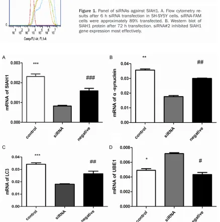
siR-NA-FAM cells were approximately 89% trans-fected in SH-SY5Y cells measured by flow cytometry. After 72 h transfection, we found
[image:2.612.88.526.185.626.2]siRNA #2 showed the greatest suppression of SIAH1 and was selected for subsequent assays (Figure 1).
Figure 1. Panel of siRNAs against SIAH1. A. Flow cytometry re
-sults after 6 h siRNA transfection in SH-SY5Y cells. siRNA-FAM cells were approximately 89% transfected. B. Western blot of SIAH1 protein after 72 h transfection. siRNA#2 inhibited SIAH1 gene expression most effectively.
Figure 2. Real time PCR analysis of mRNA levels after siSIAH1 treatment in SH-SY5Y cells. A. SIAH1. B. α-synuclein level reduced significantly (**P<0.01) (##P<0.01). C. LC3-II level reduced significantly (***P<0.001) (##P<0.01). D.
SIAH1 silencing increased cell proliferation in SH-SY5Y cells
To examine the effect of SIAH1 siRNA on cell proliferation, we measured cell proliferation wi-
[image:3.612.89.524.68.669.2]th a CCK-8 assay. We found that SIAH1 knock-down significantly increased SH-SY5Y cell pro-liferation compared with control and negative siRNA groups. This indicates that silencing SIAH1 may be beneficial to H-SY5Y cells.
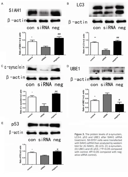
Figure 3. The protein levels of α-synuclein, LC3-II, p53 and UBE1 after SIAH1 siRNA treatment. SH-SY5Y cells were transfected with SIAH1-siRNA then analyzed by western blot for (A) SIAH1, (B) LC3, (C) α-synuclein, (D) UBE1 and (E) p53. (*P<0.05 compared
SIAH1 silencing affected cell apoptosis
We measured apoptosis of SH-SY5Y cells after SIAH1 siRNA using flow cytometry. We found
[image:4.612.92.522.70.610.2]ine the role of SIAH1 in autophagy, we analyzed expression of pathway proteins. Microtubule-associated protein 1 light chain 3 (LC3) is cleaved by Atg4 into LC3-I and LC3-II, LC3-II associated with both outer and inner mem-branes of autophagosome, and the amount of LC3-II correlates with the extent of autophagas-ome [8]. We found LC3-II mRNA and protein reduced after siRNA treatment (Figures 2C,
3B).
SIAH1 silencing the UPS pathway
The UPS pathway plays a critical role in α-synuclein degradation. To investigate the effect of SIAH1 on the UPS pathway, we mea-sured the protein level of UBE1, that important for the UPS degradation. We found UBE1 pro-tein elevated in SH-SY5Y cells after SIAH1 siRNA treatment compared with mock cells ( t = 3.480, P<0.05) or negative siRNA (t = 3.765, P<0.05) (Figure 3D). Consistent with our west-ern blot results, UBE1 mRNA increased in SH-SY5Y cells after SIAH1 siRNA transfection compared with mock cells (t = 8.739, P<0.05) or negative siRNAs (t = 9.170, P<0.05) (Figure 2D).
Crosstalk between UPS pathway and autopha-gy pathway
UPS pathway and ALP pathway are two impor-tant protein degradation systems, and growing evidence suggests that there is a crosstalk between UPS pathway and autophagy pathway. The previous study has shown that p53 has an important in mediating UPS inhibition induced autophagy activation [9]. We measured the pro-tein level of p53. We found p53 propro-tein elevat-ed in SH-SY5Y cells after SIAH1 siRNA treat-ment compared with mock cells (t = 3.326, P<0.05) or negative siRNA (4.400, P<0.05) (Figure 3E).
4B). SIAH-1, α-synuclein and LC3 lost its colo-calization in SH-SY5Y cells.
Discussion
PD pathology is characterized by the accumula-tion of α-synuclein aggregates, known as Lewy bodies, in the substantia nigra (SN) of the mid-brain [10]. The UPS and autophagy-lysosome pathway (ALP) are the two major degradation pathways that maintain protein homeostasis in cells. While the UPS clears most short-lived and soluble proteins [11], the ALP degrades longer-lived macromolecules and dysfunctional organ-elles [12]. α-synuclein exists in three forms: monomers, dimers and protofibrils [13]. How- ever, the mechanism of α-synuclein degrada-tion by the UPS or the autophagy pathway remains unknown. UPS pathway degrades monomeric α-synuclein [14]. Only a small pro-portion of soluble α-synuclein oligomers, not including monomeric α-synuclein, are degrad-ed by the 26S proteasome [15]. α-synuclein monoubiquitylation by E3 ubiquitin ligase-SIAH plays a critical role in α-synuclein aggregation and contributes to Lewy body formation [16]. Monoubiquitination of α-synuclein by SIAH1 is cleared solely by the 26S proteasome, while deubiquitinated α-synuclein is degraded mainly by autophagy [17]. Our study supports the hypothesis that SIAH1 knockdown decreases α-synuclein monoubiquitination and aggrega-tion, contributing to increasing monomers of α-synuclein.
cell proliferation and apoptosis suppression. Our results are consistent with previous studies showing inhibition of SIAH1 reduced apoptosis [22].
In our study, SIAH1 knockdown significantly inhibited α-synuclein expression, suggesting a key role in α-synuclein monoubiquitylation and aggregation. This may provide a therapeutic opportunity in treating PD. Our results are con-sistent with previous research showing SIAH1 monoubiquitylates α-synuclein and leads to aggregation, a toxic environment in cells [16]. In our experiments, RNAi-mediated downregu-lation of SIAH1 effectively decreased protein and mRNA of autophagic vacuoles marker (LC3-II). We can infer autophagy pathway de- creased after SIAH1 siRNA treatment.
In addition, we also found that UBE1 prote- in and mRNA expression levels increased. α-synuclein degradation through the UPS con-sists of two consecutive steps. First, conjuga-tion of multiple ubiquitin moieties to the sub-strate occur, then 26S proteasome complex degrades it [23]. Ubiquitylation occurs via a three-step cascade catalyzed by ubiquitin-acti-vating enzyme (E1), ubiquitin-conjugating en- zyme (E2), and ubiquitin-ligase enzyme (E3). After the initial ubiquitin is attached, polyubiq-uitylation of the substrate adds ubiquitin mole-cules, forming ubiquitin chains [24, 25]. In our study, we found the expression of E1 increased, indicating the UPS degradation pathway was activated after SIAH1 siRNAs transfection.
In conclusion, our results show SIAH1 knock-down inhibited cellular apoptosis and increased cell proliferation. In addition, knockdown of SIAH1 decreased α-synuclein monoubiquitina-tion and aggregamonoubiquitina-tion. These findings argue that the UPS degradation pathway was activated upon SIAH1 inhibition. Our data suggest SIAH1 may be a promising candidate for PD treat- ment.
Materials and methods
Cell culture
SH-SY5Y neuroblastoma cells were a kind gift from the Institute of Neuroscience. SH-SY5Y cells were cultured at 37°C and 5% CO2 in DMEM/F12 medium (Gibco, USA)
supplement-ed with 10% heat-inactivatsupplement-ed fetal bovine serum (Hyclone, USA). The culture medium was changed every two days and the cells were pas-saged when they reached 80 to 90% confluen-cy. SH-SY5Y cells were divided into three groups as follows: i) a blank control group; ii) an experi-mental group transfected with siRNA-SIAH1; iii) a negative control group transfected with a null siRNA.
CCK-8 assay
Cell viability was assayed using the Cell Counting Kit-8 (CCK-8) (Dojindo, Japan). After the control siRNA and SIAH1 specific siRNAs were transfected into SH-SY5Y cells in 96-well-plate 48 h, 10 μL of the CCK-8 reagent was added and incubated at 37°C for 2 h. The opti-cal density (OD) was measured at 450 nm. Cell viability was calculated using the data and pre-sented relative to controls. Results of transfec-tion were read relative to controls, assuming the absorbance of controls was 100%. All val-ues were calculated using mean ± S.D.
Apoptosis assay
Measured SH-SY5Y cells apoptosis after trans-fection control siRNA and SIAH1-siRNA for 72 h using Annexin V/FITC kit (BD Pharmingen, USA) in accordance with the manufacturer’s instruc-tions and detected by flow cytometry.
siRNA design and transfection
of the protein extraction kits (Beyotime Bio- technology, China). Protein concentrations de- termine using BCA protein assay kits (Bioworld, USA). Equal quantities of protein extracts were loaded onto 12% SDS-PAGE gels and wet-trans-ferred to PVDF membranes. After blocking with 5% powdered skim milk for 1 h, the membranes were incubated overnight at 4°C with anti-SIAH (1:100 SantaCruz, America), anti-α-synuclein (1:500, Abcam, USA), anti-LC3 (1:2000, Abcam, USA), UBE1 (1:1000, Cell Signaling, USA), p53 (1:1000 Abcam USA). β-actin (1:1000 Abcam USA). The membranes were incubated with HRP-conjugated secondary antibodies for 2 h at room temperature, and visualized using ECL kits (Bioworld, USA). Determined the band intensities with Quantity One Software (Tanon 5200, China).
Real time quantitative reverse transcription-PCR (qRT-transcription-PCR)
Total RNA was isolated from cells using Trizol reagent (Life Invitrogen) according to the manu-facture’s protocol and was converted to cDNA using PrimeScript RT Master Mix (Perfect Real Time) (Takara, Japan). cDNA were amplified by real-time PCR using SYBR 7500 Fast Real-time PCR System (Applied Biosystems). The follow-ing primers were used: SIAH sense primer 5’CTGTCGCCCCAAACTTACAT-3’ and antisense primer 5’-CCACATGGATACCATCAAACTG-3’. α-sy- nuclein sense primer 5’-CCTCAGCCCAGAGC- CTTTC-3’ and antisense primer 5’-CCTCTGC- CACACCCTGCTT-3’. LC3 sense primer 5’-GAG- TGGAAGATGTCCGGCTC-3’, antisense primer: 5’-CCAGGAGGAAGAAGGCTTGG-3’; UBE1 sense primer 5’-CCCTACATGACCAAGGCACT-3’, anti-sense primer 5’-CCAGGAGGAAGAAGGCTTGG-3’. β-actin sense primer: 5’-TCAGGTCATCACTATC- GGCAAT-3’; antisense primer: 5’-AAAGAAAGG- GTGTAAAACGCA-3’. PCR products were subject-ed to a melting-curve analysis to confirm the PCR specificity.
(1:250 dilution) and anti-α-synuclein (1:100 dilution) overnight at 4°C. Cells were treated with the appropriate secondary antibody treat-ment for 1 h and DAPI (1:100 dilution) was added for 5 min to stain nucleus. Cells evalu-ated under a Confocal microscope (ZEISS, LSM710, Germany).
Statistical analysis
Data are presented as the mean ± standard deviation. Two group comparisons were per-formed using Student’s t-test. Multiple group comparisons were performed using one-way analysis of variance and Fisher’s least signifi-cant difference. Values of P < 0.05 were set as statistically significant.
Acknowledgements
We are grateful to Professor Xu Yun and her helps to this study; we also thank the Department of Neurology, Affiliated Drum Tower Hospital of Nanjing University and the Institute of Neuroscience at Soochow University for their help in this study. We thank Dr. Austin Cape for careful reading and insightful sugges-tions. This project was supported by the China Postdoctoral Science Foundation (No. 1630), the natural science foundation of Jiangsu Province (No. BK2011402), the Ji-angsu Province Postdoctoral Research Funding (No. 1301174C), and Jiangsu Province Health Department Funding (No. H201361).
Disclosure of conflict of interest
None.
Address correspondence to: Dr. Zeng-Lin Cai, Department of Neurology, Affiliated Lianyungang Hospital of Xuzhou Medical College, Lianyungang,
References
[1] Yang YX, Wood NW, Latchman DS. Molecular basis of Parkinson’s disease. Neuroreport
2009; 20: 150-156.
[2] Deng H, Liang H, Jankovic J. F-box only protein 7 gene in parkinsonian-pyramidal disease.
JAMA Neurol 2013; 70: 20-24.
[3] Yuan L, Song Z, Xu H, Gu S, Zhu A, Gong L,
Zhao Y, Deng H. EIF4G1 Ala502Val and
Arg1205His variants in Chinese patients with
Parkinson disease. Neurosci Lett 2013; 543:
69-71.
[4] Ozansoy M, Basak AN. The central theme of Parkinson’s disease: alpha-synuclein. Mol
Neurobiol 2013; 47: 460-465.
[5] Rott R, Szargel R, Haskin J, Shani V, Shainskaya
A, Manov I, Liani E, Avraham E, Engelender S.
Monoubiquitylation of alpha-synuclein by sev -en in abs-entia homolog (SIAH) promotes its
ag-gregation in dopaminergic cells. J Biol Chem
2008; 283: 3316-3328.
[6] Engelender S. Ubiquitination of alpha-synucle
-in and autophagy -in Park-inson’s disease. Autophagy 2008; 4: 372-374.
[7] Lee JT, Wheeler TC, Li L, Chin LS. Ubiquitination of synuclein by Siah-1 promotes alpha-synuclein aggregation and apoptotic cell
death. Hum Mol Genet 2008; 17: 906-917. [8] Kabeya Y, Mizushima N, Ueno T, Yamamoto A,
Kirisako T, Noda T, Kominami E, Ohsumi Y, Yoshimori T. LC3, a mammalian homologue of yeast Apg8p, is localized in autophagosome membranes after processing. EMBO J 2000;
19: 5720-5728.
[9] Du Y, Yang D, Li L, Luo G, Li T, Fan X, Wang Q, Zhang X, Wang Y, Le W. An insight into the mechanistic role of p53-mediated autophagy
induction in response to proteasomal
inhibi-tion-induced neurotoxicity. Autophagy 2009; 5:
663-675.
[10] Braak H, Del TK, Rub U, de Vos RA, Jansen SEN, Braak E. Staging of brain pathology re
-lated to sporadic Parkinson’s disease.
Neu-robiol Aging 2003; 24: 197-211.
[11] Goldberg AL. Protein degradation and
protec-tion against misfolded or damaged proteins.
Nature 2003; 426: 895-899.
[12] Klionsky DJ. Autophagy: from phenomenology
to molecular understanding in less than a
de-cade. Nat Rev Mol Cell Biol 2007; 8: 931-937.
[13] Winslow AR, Chen CW, Corrochano S, Acevedo-Arozena A, Gordon DE, Peden AA, Lichtenberg M, Menzies FM, Ravikumar B, Imarisio S, Brown S, O’Kane CJ, Rubinsztein DC. alpha-Synuclein impairs macroautophagy: implica
-tions for Parkinson’s disease. J Cell Biol 2010;
190: 1023-1037.
[14] Webb JL, Ravikumar B, Atkins J, Skepper JN, Rubinsztein DC. Alpha-Synuclein is degraded by both autophagy and the proteasome. J Biol
Chem 2003; 278: 25009-25013.
[15] Emmanouilidou E, Stefanis L, Vekrellis K. Cell-produced alpha-synuclein oligomers are tar -geted to, and impair, the 26S proteasome. Neurobiol Aging 2010; 31: 953-968.
[16] Rott R, Szargel R, Haskin J, Shani V, Shainskaya
A, Manov I, Liani E, Avraham E, Engelender S.
Monoubiquitylation of alpha-synuclein by sev -en in abs-entia homolog (SIAH) promotes its
ag-gregation in dopaminergic cells. J Biol Chem
2008; 283: 3316-3328.
[17] Rott R, Szargel R, Haskin J, Bandopadhyay R, Lees AJ, Shani V, Engelender S. alpha-Synucle
-in fate is determ-ined by USP9X-regulated monoubiquitination. Proc Natl Acad Sci U S A
2011; 108: 18666-18671.
[18] Reed JC, Ely KR. Degrading liaisons: Siah structure revealed. Nature Struct Biol 2002; 9:
8-10.
[19] Genbacev O, Zhou Y, Ludlow JW, Fisher SJ. Regulation of human placental development by oxygen tension. Science (New York, N.Y.)
1997; 277: 1669-1672.
[20] Tang AH, Neufeld TP, Kwan E, Rubin GM. PHYL acts to down-regulate TTK88, a transcriptional repressor of neuronal cell fates, by a
SINA-dependent mechanism. Cell 1997; 90: 459-467.
[21] Della NG, Senior PV, Bowtell DD. Isolation and characterisation of murine homologues of the Drosophila seven in absentia gene (sina). Development (Cambridge, England) 1993;
117: 1333-1343.
[22] He HT, Fokas E, You A, Engenhart-Cabillic R, An HX. Siah1 proteins enhance radiosensitivity of human breast cancer cells. BMC Cancer 2010;
10: 403.
[23] Glickman MH, Ciechanover A. The ubiquitin-proteasome proteolytic pathway: destruction for the sake of construction. Physiol Rev 2002;
82: 373-428.
[24] Layfield R, Tooth D, Landon M, Dawson S, Mayer J, Alban A. Purification of poly-ubiquiti
-nated proteins by S5a-affinity chromatography.
Proteomics 2001; 1: 773-777.
[25] Gallastegui N, Groll M. The 26S proteasome:
assembly and function of a destructive ma
-chine. Trends Biochem Sci 2010; 35: