A STUDY OF CATEGORIES AND METHODS OF IMAGE SEGMENTATION
*,1
Mr. Chetan S. More,
2Shraddha S. Kavathekar,
1,2,3
Department of Electronics and Tele Communication, Dr. J. J. Magdum College of Engineering,
4
Department of Electronics, Dr. J. J. Magdum College
ARTICLE INFO ABSTRACT
Image segmentation is the most important part in digital image processing. In image segmentation, digital image is divided into multiple set of pixels.
it has become important to categorise the research outcomes and provide readers with an overview of the existing segmentation techniques in each category.
cut out region of interest (ROI) from a available for image segmentation
emphasis on developed technologies and image properties used by them. In this article, and Method
Copyright © 2015 Chetan S. More et al. This is an open access article distributed under the Creative Commons Att use, distribution, and reproduction in any medium, provided the original work is properly cited.
INTRODUCTION
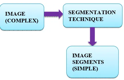
One of the most important and ubiquitous tasks in image analysis is segmentation. Image segmentation is a critical step as it directly influences the overall success to understand the image. Image segmentation refers to the process of partitioning an image into N number of parts. In terms of mathematical formulae, Image segmentation divides an image f(x, y) into continuous, disconnect and nonempty subsets, f1, f2,…..…,fn. From these subsets higher level information can be easily extracted. These several parts obtained by segmentatio something more meaningful and easier for further process. paper is arranged in five parts. Part II provides an introduction to image segmentation. Part III explains the categories and methods of image segmentation. Part IV covers conclusion.
Image Segmentation
In the field of computer vision, images are considered as one of the most important medium of conveying information . objective of computer vision and digital image processing is to automate different tasks and image segmentation is important step in it. The information extracted from them can be used for other tasks for example: navigation of robots, extracting malign tissues from body scans, detection of
*Corresponding author: Mr. Chetan S. More
Department of Electronics and Tele Communication, Dr. J. J. Magdum College of Engineering, Jaysingpur, India.
ISSN: 0975-833X
Article History:
Received 17th December, 2014
Received in revised form 28th January, 2015
Accepted 07th February, 2015
Published online 17th March,2015
Key words:
Image Segmentation, Thresholding, Edge Detection, Discontinuity, Similarity, Region Based Segmentation.
RESEARCH ARTICLE
A STUDY OF CATEGORIES AND METHODS OF IMAGE SEGMENTATION
Shraddha S. Kavathekar,
3Prof. Mrs. S. R. Mahadik
4Prof. Mrs. Anita P. Patil
Electronics and Tele Communication, Dr. J. J. Magdum College of Engineering,
Jaysingpur, India
Electronics, Dr. J. J. Magdum College of Engineering, Jaysingpur, India
ABSTRACT
Image segmentation is the most important part in digital image processing. In image segmentation, digital image is divided into multiple set of pixels. With the growing research on im
it has become important to categorise the research outcomes and provide readers with an overview of the existing segmentation techniques in each category. Image segmentation is generally required to cut out region of interest (ROI) from an image. Currently there are many different algorithms available for image segmentation The state of art research on each category is provided with emphasis on developed technologies and image properties used by them. In this article,
and Methods of Image Segmentation is briefly explain with suitable practical application.
is an open access article distributed under the Creative Commons Attribution License, which use, distribution, and reproduction in any medium, provided the original work is properly cited.
and ubiquitous tasks in image Image segmentation is a critical step as it directly influences the overall success to understand the Image segmentation refers to the process of partitioning n terms of mathematical formulae, Image segmentation divides an image f(x, y) into continuous, disconnect and nonempty subsets, f1, f2,…..…,fn. From these subsets higher level information can be easily These several parts obtained by segmentation are something more meaningful and easier for further process. This paper is arranged in five parts. Part II provides an introduction to image segmentation. Part III explains the categories and methods of image segmentation. Part IV covers conclusion.
In the field of computer vision, images are considered as one of the most important medium of conveying information .Main objective of computer vision and digital image processing is to automate different tasks and image segmentation is an The information extracted from them can be used for other tasks for example: navigation of robots, extracting malign tissues from body scans, detection of
le Communication, Dr. J. J. Magdum College
cancerous cells, and identification of an airport from remote sensing data. Image segmentation is a method which fulfill above requirements, and with the help of which, we can understand images and extract information or objects. Thus, image segmentation is the first step in image analysis.
An Image is basically a two dimensional function of spatial coordinates, f(x, y), and amplitude of this function at a given coordinate gives the intensity value of the image. The product of functions of illumination and reflection, expresses the image as shown in expression below:
f(x, y) = i(x, y). r(x, y)
Where i(x, y) is the function of intensity and r(x, y) is the function of reflectivity.
In general Image segmentation is defined as a ‘process of partitioning an image into homogenous groups or parts such that each region is homogenous but the union of two adjacent regions is not homogenous’.
analyze images and extract data from them. regions of interest in a scene or annotate the data.
Available online at http://www.journalcra.com
International Journal of Current Research
Vol. 7, Issue, 03, pp.13251-13257, March, 2015
INTERNATIONAL
z
A STUDY OF CATEGORIES AND METHODS OF IMAGE SEGMENTATION
Prof. Mrs. S. R. Mahadik and
Electronics and Tele Communication, Dr. J. J. Magdum College of Engineering,
f Engineering, Jaysingpur, India
Image segmentation is the most important part in digital image processing. In image segmentation, With the growing research on image segmentation, it has become important to categorise the research outcomes and provide readers with an overview of Image segmentation is generally required to n image. Currently there are many different algorithms The state of art research on each category is provided with emphasis on developed technologies and image properties used by them. In this article,Categories
s of Image Segmentation is briefly explain with suitable practical application.
ribution License, which permits unrestricted
cancerous cells, and identification of an airport from remote sensing data. Image segmentation is a method which fulfill above requirements, and with the help of which, we can nderstand images and extract information or objects. Thus, image segmentation is the first step in image analysis.
An Image is basically a two dimensional function of spatial f(x, y), and amplitude of this function at a given coordinate gives the intensity value of the image. The product of functions of illumination and reflection, expresses the image as shown in expression below:
the function of intensity and r(x, y) is the
In general Image segmentation is defined as a ‘process of partitioning an image into homogenous groups or parts such that each region is homogenous but the union of two adjacent s is not homogenous’. It is the fundamental step to analyze images and extract data from them. It can identify the regions of interest in a scene or annotate the data.
Figure 1. Permanent Sterilization Method, 2007-08
If R represents an image, then the image segmentation is simply division of R into sub regions R1, R2…..Rn, such that
and is governed by following set of rules: a) Ri is a connected set, i=1,2,…..n. b) Ri ∩ Rj = Ø for all i and j, i ≠ j c) Q(Ri) = True for i= 1,2,…n.
d) Q(Ri U Rj) = False for adjoint regions, Ri and Rj
Where Q(Rk) is a logical predicate.The rules described above mentions about continuity, one-to-one relationship, homogeneity and non-repeatability of the pixels after segmentation respectively. There are two main objectives of Segmentation. To decompose the image into parts for further analysis is the first objective. In simple cases, the environment might be well enough controlled so that the segmentation process reliably extracts only the parts that need to be analyzed further. In complex cases, the segmentation problem can be very difficult and might require application of a great deal of domaina building knowledge. To perform a change of representation is the second objective of segmentation. The pixels of the image must be organized into higher-level units that are either more meaningful or more efficient for further analysis (or both). A critical issue is whether or not segmentation can be performed for many different domains using general bottom-up methods that do not use any special domain knowledge.
Before the segmentation sometime image denoising is done to avoid from the false contour selection for segmentation to segment the image without loss of information for medical diagnosing purpose is a challenging job. Different interpretation of image segmentation has been used for different applications. For example, it is viewed as a bridge between low level and high level vision subsystems in machine vision applications, as a tool to delineate anatomical structure and other regions of interest whose a priori knowledge is generally available, in medical imaging and it is posed as a stochastic estimation problem, with assumed prior distributions on image structure, which is widely used in remote sensing, in statistical analysis.
Categories and Methods of Image Segmentation
All methods are not equally good for a particular type of image; there is not a single method that can be considered good for different images. And therefore selection of segmentation method remains a challenging problem in image processing and computer vision, and thus, there is no universally accepted method for image segmentation. Currently based on different technologies, image segmentation approaches are divided into following categories, based on two properties of image
3.1Detecting Discontinuities
It means to partition an image based on abrupt changes in intensity, this includes image segmentation algorithms like edge detection.
3.1.1 Edge Detection Method
Edges are usually found by applying masks over the image. The gradient or the zero crossing techniques are employed to find the edges in the given image. The convolution operation between the mask and the image determines the edge set for the image.
Edge detection operators can be broadly classified into two categories as: First order derivative operators and Second order derivative operators
A. First order derivative operators
There are two methods for first order derivative edge detection.
1) One of the methods is evaluating the gradients generated along two orthogonal directions.
2) The second approach is utilizing a set of discrete edge templates with different orientations.
The first derivative operator uses gradient method to find the edges by using the maximum and minimum value of the gradient. The gradient is a measure of change in a function.
Direction of gradient is given by
α is measured with respect to x axis.
The operators used in this category are Robert‟s operator, Sobel‟s operator and Prewitt‟s operator.
i. Robert’s operator
The Roberts operator performs a simple, 2 dimensional spatial gradient measurement on a given image. Pixel values at each
IMAGE (COMPLEX)
SEGMENTATION TECHNIQUE
IMAGE SEGMENTS (SIMPLE)
point in the output show the estimated absolute magnitude of the spatial gradient of the image input at that given point. The operator consists of a pair of 2×2 convolution kernels.
These kernels are designed to respond maximally to edges running at 45° to the pixel grid, one kernel for each of the two perpendicular orientations. The kernels can be applied separately to the input image, to produce separate measurements of the gradient component in each orientation (call these Gx and Gy). These can then be combined together to find the absolute magnitude of the gradient at each point and the orientation of that gradient. The gradient magnitude is given by:
(mod G) =sqr rt ( (Gx2) +( Gy2))
an approximate magnitude is computed using:
mod G = mod Gx +mod Gy
Which is much faster .The angle of orientation of the edge giving rise to the spatial gradient is given by:
Gx Gy
ii. Sobel’s Operator
The operator for Sobel consists of a pair of 3×3 convolution kernels. Kernels are nothing but the ‘information available. These kernels are designed to respond maximally to edges running vertically and horizontally relative to the pixel grid, one kernel for each of the two perpendicular orientations. The kernels can be applied separately to the input image, to produce separate measurements of the gradient component in each orientation (call these Gx and Gy). These can then be combined together to find the absolute magnitude of thegradient at each point and the orientation of that gradient.
The gradient magnitude is given by
(mod G) =√ (Gx)2 +(Gy)2
Typically, an approximate magnitude is computed using:
(mod G) =(mod Gx) +(mod Gy) which is much faster to compute.
The angle of orientation of the edge (relative to the pixel grid) giving rise to the spatial gradient is given by:
Theta= arc tan (Gy/ Gx)
Gx Gy
iii. Prewitt’s operator
This is modification of sobel's operator by changing the centre coefficient to 1.This operator uses 3 X 3 mask to find the edges. The mask used along x and y direction correspondingly are
-1 -1 -1 -1 0 1
0 0 0 -1 0 1
1 1 1 -1 0 1
Gx Gy
B. Second Order Derivative operators
These operators work on zero crossing detection of the second derivative of the gradient. It detects the local maxima in gradient values and treats them as edges. The Laplacian operator is used with the second derivative operator. The Laplacian operator for any function f(x, y) is given by
Where
The frequent used second derivative operators for edge detection are Laplacian of Gaussian (LoG) operator and Canny edge operator.
i. Laplacian of Gaussian Operator
The Laplacian of an image highlights regions of rapid intensity change. The operator normally takes a single gray level image as input and produces another gray level image as output. The kernels used for approximation of second derivatives Laplacian operations are
Gx Gy
Gz
13253 International Journal of Current Research, Vol. 7, Issue, 03, pp.13251-13257, March,2015
ii. Canny Edge Operator
Canny edge operator is considered as superior edge detection operator among the available operators based on the experimental results as it determines strong and weak edges in the image. It is preferred over all other edge detection techniques. It is because this technique filters the noise present in the given image in more efficient way.
Original Image
Roberts Edge Sobel Edge
Priwitt's Edge
Laplacian of Gaussian Operator Canny Edge
Fig.2. Edge Detection Method
13254 Mr. Chetan S. More et al.
Canny edge operator is considered as superior edge detection operator among the available operators based on the experimental results as it determines strong and weak edges in the image. It is preferred over all other edge detection chniques. It is because this technique filters the noise present
Roberts Edge Sobel Edge
Canny Edge
Detection Method
3.2 Detecting Similarities
It means to partition an image into regions that are similar according to a set of predefined criterion, this includes image segmentation algorithms like thresholding, region based Segmentation.
3.2.1 Region Based Segmentation
Segmentation methods based on regions
and are more immune to noise as compared to segmentation based on edge detection. Region based segmentation algorithms segment an image into regions that are similar according to a set of predefined criteria, opposing to edge based Segmentation techniques that segment image based on the abrupt changes in the intensities of neighboring pixel. Region based segmentation include:
1) Region Growing
This method group pixels in an entire image into sub regions or large regions based on predefined criterion. In other words, the basic idea is to group a collection of pixels with similar properties to form a region. Region growing can be processed into four steps:
(i) Select a group of seed particles in original image
(ii) Select a set of criteria for determining similar seeds based on properties such as grey level intensity or color and then set up a stopping rule.
(iii)Grow the region by adding to each seed those neighboring pixels that have predefined properties similar to th pixel.
(iv)Stop the region growth when there are no more pixels that match the criterion for inclusion in that region.
Fig.3. Results of region growing algorithms for images
Mr. Chetan S. More et al. A study of categories and methods of image segmentation
It means to partition an image into regions that are similar according to a set of predefined criterion, this includes image segmentation algorithms like thresholding, region based
3.2.1 Region Based Segmentation
on regions are relatively simple and are more immune to noise as compared to segmentation based on edge detection. Region based segmentation algorithms segment an image into regions that are similar according to a set of predefined criteria, opposing to edge egmentation techniques that segment image based on the abrupt changes in the intensities of neighboring pixel.
Region based segmentation include:
This method group pixels in an entire image into sub regions edefined criterion. In other words, the basic idea is to group a collection of pixels with similar properties to form a region. Region growing can be processed
Select a group of seed particles in original image
Select a set of criteria for determining similar seeds based on properties such as grey level intensity or color and then
Grow the region by adding to each seed those neighboring pixels that have predefined properties similar to the seed
Stop the region growth when there are no more pixels that match the criterion for inclusion in that region.
2) Region Splitting and Merging
Previous mentioned techniques, region grows by selecting a set of seed points. However, in this technique, the image is subdivided into a set of arbitrary unconnected regions and merge/split the region according to the condition of the a tree in which each node has exactly four branches. This include following steps:
(i) Start splitting the region into four branches.
(ii) Merge any region when no further splitting is possible. (iii)Stop when no further merging is possible.
3.2.2 Threshold based segmentation
The simplest method of image segmentation is called the thresholding method. This method is based on a clip-level (or a threshold value) to turn a gray-scale image into a binary
image. Thresholding is probably the most frequently used
technique to segment an image. The thresholding operation is a grey value remapping operation gdefined by:
where v represents a grey value, and t is the threshold value. Thresholding maps a grey-valued image to a binary image. After the thresholding operation, the image has been segmented into two segments, identified by the pixel values 0 and 1 respectively. Image segmentation by using threshold method is quite simple but very powerful approach for segmenting images based on image-space region i.e. characteristics of the image. This method is usually used for images having light object on darker background or vice versa. Thresholding algorithm will choose a proper threshold value T to divide image’s pixels into several classes and separate objects from the background. Any pixel (x, y) for which f(x, y)>=T is considered to be foreground while any pixel (x, y) which has value f(x, y) <T is considered to be background.
Based on the selection of threshold value, there are two types of thresholding method that are in existence:
1) Global Thresholding
Global (single) thresholding method is used when there the intensity distribution between the objects of foreground and background are very distinct. When the difference between foreground and background objects are very distinct, a single value of threshold can simply be used to differentiate both objects apart. Thus, in this type of thresholding, the value of threshold T depends solely on the property of the pixel and the grey level value of the image. Some most common used global thresholding methods are Otsu method, entropy based thresholding, etc.
2) Local thresholding
This method divides an image into several sub regions and then choose various thresholds Ts for each sub region respectively. Thus, threshold depends on both f(x, y) and p(x,
y). Some common used Local thresholding techniques are simple statistical thresholding, 2-D entropy-based thresholding histogram transformation thresholding etc.
Note that
1. Although pixels in a single thresholded category will have similar values (either in the range 0 to t, or in the range (t + 1) to 255), they will not usually constitute a single connected component. This is not a problem in the soil image because the object (air) is not necessarily connected, either in the imaging plane or in three-dimensions. In other cases, thresholding would be followed by dividing the initial categories into sub-categories of connected regions. 2. More than one threshold can be used, in which case more
than two categories are produced. 3. Thresholds can be chosen automatically.
Example of Thresholding Based Segmentation
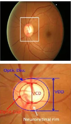
[image:5.595.361.510.454.711.2]Glaucoma detection in medical image analysis is one of the example of thresholding based segmentation. Glaucoma is a chronic eye disease in which the optic nerve is progressively damaged. The optic nerve head or the optic disc is the location where ganglion cell axons exit the eye to form the optic nerve. The disc can be divided into two distinct zones; namely, a central bright zone called the optic cup (in short, cup) and a peripheral region called the neuroretinal rim. Fig. 1 shows the major structures of the disc. The CDR is computed as the ratio of the vertical cup diameter (VCD) to vertical disc diameter (VDD) clinically. Accurate segmentations of disc and cup are essential for CDR measurement. Based on the segmented disc and cup, CDR is computed for glaucoma screening. A larger CDR indicates a higher risk of glaucoma.
Fig. 4. Major structures of the optic disc. The region enclosed by the blue line is the optic disc; the central bright zone enclosed by the red line is the
optic cup; and the region between the red and blue lines is the neuroretinal rim
13255 International Journal of Current Research, Vol. 7, Issue, 03, pp.13251-13257, March,2015
Conclusion
This paper summarizes various segmentation techniques, like segmentation based on Edge, Region and thresholding. Thus segmentation is done to estimate the surfaces. Segmentation can be applied to any type of image. Also the comparing of all methods of image segmentation is done. Now it is easy to conclude that thresholding method is the simplest and computationally fast. Depending on the application technique varies.
REFERENCES
Arushi Chhabra, Ankit Gupta and Akila Victor 2013. "Comparison of Image Segmentation Algorithms" Published by IJETTCS, Volume 2, Issue 3, May – June 2013 ISSN 2278-6856.
Canny, J., "A Computational Approach to Edge Detection", IEEE Transactions on Pattern Analysis and Machine Intelligence,Vol.8, No. 6, Nov. 1986.
Caroline Pantofaru, Martial Hebert "A Comparison of Image Segmentation Algorithms" Carnegie Mellon University Research Showcase @ CMU.
Chan T. and L. Vese, 2001. Active contours without edges, IEEE transactions on image processing 10(2), pp. 266-277.
Dey V. A, Y. Zhang, M. Zhong 2010. "A review on image segmentation techniques with remote sensing perspective" Published by ISPRS TC VII Symposium – 100 Years ISPRS, Vienna, Austria, July 5–7, 2010, IAPRS, Vol. XXXVIII, Part 7A.
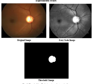
Experimental results
Original Image Gray Scale Image
[image:6.595.104.500.57.416.2]Threshold Image
Fig. 5. Thresholding Technique is apply to optic disc
4. Comparison of Segmentation Methods
Table 1. Image Segmentation Methods: A Comparative Study
Parameter Edge Based Segmentati-on Region Based Segmentati-on Threshold Based Segmentati-on
Nature of the Output Image Black-White Black-White Black-White
Spatial Information Neglected Considered Neglected
Region-Continuity Moderate High Moderate
Computation Complexity Moderate High Less
Speed Moderate Slow Fast
Noise Immunity Less Less Less
Detection of Multiple objects Poor Moderate Poor
Automaticity Interactive Interactive (Semi Automatic) Interactive (Semi Automatic)
Accuracy Moderate Good Moderate
[image:6.595.62.539.467.564.2]He, L. and S. Osher, 2007. Solving the Chan-Vese Model by a Multiphase Level Set Algorithm Based on the Topological Derivative, Lecture notes in Computer Science, no. 4485, 2007, pp. 777-788.
Rohan Kandwal, Ashok Kumar and Sanjay Bhargava 2014. "Review: Existing Image Segmentation Techniques" Published in International Journal of Advanced Research in Computer Science and Software Engineering, Volume 4, Issue 4, April 2014 ISSN: 2277 128X.
Xia Yong, Dagan Feng and Zhao Rongchun "Optimal Selection of Image Segmentation Algorithms Based on Performance Prediction" Patter Recognition, 32(2): 823-582.
Yogamangalam R. and B .Karthikeyan"Segmentation
Techniques Comparison in Image Processing"
R.Yogamangalam et al. / International Journal of
Engineering and Technology (IJET).
*******
13257 International Journal of Current Research, Vol. 7, Issue, 03, pp.13251-13257, March,2015