LIVE MICROSCOPY OF RNA GRANULE
SORTING IN HIPPOCAMPAL NEURONS IN
SPACE AND TIME
Karl Emory Bauer
Dissertation
Supervisor
Prof. Dr. Michael A. Kiebler
BioMedizinisches Centrum/ Lehrstuhl Zellbiologie
Ludwig-Maximilians-Universität München
First Reviewer:
Prof. Dr. Michael A. Kiebler
Second Reviewer:
Dr. Oliver Griesbeck
External Reviewer:
Prof. Dr. Gunter Meister
Table of Contents
ABBREVIATIONS IV
1. INTRODUCTION 1
1.1 OVERVIEW 1
1.2 MRNA AND RBPS IN LEARNING AND MEMORY FORMATION 2 1.2.1 MRNA TRANSPORT, LOCALIZED TRANSLATION AND SYNAPTIC PLASTICITY 2 1.2.2 MRNA TRANSPORT IS A MOTOR-DRIVEN PROCESS 5 1.2.3REGULATION OF MRNA LOCALIZATION BY RNA-BINDING PROTEINS 6 1.3 THE STAUFEN2 PROTEIN IN MRNA TRANSPORT, LEARNING AND MEMORY FORMATION 10 1.3.1STAUFEN STRUCTURE AND RNA-BINDING 10 1.3.2THE ROLE OF STAUFEN IN MRNA LOCALIZATION 13 1.3.3THE ROLE OF STAUFEN2 IN LEARNING AND MEMORY 14 1.4 RGS4 AND RECEPTOR MEDIATED SIGNALING IN THE CENTRAL NERVOUS SYSTEM 14 1.4.1G-PROTEIN COUPLED RECEPTOR SIGNALING IN THE CENTRAL NERVOUS SYSTEM 14 1.4.2STAUFEN2 REGULATES REGULATOR OF G-PROTEIN SIGNALING 4 MRNA 17 1.5 P-BODIES AND THE RNA-HELICASE RCK IN THE CENTRAL NERVOUS SYSTEM 18
1.5.1P-BODIES AND MRNA REGULATION 18
1.5.2THE RCK PROTEIN AND ITS FUNCTION IN THE CENTRAL NERVOUS SYSTEM 19 1.6 RNA LIVE CELL IMAGING AND THE MS2 SYSTEM 20 1.6.1AN OVERVIEW OF MRNA IMAGING TECHNIQUES AND CHALLENGES 20 1.6.2THE MS2 SYSTEM FOR MRNA LIVE CELL IMAGING 22 1.6.3ADVANCEMENT OF THE MS2RNA IMAGING SYSTEM 24
1.7 AIMS 27
2. MANUSCRIPT 1: 28
LIVE CELL IMAGING REVEALS 3´-UTR DEPENDENT MRNA SORTING TO SYNAPSES 28
2.2 INTRODUCTION 31
2.3 RESULTS 33
2.3.1THE RGS43´-UTR LOCALIZES AN MS2 REPORTER MRNA TO DISTAL DENDRITES 33 2.3.2THE RGS43´-UTR MEDIATES AN ANTEROGRADE TRANSPORT BIAS TO DISTAL DENDRITES 35 2.3.3INHIBITION OF NEURONAL ACTIVITY ABOLISHES THE RGS43´-UTR DEPENDENT
2.3.6STAU2 REGULATES THE TRANSPORT OF RGS43´-UTR MRNA IN DENDRITES 48
2.4 DISCUSSION 52
2.4.1LOCALIZED MRNAS TRAVERSE THE DENDRITE IN A SUSHI-BELT-LIKE FASHION 52 2.4.2THE RGS43´-UTR MEDIATES AN ANTEROGRADE TRANSPORT BIAS DEPENDENT ON
NEURONAL ACTIVITY AND THE STAU2 PROTEIN 53
2.4.3RGS4 MRNA IS RECRUITED TO SYNAPSES DEPENDENT ON ITS SPECIFIC 3´-UTR AND
NEURONAL ACTIVITY 54
2.4.4A MODEL OF DYNAMIC DENDRITIC RGS4 MRNA SORTING AND SYNAPTIC RECRUITMENT 55
2.5 METHODS 57
2.5.1NEURONAL CELL CULTURE,TRANSFECTION AND TRANSDUCTION 57
2.5.2PLASMIDS 57
2.5.3LENTIVIRUS PRODUCTION 58
2.5.4SINGLE MOLECULE FLUORESCENT IN SITU HYBRIDIZATION 58
2.5.5IMMUNOSTAINING 59
2.5.6CHEMICAL TREATMENTS 59
2.5.7MICROSCOPY 59
2.5.8IMAGE DATA ANALYSIS 60
2.5.9STATISTICAL ANALYSIS 62
2.6 ACKNOWLEDGMENTS 63
2.7 SUPPLEMENTARY MATERIALS 65
3. MANUSCRIPT 2: 82
NEURONAL ACTIVITY GOVERNS RCK GRANULE SIZE 82
DURING MATURATION OF HIPPOCAMPAL NEURONS IN CULTURE 82
3.1 ABSTRACT 83
3.2 INTRODUCTION 84
3.3 RESULTS 86
3.3.1SOMATIC RCK GRANULES PARTIALLY DISASSEMBLE DURING HIPPOCAMPAL NEURONAL
MATURATION IN CULTURE 86
3.3.2SOMATIC RCK GRANULES IN MATURE NEURONS REVERSIBLY REASSEMBLE AFTER INHIBITION
OF NEURONAL ACTIVITY 89
3.3.3TRANSLATION ACTIVITY CONTROL RCK GRANULE SIZE UPSTREAM OF NEURONAL ACTIVITY 91 3.3.4DISASSEMBLY OF RCK GRANULES IN MATURE NEURONS IS INDEPENDENT OF STAU2 93 3.3.5THE EXPRESSION OF A HELICASE DEFICIENT RCK MUTANT DISRUPTS ENDOGENOUS RCK
3.4 DISCUSSION 97
3.5 MATERIALS AND METHODS 100
3.5.1NEURONAL CELL CULTURE,TRANSFECTION AND TRANSDUCTION 100
3.5.2CRYOSECTIONS 100
3.5.3PLASMIDS 100
3.5.4CHEMICAL TREATMENTS 101
3.5.5IMMUNOSTAINING 101
3.5.6MICROSCOPY 101
3.5.7IMAGE DATA ANALYSIS 102
3.5.8STATISTICAL ANALYSIS 102
3.6 ACKNOWLEDGMENTS 103
3.7 SUPPLEMENTARY MATERIALS 105
4. DISCUSSION 111
4.1 SUMMARY 111
4.2 ADVANCEMENT OF THE MS2 SYSTEM 112
4.3 HOW IS ANTEROGRADE TRANSPORT REGULATED VIA THE 3´-UTR? 113 4.4 WHAT ARE THE MOLECULAR MECHANISMS GOVERNING MRNA LOCALIZATION AND LOCAL
TRANSLATION? 115
4.5 HOW DOES NEURONAL ACTIVITY REGULATE MRNA AND PROTEIN SORTING? 117
4.6 OUTLOOK 119
REFERENCES 120
APPENDICES 142
ACKNOWLEDGMENTS 142
Abbreviations
Arc Activity-regulated cytoskeleton-associated protein ALS Amyotrophic lateral sclerosis
BicD Bicaudal-D
Btz Barentsz
Calm Calmodulin
CamKII Calcium/Calmodulin-dependent protein kinase II CLIP Cross-linking and immunoprecipitation
CPE Cytoplasmic polyadenylation elements
CPEB1 Cytoplasmic polyadenylation element binding protein 1 DEAD Asp-Glu-Ala-Asp
DIV days in vitro
dsRBD double-stranded RNA-binding domain dsRBP double-stranded RNA-binding protein E17 embryonic day 17
eGFP enhanced green fluorescent protein
Egl Egalitarian
FISH Fluorescent in situ hybridization FMRP Fragile X Mental Retardation Protein FP fluorescent protein
GPCR G-protein coupled receptor
hnRNP Heterogeneous Nuclear Ribonucleoprotein
IP Immunoprecipitation
ISH In situ hybridization
KIF1A Kinesin family member 1A KIF5 Kinesin family member 5
KD knock-down
LTP Long-term potentiation
MAP Microtubule-associated protein MCP MS2-coat protein
mGluR metabotropic glutamate receptor RNA ribonucleic acid
miRNA micro RNA
mRNA messenger RNA
NLS nuclear localization signal ORF open reading frame P-bodies Processing-bodies
PSF polypyrimidine tract binding protein-associated splicing factor Pur Purine-rich element binding protein
qRT-PCR quantitative reverse transcriptase (real-time) PCR RBD RNA-binding domain
RBP RNA-binding protein RFP red fluorescent protein
Rgs Regulator of G-protein signaling RISC RNA-induced silencing complex RNP Ribonucleoprotein particle shRNA short hairpin RNA
smFISH single molecule fluorescence in situ hybridization
smiFISH single molecule inexpensive fluorescence in situ hybridization SMN survival of motor neuron protein
Stau Staufen
SYNCRIP Synaptotagmin Binding Cytoplasmic RNA Interacting Protein tdMCP tandem MS2-coat protein
1. Introduction
1.1 Overview
The ability to learn and form memory is of vital importance, not only for basic survival, but also as a basis for higher cognitive functions. This ability is attributed to the cells of the nervous system that comprise the brain, i.e. neurons and glial cells. It is well established today that neurons possess the ability to form special functional connections, termed synapses, that are used to transmit directional signals such as action potentials form one cell to the next (Albright et al., 2000). In addition, glia have a vital role in the maintenance and regulation of synaptic strength (Allen and Lyons, 2018; Eroglu and Barres, 2010). Various anatomical regions of the brain and the neuronal connections between them have been linked to learning and memory. A core role is attributed to the hippocampus, which has been shown to be essential for the retention of new information and the formation of long-term memory (Albright et al., 2000). Long-term memory goes hand in hand with long lasting synaptic strengthening or weakening based on previous electrophysiological activity patterns, called long-term potentiation (LTP) and long-term depression (LTD). The ability of synapses to change their transmission strength is termed synaptic plasticity. On a molecular level, we can distinguish (at least) two phases in this process: an early phase over the first few hours (1-3 h), which is protein-synthesis independent and a late phase (up to 24 h), which depends on new protein synthesis (Bailey and Chen, 1983; Bailey et al., 2015; Frey et al., 1988; Stanton and Sarvey, 1984; Sutton and Schuman, 2006). Therefore, the synthesis of new protein is essential for the development of long-term memory. Moreover, it is understood today that new protein synthesis can occur locally at active synapses (Kang and Schuman, 1996; Yoon et al., 2016). This is accomplished by the translation of locally available messenger ribonucleic acid (mRNA), which would allow the synthesis of the encoded protein and its direct integration into the synapse, changing the synaptic proteome, and directly affecting synaptic plasticity (Doyle and Kiebler, 2011).
neuronal activity and RNA-binding proteins (RBPs) regulate the subcellular localization of two different types of RNA granules (RNA-protein complexes), i.e. dendritic transport granules and processing bodies (P-bodies). These processes are the foundation of synaptic plasticity, and therefore, essential for learning and memory formation.
1.2 mRNA and RBPs in learning and memory formation
1.2.1 mRNA transport, localized translation and synaptic plasticity
The translation of localized mRNA is a critical process, by which cells can target protein expression to certain intracellular subcompartments (Buxbaum et al., 2015a; Medioni et al., 2012). Thereby, a protein can be synthesized by ribosomes at a defined subcellular location, restricting its function both in space and time. This is an essential mechanism for many biological processes, such as embryonic axis formation, cell division or cell migration (St Johnston, 2005). In neurons, mRNA localization has been implicated in several processes such as axonal outgrowth and regeneration, dendritic branching, synapse morphology and in higher order functions such as learning and the formation of memory (Klann and Dever, 2004; Sahoo et al., 2018; Willis et al., 2005; Yoon et al., 2016).
coincides with a similar localization pattern of the equivalent protein, and the disruption of mRNA localization in turn also disrupts the localization of the protein (Lecuyer et al., 2007; Riechmann and Ephrussi, 2004). Therefore, initial protein localization is often directly linked to the localization of its mRNA. After translation however, a protein can undergo its own transport, or be degraded, altering its distribution independent of the original transcript.
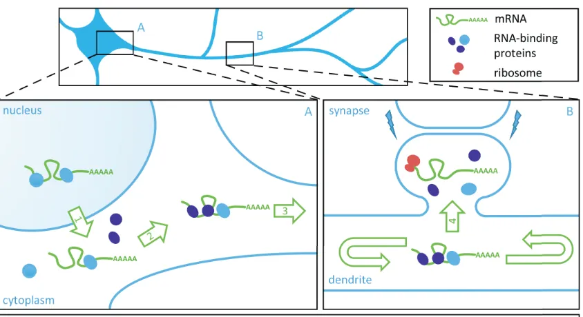
Figure 1.1: Model of neuronal mRNA transport and local translation
at synapses. Insets A and B represent the soma (A) and dendritic
compartment (B) of a schematic neuron. Neuronal mRNA transport,
processing and translation is illustrated step by step by numbered green arrows. In B, synaptic signaling is signified via lightning bolts. This model is based on Doyle and Kiebler, EMBO Journal, 2011.
may be released from associated factors to permit translation by localized ribosomes. After translation the mRNA might eventually be repackaged and transported to a new destination for another round of translation. This model of mRNA transport and translation would be an efficient and economical way for a cell to rapidly deal with local protein demand at sites far from the soma.
The following chapters will take a closer look at relevant scientific insight into mRNA transport and translation, and its regulation.
1.2.2 mRNA transport is a motor-driven process
kinesin and dynein have been implicated in this process (Gagnon and Mowry, 2011; Kanai et al., 2004; Ma et al., 2011; McClintock et al., 2018; McKenney et al., 2014). For instance, knock-down of the kinesin heavy chain KIF5B disrupts the transport of MBP mRNA into processes of oligodendrocytes (Ainger et al., 1993), or Arc and CaMKIIα mRNAs in dendrites (Kanai et al., 2004), while KIF5B overexpression has the opposing effect, i.e. an increase in dendritic mRNA localization (Kanai et al., 2004). Furthermore, KIF5B is found in RNA granules together with the dendritic mRNAs Arc and CaMKIIα. In addition, the observed velocities of RNPs in various publications are all consistent with motor-driven transport (Köhrmann et al., 1999 6.4 µm/min; Park et al., 2014, 1.3 µm/s). If dependent on motor proteins, the directed displacement of RNPs along microtubules is an ATP dependent process and must be regulated on a cellular level. To date it is yet unknown, how a motor protein is linked to an RNP and which factors mediate the association. The study of this process is complicated in dendrites, which have microtubules of mixed polarity, making it difficult to easily discern plus- or minus-end directed transport (Baas et al., 1988; Burton, 1988; Kanai et al., 2004).
1.2.3 Regulation of mRNA localization by RNA-binding proteins
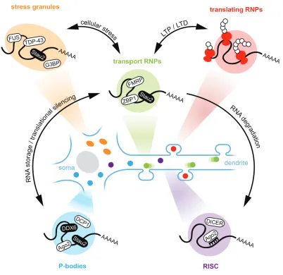
Figure 1.2: Schematic overview of representative examples of known mRNA granules in neurons. For simplicity, only soma, a dendrite and mushroom-shaped spines are shown. Types of mRNA granules are color coded: stress granules (orange), transport RNPs (green), translating RNPs (red), P-bodies (blue) and RISC (purple). Physiological relevant interplay resulting in mRNA or protein reorganization between granule types are indicated, with a focus on transport RNPs. RNPs = ribonucleoprotein particles, LTP = long-term potentiation, LTD = long-long-term depression, RISC = RNA-
composition. Importantly, in neurons, neuronal activity can have an effect on RNP composition (Fontes et al., 2017; Schanzenbacher et al., 2018).
Certain RBPs regulate the transport and localization of their target mRNAs, as mentioned above for Arc, CaMKIIα and ß-actin mRNA (Kanai et al., 2004). To mediate localization, RBPs recognize and bind RNA sequences or structures termed cis-elements or zip codes, often, but not exclusively located in the 3´-untranslated region (3´-UTR) of mRNA (Jambhekar and Derisi, 2007). For instance, the cytoplasmic polyadenylation element binding protein 1 (CPEB1) binds the cytoplasmic polyadenylation elements (CPEs) located in the 3´-UTR of CaMKIIα mRNA to mediate dendritic localization (Huang et al., 2003). Neurons cultured from mice deficient for CPEB1 display a decrease in dendritic localization of mRNA reporters carrying CPEs in their 3´-UTRs. Additionally, CaMKIIα and Microtubule-associated protein 2 (MAP2) mRNAs are reduced in synaptosomal preparations of neurons expressing a dominant negative CPEB protein. Such RNA zip codes or localization elements have been identified for other mRNAs as well (Heraud-Farlow et al., 2013; Rook et al., 2000; Zhang et al., 2001).
The interaction of specific RBPs with their target mRNAs might be an essential part of correct RNP assembly. Little is still known about the regulation and maintenance of RNP assembly. However, examples of individual proteins and mRNAs suggest this process is essential, as in the case of survival of motor neuron protein (SMN) and ß-actin mRNA. SMN is involved in the assembly of spliceosomal RNPs (Monani, 2005). The neuromuscular disorder spinal muscular atrophy is a consequence of the hereditary loss of SMN. A mouse model of this disease shows the mislocalization of ß-actin mRNA in axonal growth cones (Rossoll et al., 2003).
considered the basis for learning and memory formation (Sutton and Schuman, 2006).
In conclusion, previous research has established that RBPs play a key role in the processing and regulation of their target RNA, and that their absence or dysfunction can have severe consequences for an organism, including learning and memory impairments. As highlighted here, one aspect of this regulation is the targeting and transport of RNA to a specific subcellular location. However, how specific RBPs achieve this and which other factors determine their function are less known.
1.3 The Staufen2 protein in mRNA transport, learning and memory formation
1.3.1 Staufen structure and RNA-binding
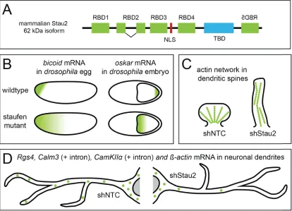
Figure 1.3: The role of the double-stranded RNA-binding protein Staufen2 (Stau2) in mRNA localization and dendritic spine
morphogenesis. (A) Schematical representation of the domains of the
62 kD Stau2 isoform. RBD = RNA-binding domain, NLS= nuclear localization signal, TBD = tubulin binding domain. Mirrored RBD5
indicates inversion of mammalian Stau2 compared to Drosophila
staufen. (B) Illustration of staufen dependent bicoid and oskar mRNA
localization (green gradient) in the development of Drosophila. Based
on Ferrandon et al., Cell, 1994. (C) Illustration of Stau2 dependent
dendritic spine morphology and actin (green dots) network
remodeling. Based on Goetze et al., JCB, 2006. (D) Illustration of
Stau2-dependent Rgs4, Calm3 (intron containing, + intron),
CaMKII (intron containing, + intron) and ß-actin mRNA granules
cause Stau2 to accumulate almost exclusively in the nucleolus (Macchi et al., 2004). Mammalian Stau2 expresses four isoforms of 52 kD, 56 kD, 59 kD, and 62 kD, that vary in their N- and C-terminal domains, i.e. truncations of RBD1 and RBD5, due to alternative splicing. It is thought that these isoforms have different functions in the cell. A study investigating the developing chicken eye found that the knock-down of Stau2 leads to a reduction in eye size (Cockburn et al., 2012). Interestingly, different Stau2 isoforms rescued the phenotype to a varying degree, where the longest 62 kD isoform performed a full rescue and the smallest 51 kD isoform a partial rescue. Together, these findings indicate possible different regulatory roles for the isoforms during eye development.
cross-linking and immunoprecipitation), to reliably identify dsRNA-binding sites. However, the apparent lack of sequence specificity and the possibility of long-range RNA duplexes make the prediction of local binding sites difficult.
1.3.2 The role of Staufen in mRNA localization
1.3.3 The role of Staufen2 in learning and memory
Importantly, Stau2 has been implicated in neuronal signaling, synaptic plasticity and memory. Stau2 is required for DHPG-induced protein synthesis depended long-term depression (LTD) via mGluR, linking it directly to learning and memory (Lebeau et al., 2011). Here, the lack of Stau2 results in transport defects of Map1b mRNA via its 3´-UTR, and a reduction of the encoding protein necessary for the maintenance of metabotropic Glutamate Receptor (mGluR)-LTD. Furthermore, Stau2 knock-down results in a reduction of the amplitude of miniature excitatory postsynaptic currents (mEPSCs) in young neurons, along with a reduction in the number of dendritic spines and synapses, and an altered dendritic actin network (Fig. 1.3C) (Goetze et al., 2006). A recent study observed additional electrophysiological defects in vivo, including favored LTP and impaired LTD in Stau2 deficient rats (Berger et al., 2017). The role of Stau2 in synaptic plasticity and memory formation in vivo is corroborated by behavioral studies in rats and mice. The forebrain specific knock-down of Stau2 in a transgenic rat resulted in defects in spatial working memory, spatial novelty detection and associative learning and memory (Berger et al., 2017). Furthermore, mice with a reduction in Stau2 protein levels display reduced locomotion, and the inability to distinguish between familiar and novel objects (Popper et al., 2018). Another study investigating olfactory memory identified transcriptionally regulated genes, including staufen, during memory formation in Drosophila (Dubnau et al., 2003), raising the question if the Staufen proteins might have a conserved role in memory. Together, these studies show that Stau2 is not only essential in the expression and spatial regulation of its target mRNAs, but also has a fundamental impact on synaptic plasticity, learning and memory formation.
1.4 Rgs4 and receptor mediated signaling in the central nervous system
1.4.1 G-protein coupled receptor signaling in the central nervous system
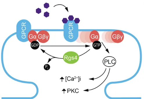
protein Stau2 in the E17 rat brain (Heraud-Farlow et al., 2013). This was accomplished by immunoprecipitation of endogenous Stau2-containing RNA granules followed by microarray analysis and independent verification via qRT-PCR. Further analysis revealed that Rgs4 was one of eight significantly enriched Stau2 target-mRNAs that function in the G protein-coupled receptor (GPCR) signaling pathway. This data indicates that Stau2 might regulate the production of proteins of common function. At neuronal synapses, the GPCR signaling pathway regulates neurotransmitter release and synaptic transmission, playing a role in both pre- and postsynaptic regulation (Betke et al., 2012; Rojas and Dingledine, 2013; Tedford and Zamponi, 2006). The ligand-dependent activation of the receptor of heterotrimeric G proteins (Gαβγ) leads to the exchange of GDP to GTP at the Gα subunit, which results in the release of Gα from Gβγ, both of which can affects downstream second messengers (Fig. 1.4) (Bourne et al., 1990; Hamm, 1998; Simon et al., 1991). Importantly, Gβγ activates G protein-coupled inwardly rectifying potassium (GIRK) channels, which leads to cell hyperpolarization, affecting the excitability of the neuron (Dascal, 1997). The Rgs4 protein and other members of the regulator of G protein family act as negative regulators of the GPCR pathway (Abramow-Newerly et al., 2006; Gerber et al., 2016). Here they function as GTPase activating proteins (GAPs), facilitating the hydrolysis of GTP to inactive GDP on the Gα subunit (Gq and Gi
Figure 1.4: Schematically representation of G-protein coupled receptor (GPCR) activation and its regulation by Rgs4 at the postsynaptic site. Ligand activation (purple hexagons) of GPCRs results in the exchange of guanosin diphosphate (GDP) to guanosin triphosphate (GTP) at the G-protein α-subunit, leading to the dissociation of Gα and Gβγ
and the activation of downstream signaling pathways. Rgs4
inhibits Gαβγ dissociation at group I metabotropic glutamate
receptors (mGluRs) by activating Gα GTPase activity and the
1.4.2 Staufen2 regulates Regulator of G-protein signaling 4 mRNA
Rgs4 is a ~24 kD protein with a single RGS domain and is expressed in multiple brain regions including the hippocampus (Gold et al., 1997; Heraud-Farlow et al., 2013; Saugstad et al., 1998). Among the RGS proteins, Rgs4 has the highest expression in the brain (Larminie et al., 2004). Investigation into the subcellular localization of Rgs4 mRNA in hippocampal neurons via fluorescence in situ hybridization (FISH) shows that endogenous Rgs4 mRNA is present in distinct granules in both the cell body and neurites (Heraud-Farlow et al., 2013). Knock-down of Stau2 results in a reduction of total Rgs4 mRNA levels, both in vitro and in vivo (Berger et al., 2017; Heraud-Farlow et al., 2013). FISH experiments revealed that Stau2 silencing induced a strong reduction in fluorescence intensity in the cell body and a near complete depletion of Rgs4 mRNA granules from dendrites (Fig. 1.3) (Heraud-Farlow et al., 2013). Importantly, the depletion of Stau2 from primary cortical neurons, results in a significant down regulation of Rgs4 mRNA as well. Additionally, a translation assay using an Rgs4 3´-UTR luciferase reporter in cortical neurons revealed that Stau2 regulates Rgs4 mRNA via its 3´-UTR. Stau2 knock-down significantly decreases the expression of the luciferase reporter, indicating that Stau2 may indeed regulate Rgs4 mRNA stability via its 3´-UTR.
1.5 P-bodies and the RNA-helicase Rck in the central nervous system
1.5.1 P-bodies and mRNA regulation
Transport RNPs, which deliver transcripts to specific subcellular locations in a translationally silent state, interact with other types of mRNA granules and share functional protein components with them. Such mRNA granules include P-bodies, stress granules or the RNA-induced silencing complex (RISC) (Fig. 1.2) (Balagopal and Parker, 2009; Fernandez-Moya et al., 2014). Although such RNA granules have overlapping components and functions, they can be categorized by the presence or absence of specific proteins, which will determine whether they are marked for transport, translation, storage or degradation. In their physiological state, all such granules can be both stable or have a high turnover of their components (Barbee et al., 2006; Kedersha et al., 2000). This dynamicity is related to the phenomenon of phase separation, as researched extensively in previous years (Brangwynne et al., 2009; Hyman et al., 2014; Molliex et al., 2015). By phase separation a cell can separate molecular processes in membrane-less droplets with liquid properties, based on the local concentration of the involved components. Physiological phase separation can be perturbed by the formation of aberrant solid-state aggregates, which have been linked to neurological pathologies such as amyotrophic lateral sclerosis (ALS) or frontotemporal dementia (FTD).
for P-bodies in neuronal development, synaptic plasticity, learning and memory (Ashraf et al., 2006; Schratt et al., 2006). Previous research has shown that P-bodies are localized to dendrites of rat hippocampal neurons, but are distinct from transport mRNPs and are not co-transported together (Vessey et al., 2006; Zeitelhofer et al., 2008). However, the observation of docking events between the P-body marker DCP1a and the mRNP marker Stau2 suggests an interaction between these two RNA-carrying granules. Importantly in the context of neurons, chemical stimulation of neuronal activity results in a reduction in dendritically localized P-bodies (Zeitelhofer et al., 2008). Taken together, this data shows that P-bodies are an essential RNA granule and are expected to have a profound impact on other types of RNA granules and their regulation in the cell.
1.5.2 The Rck protein and its function in the central nervous system
1.6 RNA live cell imaging and the MS2 system
1.6.1 An overview of mRNA imaging techniques and challenges
The imaging of RNA expression in fixed cells or tissue is traditionally achieved by in situ hybridization (ISH) of the endogenous RNA with a labeled-antisense probe (Hougaard et al., 1997). The method can be applied as whole-mount ISH on small organisms, on tissue slices or single cells. The sensitivity of this approach was further improved by methods such as fluorescence in situ hybridization (FISH), single molecule fluorescence in situ hybridization (smFISH) or single molecule inexpensive fluorescence in situ hybridization (smiFISH) (Levsky and Singer, 2003), that all allow the quantification of individual RNA molecules. Additional adaptations of these methods have made it possible not only to image endogenous mRNAs, but also miRNAs or long-noncoding RNAs (Tsanov et al., 2016). The exposure of biological replicates to different conditions (e.g. chemical treatments) or the fixation of samples at different time-points has made this method a powerful tool in the field of cell and molecular biology.
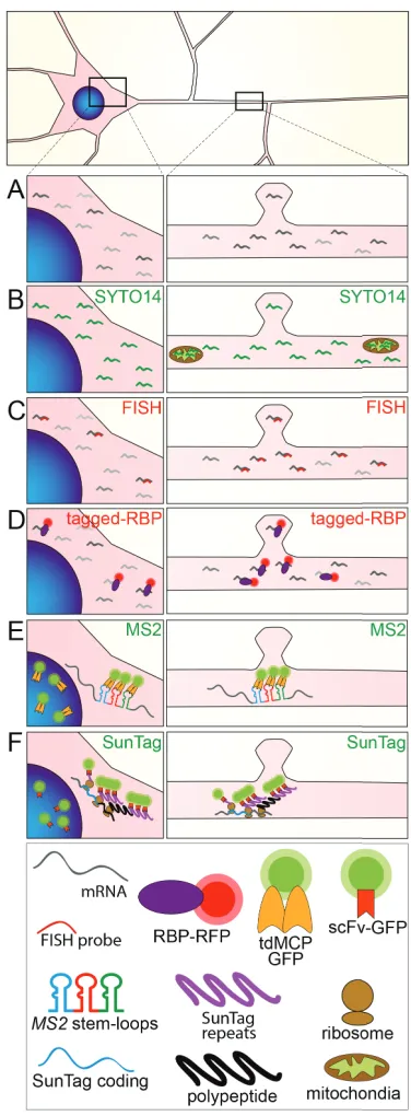
Figure 1.5: Methods to study mRNA granule transport and local translation. Schematic representation of a neuron, showing the soma (left panels) and a
dendritic segment (right panels), indicating cytoplasmic mRNA molecules (grey) (A)
that can be detected with a sequence unspecific dye for nucleic acids such as SYTO 14 (B) or, alternatively, by sequence specific fluorescent in situ hybridization (FISH)
(C). (D) Schematic representation of the MS2-MCP system to visualize pre-labeled
mRNAs. (E) Schematic representation of RNA granule visualization by FP-tagged
RBP (in red). (F) Schematic representation of the SunTag system to visualize local
are extremely susceptible to cellular stress. Methods that rely on the microinjection of individual cells to deliver probes or other agents can be tedious and harmful to neurons (Tübing et al., 2010). Conversely, cell permeable nucleic acid dyes are not specific in labeling single mRNA transcripts. A more suitable approach for neurons is the use of genetically encoded systems, such as the MS2-, PP7- or λN/BoxB-system, which rely on similar principles (Bauer et al., 2017; Mikl et al., 2010). Such methods allow the use of classical transfection based delivery methods to generate transient expression in cells, such as calcium phosphate co-precipitation or viral transduction, or even the generation of transgenic cell lines or animals. This approach based on genetically encoded systems causes low cellular stress, while still labeling specific single transcripts. Moreover, it allows further flexibility in addressing biological questions by e.g. introducing mutations to the genetically encoded reporter. The MS2 system has been well established and greatly improved upon in the last decades (Bauer et al., 2017; Bertrand et al., 1998). To date, the system has been modified to address multiple biological questions, presenting a flexible toolbox to inquire multiple aspects of mRNA metabolism in the living cell.
1.6.2 The MS2 system for mRNA live cell imaging
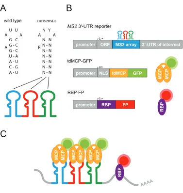
Figure 1.6: The MS2-system for RNA live-cell imaging. (A) Wilde-type and consensus sequence of the 19 nucleotide MS2 RNA stem-loop. A = Adenosine, U = Uracil, C = Cytosine, G = Guanine, N = nucleotide, R =
Purine, Y = Pyrimidine. Modified from Schneider et al., J. Mo. Bio.,1992. (B)
Schematic representation of conventional DNA expression cassettes making use of the MS2-system for RNA live-cell imaging and the corresponding mRNA MS2 reporter and fluorescently labeled proteins (MCP-GFP for mRNA, and RBP-FP for protein visualization). ORF = open reading frame, UTR = untranslated region, NLS = nuclear localization signal, tdMCP = tandem MS2 coat protein, RBP = RNA-binding protein, (G)FP = (green)
fluorescent protein. (C) Illustration of the MS2-system for simultaneous
MCP-GFP recruitment and therefore in an improved detectable signal. Importantly, the MCP has a high specificity in recognizing the RNA stem-loop and a high affinity for binding to it (Horn et al., 2004; Schneider et al., 1992; Stockley et al., 1995). As both the stem-loop and the MCP originate from the MS2 phage, the MCP should not bind to other nucleotide sequences in e.g. mammalian cells, which are often the focus of research.
The MS2-system was first used in 1998 to investigate the intracellular transport of an ASH1 mRNA reporter in the budding yeast Saccharomyces cerevisiae (Bertrand et al., 1998). Using this method, the authors were able to demonstrate the 3´-UTR dependent transport of this mRNA from the mother to the daughter cell during bugging. In addition, this transport was impaired in strains deficient for the She1 and Myo4 proteins. This example nicely illustrates the strength of live cell imaging in addressing relevant biological questions, which cannot be answered by other means. Over the years, additions and modifications have been made to the components of the MS2 system, to enhance detectability or address new biological questions. The most important of these adaptations will be discussed for the MCP and the stem-loop array below.
1.6.3 Advancement of the MS2 RNA imaging system
On the RNA side, the array of repetitive RNA stem-loops is used to recruit multiple GFP-molecules to the reporter mRNA via the GFP-tagged MCP. To increase the GFP signal bound to the mRNA the number of stem-loops can simply be increased, thereby providing additional binding sites for MCP-GFP. Following this strategy, various numbers of stem-loops, including 6, 24, 32 or even 132 MS2 hairpins have been integrated into the reporter mRNA, ultimately increasing the GFP signal intensity and signal to noise ratio (Bertrand et al., 1998; Park et al., 2014; Pichon et al., 2016; Tantale et al., 2016; Wu et al., 2015). However, it is important to note that a full coverage of all stem-loops cannot be expected. For instance, when 24 stem-loops are used, on average only 13 were found to be bound by tdMCP in the cell (Wu et al., 2012).
1.7 Aims
Based on current scientific knowledge and the extensive previous work by the Kiebler lab as outlined in preceding chapters, two separate projects were designed to address the intracellular sorting of both mRNA and RBPs. The first project aimed to investigate intracellular 3´-UTR dependent mRNA transport via the neuronal Stau2-target mRNA Rgs4. The second project is based on my own previous work in the Kiebler lab (master’s thesis available from the University of Vienna; http://othes.univie.ac.at/29195/), where a number of RBPs were screened and their localization was investigated during aging/maturation in cell culture. Investigating RBPs allows to address how different types of RNA granules behave in the cell, complementing the approach taken in the first project. Therefore, this second project aspired to unravel changes in subcellular RNP granule localization of the RBP Rck, which is also found in neuronal Stau2 granules and P-bodies.
Specific aims for each project are defined as follows.
Aims Project 1
1.1 Is mRNA transported in a sushi-belt like fashion?
1.2 Does the 3´-UTR of Rgs4 mediate sequence specific mRNA transport dynamics and localization?
1.3 Does neuronal activity influence Rgs4 3´-UTR mediated transport dynamics and localization?
1.4 Does Staufen2 regulate Rgs4 transport via its 3´-UTR?
Aims Project 2
2.1 Does the localization of Rck change during neuronal maturation in culture?
2.2 Does neuronal activity play a role in age-dependent alterations in Rck localization?
2. Manuscript 1:
Live cell imaging reveals 3´-UTR dependent mRNA sorting to synapses
(Currently under revision at Nature Communications, April 24th 2019)
Karl E Bauer1, Inmaculada Segura1, Imre Gaspar2,4, Volker Scheuss1, Christin Illig1,
Georg Ammer1,5, Saskia Hutten1, Eugénia Basyuk3,6, Sandra M Fernández-Moya1, Janina Ehses1, Edouard Bertrand3 and Michael A Kiebler1,*
1 BioMedical Center, Medical Faculty, Ludwig Maximilians University, Großhaderner
Str. 9, 82152 Planegg-Martinsried, Germany
2 EMBL, Meyerhofstraße 1, 69117 Heidelberg, Germany
3 Institut de Génétique Moléculaire de Montpellier, CNRS UMR5535, 1919 route de
Mende, 34293 Montpellier, France
4 Current address: Institute of Molecular Biotechnology, Dr. Bohr-Gasse 3,
1030 Vienna, Austria
5 Current address: MPI of Neurobiology, Am Klopferspitz 18, 82152 Martinsried,
Germany
6 Current address: Institut de Génétique Humaine de Montpellier, CNRS UMR9002,
141 rue de la Cardonille, 34396 Montpellier, France
* Correspondence: [email protected] (MAK)
No. of Figures: 6 Figures
6 Supplementary Figures
Key words: mRNA granules, Staufen2, MS2 system, Rgs4, 3´-UTR, dendritic mRNA transport, mRNA localization, synaptic activity-dependent recruitment, sushi-belt model
2.1 Abstract
2.2 Introduction
Messenger RNAs (mRNAs) display a variety of subcellular localization patterns in a plethora of model systems (Buxbaum et al., 2015b; Holt and Bullock, 2009; Palacios and St Johnston, 2001), including the dendritic compartment of the hippocampus (Cajigas et al., 2012). Several distinct mechanisms have been proposed to explain how the sorting of specific mRNAs to subcellular locations can be achieved (Andreassi and Riccio, 2009; Holt and Bullock, 2009; Palacios and St Johnston, 2001), from simple diffusion to the more complex sushi-belt model of dendritic mRNA trafficking (Doyle and Kiebler, 2011). The latter proposes that mRNA granules patrol dendrites in a highly dynamic multidirectional fashion, without being irreversibly anchored at a single specific location. Multiple approaches demonstrated that specific transcripts can be actively transported along cytoskeletal structures (Dynes and Steward, 2007; Saxton, 2001; Tübing et al., 2010). Such active and directed transport has been hypothesized to be the driving force that mediates mRNA sorting to specific distal locations in neurons, such as postsynaptic sites or axonal growth cones, where it may become available for local translation (Dictenberg et al., 2008; Dynes and Steward, 2012; Eliscovich et al., 2017; Terenzio et al., 2018; Wu et al., 2016; Yoon et al., 2016). This allows the tightly regulated production of the resulting protein, both spatially and temporally. Localization of mRNA and subsequent local translation are particularly important in neurons, where synapses containing a specific proteome can be located at distal dendrites far from the site of transcription. Ultimately, local protein synthesis at synapses is fundamental for learning and the formation of long-term memory (Doyle and Kiebler, 2011; Jung et al., 2014; Palacios and St Johnston, 2001; St Johnston, 2005).
Sorting signals, usually within the 3´-untranslated region (3´-UTR) of the mRNA, play a crucial role in mRNA localization (Holt and Bullock, 2009; Martin and Ephrussi, 2009; Mayr, 2017; Meer et al., 2012). Such signals are able to interact with specific RNA binding proteins (RBPs), such as ZBP1, FMRP or Staufen2 (Stau2), to form neuronal RNA granules (Dictenberg et al., 2008; Fernandez-Moya et al., 2014; Kiebler and Bassell, 2006). Thereby, Stau2 and ZBP1 regulate the dendritic localization of Calm3 and ß-actin mRNA, respectively (Eom et al., 2003; Sharangdhar et al., 2017). Through these processes RBPs significantly contribute to synaptic function (Dictenberg et al., 2008; Goetze et al., 2006; Kao et al., 2010).
The negative regulator of G protein signaling 4 (Rgs4) mRNA is a previously identified physiological target mRNA of Stau2 in the brain (Heraud-Farlow et al., 2013). It encodes a GTPase activating protein of the G protein-coupled receptor (GPCR) pathway, and therefore modulates receptor mediated neuronal signaling at the synapse (Gerber et al., 2016; Pacey et al., 2011; Saugstad et al., 1998). Fluorescent in situ hybridization (FISH) has shown that Rgs4 mRNA is present in cytosolic RNA granules localized in distal dendrites. Further analysis confirmed the presence of Rgs4 mRNA in Stau2 granules (Heraud-Farlow et al., 2013). As the long Rgs4 3´-UTR contains in vivo cross-linking sites for Stau2 (Sharangdhar et al., 2017), it might provide key binding sites for a direct interaction with Stau2. Silencing of Stau2 induces a reduction of endogenous Rgs4 mRNA both in vitro and in vivo, suggesting an involvement of Stau2 in the regulation of Rgs4 mRNA levels (Berger et al., 2017; Heraud-Farlow et al., 2013).
2.3 Results
2.3.1 The Rgs4 3´-UTR localizes an MS2 reporter mRNA to distal dendrites
ß
Figure 2.1: Reporter mRNAs display directed dendritic transport dynamics in primary hippocampal neurons. (A) Scheme of both MS2 only and MS2+Rgs4 MS2 reporter constructs and tdMCP-GFP expression cassettes (upper) and the MS2 system (lower). Abbreviations: pRSV = Rous sarcoma virus promoter, pUBC = Ubiquitin C promoter, ORF = open reading frame, NLS = nuclear localization signal, tdMCP = tandem MS2 coat protein, UTR = untranslated region. (B) Phase contrast, GFP fluorescence (reporter), MS2 single molecule FISH and overlay in a rat hippocampal neuron expressing both tdMCP-GFP and MS2+Rgs4 MS2 reporter mRNA (scheme). Arrowheads indicate overlapping tdMCP-GFP bound MS2 reporter mRNA and MS2 smFISH. Fluorescent images were deconvolved to assess overlap (for unprocessed images see Supplementary Fig. 1C). Scale bar 20 µm. Boxed region is magnified in right panels. (C-F) Representative kymographs (left) and extracted tracks (right) illustrating differences in unidirectional MS2+Rgs4 3´-UTR mRNA granule transport speed, displacement and directionality (C), as well as interrupted (D) and multidirectional transport (E). Anterograde and retrograde transport are indicated in green or red arrowheads and lines, respectively. (F) Quantification of relative transport dynamics of MS2 only and MS2+Rgs4 3´-UTR reporter mRNAs in 1 and 10 minute time-series acquisitions, respectively.
2.3.2 The Rgs4 3´-UTR mediates an anterograde transport bias to distal dendrites
ß
Figure 2.2: Rgs4 3´-UTR mediates an anterograde transport bias. (A) Representative phase contrast and GFP fluorescence of hippocampal neuronal culture co-transfected with the MS2+Rgs4 3´-UTR reporter and tdMCP-GFP constructs. Scale bar 20 µm. Asterisk denotes GFP positive cell. (B) Time series of the dendritic boxed region in A. Representative anterograde (black arrowheads) and retrograde (white arrowheads) moving mRNA granules are indicated. (C) Kymograph of the dendritic region in B. Arrowheads indicate mRNA granules signified in B. (D-I) Dot plots (D,F,H) and histograms (E,G,I) displaying percentage of anterograde moving granules (D-E), percentage of total anterograde travel distance (F-G) and average speed (H-I) for MS2 only or MS2+Rgs4 3´-UTR reporter mRNAs, detected by tdMCP-GFP. In (I), positive values indicate anterograde and negative values indicate retrograde transport. Data represents mean ± standard deviation of three independent experiments (individual experiments shown as gray dots). Asterisks represent p-values obtained by Student’s t-test (*p < 0.05, **p < 0.01). Data was obtained from 40 µm dendritic segments at a minimal distance of 20 µm from the cell body. At least 10 dendrites/condition/experiment were analyzed. Total number of dendrites (nd) and tracks (nt) analyzed per condition are indicated.
Only displacements ≥ 1.5 µm were considered for analysis.
contrast, the mobile fraction of granules traversed the dendrites in a highly dynamic way, including unidirectional, interrupted and multidirectional movements, independent of the 3´-UTR present (Fig. 2.1F). Increasing the acquisition time to 10 minutes reduced the fraction of stationary and unidirectional granules in favor of interrupted and multidirectional movements, demonstrating that a large fraction of RNA granules indeed undergo transport in a sushi-belt like fashion. The fraction of the stationary population is consistent with previously published data for the ß-actin mRNA as well as for Staufen1 granules in the time frames analyzed (Köhrmann et al., 1999; Yoon et al., 2016). Statistical analysis by Chi-squared test could not establish any difference in the frequency of these events between MS2 only or MS2+Rgs4 3´-UTR reporter mRNAs, suggesting that the 3´-UTR of the transcript does not regulate the types of motility exhibited by the reporters.
displacement events and only few particles traveled long distances at a time, often longer than the 40 µm analyzed (Supplementary Fig. 2.2B). To exclude that the NLS included in the tdMCP protein might potentially affect transport as previously reported (Salman et al., 2005), we generated a tdMCP lacking the NLS and repeated the previous experiment. Although the fluorescent signal had higher background, we still observed an anterograde transport bias mediated by the Rgs4 3´-UTR (Supplementary Fig. 2.2C,D), showing that the NLS did not affect trafficking in our hands.
In conclusion, our live cell imaging data suggests that the Rgs4 3´-UTR was responsible for the observed anterograde transport bias, affecting both anterograde moving mRNA granule number and anterograde travel distance in dendrites. Importantly, the results establish the 3´-UTR as a key determinant as the bias was not observed in the absence of the Rgs4 3´-UTR.
2.3.3 Inhibition of neuronal activity abolishes the Rgs4 3´-UTR dependent anterograde transport bias
Next, we asked whether neuronal activity might regulate dendritic mRNA transport. As mature neurons display endogenous neuronal activity in culture, we chemically silenced activity by simultaneously inhibiting AMPA receptors, NMDA receptors and voltage-gated sodium channels via bath application of CNQX, AP5 and TTX, respectively (Sharangdhar et al., 2017). Neurons transfected with either MS2 only or MS2+Rgs4 3´-UTR reporters were left untreated or pre-incubated for 1 h with either vehicle or CNQX/AP5/TTX, and then imaged during continuous treatment (Fig. 3A). No differences in speed, displacement or transport directionality were observed for the MS2 only mRNA reporter, independent of the treatment (Fig. 2.3B-C, Supplementary Fig. 2.3A,B,G,H and data not shown). However, the anterograde transport bias mediated by the MS2+Rgs4 3´-UTR was completely abolished when neuronal activity was inhibited (46.7 ± 2.3 %), compared to vehicle treated (55.1 ± 2.9 %, p = 0.00431) or untreated neurons (56.3 ± 2.3 %, p = 0.00187) (Fig. 2.3D-E; F2,9 = 0.00136). Moreover, the inhibition of neuronal activity alleviated the
ß
Figure 2.3: Chemical inhibition of neuronal activity abolishes Rgs4 3´-UTR dependent transport bias. (A) Scheme of experimental outline. (B-G) Dot plots (B,D,F) and histograms (C,E,G) displaying percentage of anterograde moving MS2 only (B-C) or MS2+Rgs4 3´-UTR (D-G) reporter mRNA granules in rat hippocampal neurons, untreated, vehicle treated (DMSO) or silenced (100µM CNQX, 50µM AP5, 1µM TTX) and after 1 hour recovery. Data represents mean ± standard deviation of 3-4 independent experiments (individual experiments shown as gray dots). Asterisks represent p-values assessed by Tukey’s test post-hoc to one-way ANOVA analysis (* p < 0.05, **p < 0.01, ***p < 0.001). Data was obtained from 40 µm dendritic segments at a minimal distance of 20 µm from the cell body. At least 10 dendrites/condition/experiment were analyzed. Total number of dendrites (nd) and
tracks (nt) analyzed per condition are indicated. Only displacements ≥ 1.5 µm were
considered for analysis.
wash-off of chemical inhibition induced recovery of the transport bias (F2,9 = 0.00093,
p = 0.01456 for silenced vs wash-off, p = 0.00074 for silenced vs untreated, Fig. 2.3F-G). Displacement of anterograde movements of MS2+Rgs4 3´-UTR mRNA granules partially recovered after 1h wash-off (F2,9 = 0.021, p = 0.424 for silenced vs
2.3.4 Rgs4 3´-UTR dependent mRNA sorting to synapses
As inhibition of neuronal activity abolished the Rgs4 3´-UTR dependent anterograde transport bias in distal dendrites, we next investigated whether mRNA was recruited to dendritic synapses. To visualize endogenous excitatory synapses in dendrites, we generated a fluorescent synaptic marker by tagging the postsynaptic density protein 95 kD (PSD-95) with tagRFP-t (PSD-95−RFP). Mature neurons were co-transfected with PSD-95−RFP, tdMCP-GFP and either MS2 only or MS2+Rgs4 3´-UTR mRNA reporters. To analyze whether synapses might affect mRNA granule transport, we co-imaged both mRNA reporters and the synaptic marker by time-lapse dual-color microscopy in single cells for one minute. We generated kymographs for each separate channel and created dual-color overlays (Fig. 2.4A; movies 2.9-2.10). We identified the positions, where mRNA granules were found to either interrupt (termed docking) or reinitiate transport (termed undocking), and measured the distance to the closest PSD-95−RFP positive cluster. Importantly, co-expression of the fluorescent reporters together with either MS2 only or MS2+Rgs4 3´-UTR reporter mRNA did not modify synaptic density (Supplementary Fig. 2.4A). Moreover, we found no difference in the ratio of mRNA granules docking or undocking between MS2 only and MS2+Rgs4 3´-UTR reporter mRNAs, respectively (Supplementary Fig. 2.4B). While both reporter mRNAs were recruited to synapses, MS2+Rgs4 3´-UTR reporter mRNAs on average docked closer than MS2 only reporter mRNAs (1.11 ± 0.01 µm for MS2 only vs. 0.67 ± 0.02 µm for MS2+Rgs4 3´-UTR; p = 4.25E-6, Fig. 2.4B-C). Similar results were obtained for mRNA granules undocking after a previous stationary phase (Supplementary Fig. 2.4C-D). Together, these results suggest a dynamic sorting process as the Rgs4 3´-UTR mediated mRNA recruitment and eventual release close to synapses.
ß
Figure 2.4: Rgs4 3´-UTR mediates mRNA recruitment to synapses dependent on neuronal activity. (A) Representative dual-color kymograph showing MS2+Rgs4 3´-UTR reporter mRNA (green) and TagRFPt tagged PSD-95 (magenta) from a dendrite of a rat hippocampal neuron. First and last frames are shown at top and bottom. Extracted track (right) for an mRNA granule docking at a PSD-95 positive area is indicated by arrowheads. (B-C) Distance between MS2 only or MS2+Rgs4 3´-UTR reporter mRNA docking events and closest PSD-95 positive cluster in co-transfected rat hippocampal neurons is displayed as dot plot (B) and density plot (C). (D) Distribution of MS2 only or MS2+Rgs4 reporter mRNA-positive (estimated RNA number ≥ 1) and -negative (RNA < 1) PSD-95-TagRFPt clusters in soma and dendrites. P-values of Chi2 tests against the control are indicated. (E) Integrated
undocking event of either MS2 only or MS2+Rgs4 reporter mRNAs at ~ 34 % vs ~ 21% of dendritic synapses, respectively (Supplementary Fig. 2.4E). This represents a ~30% significant reduction in the frequency of these events for the MS2+Rgs4 3´-UTR reporter mRNA (p < 0.0001; Supplementary Fig. 2.4F,M). We found that the number of these events showed a moderate correlation (R ~ 0.5) with the estimated mRNA copy number within PSD-95−RFP positive synapses (Supplementary Fig. 2.4G). This suggests that the number of docking/undocking events are related with the total number of mRNA particles at synapses. Analysis of the postsynaptic sites revealed that mRNA positive synapses were both larger (data not shown) and brighter in their mean PSD-95 signal intensity than mRNA negative synapses (p < 0.0001; Supplementary Fig. 2.4H). We found that the fraction of MS2+Rgs4 3´-UTR positive synapses in dendrites was significantly lower than that of MS2 only (57.1 % vs 73.9 %; p < 0.0001; Fig. 2.4D). However, when we focused on mRNA reporter positive synapses, we found a significant difference between the frequency of total docking/undocking events of MS2 only and MS2+Rgs4 3´-UTR reporter mRNAs at synapses (p = 0.002; Fig. 2.4E).
Subsequently, we analyzed the net directionality of reporter mRNA docking/undocking at postsynaptic densities. We found that the frequency of docking events was significantly higher for the MS2+Rgs4, compared to the MS2 only reporter (~0.9 min-1 vs 0.79 min-1, p = 0.005), whereas there was no substantial difference in the frequency of undocking events (~0.99 min-1 vs 0.97 min-1 in MS2 only RNA positive synapses). In agreement with these results, we found a net influx (p = 0.009, α = 0.01) of MS2+Rgs4 into mRNA positive synapses, in contrast to the MS2 only reporter mRNA (Fig. 2.4F, Supplementary Fig. 2.4I), which was not significantly different from zero (p = 0.043, α = 0.01). In general, synapses contained mRNA both at the beginning and during the experiment regardless of their location and reporter mRNA (Supplementary Fig. 2.4J-L).
2.3.5 Neuronal activity induces the recruitment of Rgs4 3´-UTR mRNA to synapses
Next, we investigated whether inhibition of neuronal activity affected recruitment of Rgs4 3´-UTR mRNA to synapses in addition to its effect on transport directionality. Therefore, we chemically inhibited neuronal activity in mature rat hippocampal neurons transiently co-transfected with either the MS2 only or the MS2+Rgs4 3´-UTR mRNA, tdMCP-GFP and the synaptic marker PSD-95−RFP. Neither synaptic density nor the ratio of docking to undocking events was altered when silenced cells were compared with vehicle or untreated controls (Supplementary Fig. 2.4P-Q). Additionally, MS2 only mRNA granules did not exhibit any significant change in their distance to PSD-95−RFP synapses upon inhibition of neuronal activity when compared to vehicle or untreated cells (Supplementary Fig. 2.4R-S). However, the inhibition of neuronal activity increased the docking/undocking distance of MS2+Rgs4 mRNA close to synapses, to values comparable to that of MS2 only mRNA (F2,6 = 0.001, p = 0.004 for silencing vs vehicle, p = 0.001 for
silencing vs untreated, Fig. 2.4G-H; F2,6 = 0.001, p = 0.003 for silencing vs vehicle, p
Figure 2.5: Local glutamate uncaging at individual dendritic spines triggers Rgs4
3´-UTR dependent mRNA recruitment. (A) Representative GFP fluorescence of a
hippocampal neuron co-transfected with the MS2+Rgs4 3´-UTR reporter and
tdMCP-GFP constructs before (left panel) and after (middle, right panels) local glutamate uncaging. Black dot denotes the uncaging spot at dendritic spine.
Arrowheads indicate GFP positive MS2 reporter mRNA granules. Scale bar 2 µm.
(B) Dot plot displaying the change in RNA granule number 40-45 min after uncaging
compared to the RNA granule number before uncaging within 5 µm of the stimulated
2.3.6 Stau2 regulates the transport of Rgs4 3´-UTR mRNA in dendrites
Finally, we investigated whether the RBP Stau2, which is known to bind Rgs4 mRNA (Heraud-Farlow et al., 2013; Sharangdhar et al., 2017), was required for the transport of MS2+Rgs4 granules. To investigate whether mRNA is co-transported with Stau2, we co-transfected the MS2+Rgs4 reporter together with tdMCP-GFP and TagRFPt tagged Stau2 (RFP-Stau2). We observed multiple instances, where the MS2+Rgs4 reporter mRNA granules were co-transported with Stau2 (Fig. 2.6A, movies 12-13). Moreover, overexpression of Stau2 resulted in an increase of dendritic MS2+Rgs4 reporter density, while the MS2 only mRNA was unaffected (Supplementary Fig. 2.6A). Additionally, we performed a pilot experiment involving MS2 RNA-mediated tethering of Stau2 to the MS2 only reporter mRNA. When we co-transfected both tdMCP-Stau2 and tdMCP-GFP together with the MS2 only reporter, we observed that the tethering of Stau2 tended to recruit the control mRNA closer to the synaptic marker vesicular glutamate transporter 1 (VGLUT1) (Supplementary Fig. 2.6B). Together, our data suggest that Stau2 might indeed regulate Rgs4 3´-UTR dependent recruitment to synapses. However, further work is clearly necessary to substantiate these findings in the future.
ß
Figure 2.6: Stau2 regulates Rgs4 3´-UTR dependent transport. (A) Representative dual-color kymograph showing MS2+Rgs4 3´-UTR reporter mRNA (green) and tagRFPt-tagged Stau2 (magenta) from a dendrite of a rat hippocampal neuron. First and last frames are shown at top and bottom. Arrowheads indicate an MS2+Rgs4 reporter and Stau2 positive RNA granule undergoing co-transport. (B-E) Dot plots (B,D) and histograms (C,E) displaying percentage of anterograde moving MS2 only or MS2+Rgs4 3´-UTR reporter mRNA granules in shNTC and shStau2 transduced hippocampal neurons. Abbreviation: NTC = non-targeting control. Data represents mean ± standard deviation of three independent experiments (individual experiments shown as gray dots). Asterisks represent p-values obtained by Student’s t-test (*p < 0.05). Data was obtained from 40 µm dendritic segments at a minimal distance of 20 µm from the cell body. Total number of dendrites (nd) and
tracks (nt) analyzed per condition are indicated. Only displacements ≥ 1.5 µm were
2.4 Discussion
Here, we show that mRNAs travel along dendrites in a dynamic and multidirectional fashion, a process that does not exclusively require a specific 3´-UTR sequence for this dendritic localization. The presence of the Rgs4 3´-UTR, however, conveyed key properties to the reporter mRNA, which fine-tune transport as (i) it introduced a transport bias directed towards distal dendrites, and (ii) regulated the spatio-temporal association to synapses. These effects were dependent on neuronal activity and the RBP Stau2. Together, our data not only provide experimental support for the sushi-belt model of dendritic mRNA transport (Doyle and Kiebler, 2011), in which mRNAs dynamically patrol synapses, but also allow us to substantially advance this model by the introduction of a regulated transport bias giving mechanistic insight into global RNA sorting processes including recruitment by individual, stimulated synapses in polarized neurons.
2.4.1 Localized mRNAs traverse the dendrite in a sushi-belt-like fashion
phases in different directions. This observation expands our current understanding of mRNA sorting beyond local recruitment, as it experimentally proves that mRNA granules are not irreversibly anchored, but are dynamic, can be targeted to specific sites upon demand, and be released from them later on. Together, we provide experimental evidence that the sushi-belt model indeed accurately describes dendritic mRNA transport in live neurons. However, we do not exclude that other factors/mechanisms might contribute as well. Interestingly, a recent computational study provided evidence that the sushi-belt model can achieve complex spatial distribution of cargo in neurons (Williams et al., 2016).
2.4.2 The Rgs4 3´-UTR mediates an anterograde transport bias dependent on neuronal activity and the Stau2 protein
A key finding of this study is that the 3´-UTR of Rgs4 mRNA mediated an anterograde transport bias in dendrites, i.e. a preferential transport directionality towards distal regions. In contrast, neither the MS2 only nor the MS2+histone-3.3 reporter mRNAs displayed this bias (Fig. 2.2D and data not shown). Previous studies have observed both anterograde and retrograde mRNA transport. Moreover, a directional bias in transport has not been so frequently reported. It has been shown for oskar mRNA in the Drosophila oocyte, and for ß-actin and Arc mRNAs in mouse hippocampal neurons (Das et al., 2018; Dynes and Steward, 2007; Park et al., 2014; Zimyanin et al., 2008). It is worth mentioning that these studies observed a bias of similar magnitude as we report here (~ 60% towards distal regions), indicating that the dendritic bias of the MS2+Rgs4 3´-UTR reporter is within a physiological range.
is specific to the Rgs4 3´-UTR and might be caused by the absence of Stau2 in Rgs4 containing mRNA granules. Therefore, we hypothesize that Stau2 might be recruited in conjunction with neuronal activity to modulate dendritic Rgs4 mRNA transport. Similarly, previous research has shown how Staufen and other RBPs are not necessary for general transport, but can facilitate or modulate it (Brendza et al., 2000; Yoon and Mowry, 2004). Along this line, Staufen has been implicated in kinesin-1 dependent posterior localization of oskar mRNA in Drosophila and has been found in a complex with kinesin-1 in Xenopus (Brendza et al., 2000; Yoon and Mowry, 2004). In line with this finding, we observed directed co-transport of MS2+Rgs4 granules together with Stau2 in neurons. Importantly, Stau2 depletion results in both morphological and physiological synaptic phenotypes (Goetze et al., 2006; Lebeau et al., 2011). Therefore, it is tempting to speculate that Stau2 might not only regulate the expression of proteins relevant at synapses as previously shown (Heraud-Farlow et al., 2013), but also the transport and recruitment of their mRNAs. In turn, deregulation of synaptic proteins, resulting in aberrant synaptic remodeling, might render a synapse incapable of proper recruiting of relevant transcripts. Together, our data gives functional insight into the regulation of 3´-UTR dependent mRNA transport. We show that it is the Rgs4 3´-UTR that facilitates dendritic localization via the anterograde transport bias. Although dendritic sorting might be affected by other factors such as regulated mRNA degradation, this transport bias might enable fast and efficient mRNA recruitment to specific regions such as synapses as needed. Future studies will have to unravel the detailed molecular mechanisms of how Stau2, neuronal activity and 3´-UTR sequences cooperate to mediate this anterograde bias in neurons.
2.4.3 Rgs4 mRNA is recruited to synapses dependent on its specific 3´-UTR and neuronal activity
Furthermore, the MS2+Rgs4 3´-UTR reporter interacted with fewer synapses and displayed more docking events at dendritic synapses than the MS2 only reporter. This led to a slower turnover of MS2+Rgs4 mRNA content at synapses compared to MS2 only, indicating MS2+Rgs4 is more stably associated and might remain longer at synapses. We propose that the presence of MS2 only mRNA at synapses might represent a state of non-specific, default localization. These effects, along with the observed differences in the brightness of PSD-95 clusters could reflect differences in either synaptic activity or in the subtype of the synapse. As the Rgs4 protein is a negative regulator of synaptic activity, we speculate that its mRNA is transported to a subtype of synapses, where it might be regulated in situ. Here, the mRNA could be unpacked and locally translated by polysomes localized close to PSD-95 clusters (Ostroff et al., 2002), where the newly synthesized Rgs4 protein would modulate G protein-coupled receptor mediated neuronal signaling. Our findings are in agreement with a study by the Singer lab that showed that endogenous ß-actin mRNA is recruited to glutamate-stimulated dendritic spines, where it is locally translated (Yoon et al., 2016). Using a similar approach, we now show that glutamate stimulation of individual spines results in 3´-UTR dependent recruitment of Rgs4 mRNA, as the MS2 only reporter displayed no variation in recruitment.
Taking together, we hypothesize that the observed anterograde transport bias contributes to synaptic recruitment of mRNAs. As both the transport bias and synaptic recruitment are modulated by the 3´-UTR and neuronal activity, anterograde transport might indeed facilitate synaptic recruitment, especially under endogenous mRNA expression levels. Future work will aim to understand how neuronal activity affects the organization of key cytoskeletal components to mediate Rgs4 mRNA recruitment and whether it affects their capture on ribosomes at synapses.
2.4.4 A model of dynamic dendritic Rgs4 mRNA sorting and synaptic recruitment
2.5 Methods
2.5.1 Neuronal Cell Culture, Transfection and Transduction
Primary rat hippocampal neuronal cell cultures were generated as previously described (Goetze et al., 2003). In short, hippocampi of embryonic day 17 (E17) embryos of timed pregnant Sprague-Dawley rats (Charles River Laboratories) were isolated, cells dissociated and plated on poly-L-lysine coated cover slips or glass bottom dishes (WillCo Wells) and cultured in NMEM+B27 medium (Invitrogen). Experiments were performed with cultured neurons between 10-16 days in vitro (DIV). Neurons were transiently transfected by calcium phosphate co-precipitation at 10-15 DIV as previously described (Goetze et al., 2004) and imaged at 11-16 DIV. For experiments involving glutamate uncaging, neurons were plated on poly-L-lysine and laminin (Invitrogen) coated 12-mm diameter glass coverslips, at a density of 600 cells/mm2. Cells were transfected at 15-17 DIV by calcium phosphate co-precipitation
and imaged the following day. To knock-down Stau2 expression, 10-11 DIV neurons were transduced overnight with lentiviral suspension, transfected with MCP-GFP and MS2 mRNA reporter constructs at 14-15 DIV and imaged at 15-16 DIV. All animals were used according to the German Welfare for Experimental Animals (LMU-Munich, Regierung von Oberbayern).
2.5.2 Plasmids
plasmid was generated by removing the NLS from the original vector. The pUBC-NLS-ha-tdMCP and pUBC-pUBC-NLS-ha-tdMCP-Stau2 vectors were generated by extracting the UBC-NLS-ha-tdMCP sequence from the original vector and cloning the 62kD isoform of mouse Stau2 in frame in the pEGFP-C1 vector (Clontech) in place of the EGFP. The CMV-PSD95-tagRFPt vector was generated by cloning the PSD-95 open reading frame into the ptagRFPt-N1 vector (Robert H. Singer, USA). The pCMV-tagRFPt-Stau2 vector was generated by cloning the open reading frame of the 62kD isoform of mouse Stau2 into the ptagRFPt-C1 vector (Robert H. Singer, USA). The pCMV-tdTomato vector was generated by cloning the open reading frame of tdTomato (Dieter Edbauer) into the ptagRFPt-C1 vector (Robert H. Singer, USA) in place of tagRFPt. The lentiviral packaging plasmids psPAX2 and pcDNA3.1-VSV-G have previously been reported (Heraud-Farlow et al., 2013). Lentiviral plasmids pFu3a-H1-sh-NTC-pCaMKIIα-tagRFP and pFu3a-H1-sh-Stau2-2-pCaMKIIα-tagRFP were generated by exchanging the UBC promoter for the CaMKIIα promoter from the previously published FUW based vectors (Heraud-Farlow et al., 2013).
2.5.3 Lentivirus production
Control sh-NTC and sh-Stau2-2 lentiviral particles were obtained from HEK293 cells co-transfected with the plasmids psPAX2, pcDNA3.1-VSV-G and either pFu3a-H1-sh-NTC-pCaMKIIα-tagRFP or pFu3a-H1-sh-Stau2-2-pCaMKIIα-tagRFP, respectively, using calcium phosphate co-precipitation. Supernatants were filtered (0.45 µm RVDF Millex-HV; Millipore), concentrated by ultracentrifugation (23,000 rpm, 140 min, SW 32 Ti rotor; Beckman Coulter) and resuspended in Opti-MEM™ (Life Technologies) (Heraud-Farlow et al., 2013).
2.5.4 Single molecule fluorescent in situ hybridization