The effect of physiotherapy on respiratory
function in ventilated children
Thesis subm itted for the
Degree of Doctor o f Philosophy
University College London
University of London
2001
Eleanor Main
The Portex Unit of Anaesthesia, Intensive Therapy and Respiratory Medicine, Institute of Child Health
ProQuest Number: 10013944
All rights reserved
INFORMATION TO ALL USERS
The quality of this reproduction is dependent upon the quality of the copy submitted.
In the unlikely event that the author did not send a complete manuscript and there are missing pages, these will be noted. Also, if material had to be removed,
a note will indicate the deletion.
uest.
ProQuest 10013944
Published by ProQuest LLC(2016). Copyright of the Dissertation is held by the Author.
All rights reserved.
This work is protected against unauthorized copying under Title 17, United States Code. Microform Edition © ProQuest LLC.
ProQuest LLC
789 East Eisenhower Parkway P.O. Box 1346
Abstract
Background: Chest physiotherapy to clear secretions is routine in many
intensive care units, although there is little evidence to substantiate its use above routine suction procedures. U ntil recently evaluation o f such treatments has been limited by a lack o f objective measures o f respiratory function. This study aimed to evaluate the performance o f a new respiratory function m onitor in assessing the effects o f therapeutic interventions such as physiotherapy. In addition the study aimed to establish whether physiotherapy offered an advantage over routine suction procedures w ith respect to selected indices o f respiratory function.
Subjects: Consent was obtained fo r 101 muscle relaxed, fully ventilated children recruited to the study between A p ril 1998 and March 2000. O f these, 56 had a primary cardiac diagnosis and 45 had primary or secondary respiratory problems and all required physiotherapy treatment.
M ethodology and equipm ent: Validation and pilot studies were undertaken to
assess the accuracy o f the “ CO2SMO Plus” . Subjects were randomly allocated to receive
either physiotherapy or nursing suction in the morning and the alternative intervention in
the afternoon. Arterial blood gases, tidal volume, respiratory mechanics, CO2 parameters
and deadspace values were recorded before and after both interventions. Group and individual responses to treatment were evaluated and compared.
Results: Validation studies demonstrated that the “ CO2SMO Plus” could provide accurate and reproducible data, provided that tracheal tube leak was <20%. Data from 89 children (tracheal tube leak <20%) were analysed, 81 o f whom had both physiotherapy and nursing suction performed on the same day. Physiotherapy treatments were significantly longer than nursing suction, involved greater saline instillations and more suction catheters (p<0.005). Physiotherapy resulted in increased alveolar and physiological deadspace (p<0.01) and reductions in Rp,, HCO3-, base excess, O2
saturation and PeC02 (p<0.05). Following nursing suction, V t e and were reduced
(p<0.05). There was considerable individual variation in response to both physiotherapy and suction.
For my father, who started out on this project with me,
but then departed this world to do a higher degree o f his own, with love always.
Acknowledgments
M y enormous gratitude goes to the children and their families who were kind enough to participate in this study despite their very difficult circumstances. Also to nursing and
physiotherapy staff fo r helping w ith patient recruitment, tolerating the intrusion o f
respiratory function measurements and helping w ith data collection.
I am indebted to Janet Stocks who single-handedly depleted national red pen stocks in
her enthusiasm to cover my thesis in ink (and w ithout whose kindness, support and expert guidance throughout this journey 1 would not have managed nearly as well). I am
also extremely grateful to Rosemary Castle, Catherine Dunne and Ammani Prasad fo r helping me do Really-Boring-but-Essential-Stuff including some proof-reading and to
Rosemary Castle, Di Newham and Tim Peters fo r proof-reading all o f the thesis.
Thanks to Peter M illigan and Angie Wade for endeavouring to share the mysteries o f statistics w ith me and to David Hatch for doing the same w ith “ deadspace” .
Also my gratitude to my family: to Callum and Daniel who were useless at p ro o f reading
but spectacularly good at keeping my feet on the ground and grim y deposits on my clothes and making me laugh, and to Colin fo r his unfailing enthusiasm fo r me to finish
this thesis.
“Beware o f the man who M/orks hard to learn something, learns it, and fin d s h im self no
wiser than before. He is fid. I o f murderous resentment o f people who are ignorant
M/ithont having come by their ignorance the h a rd way. ”
Project Supervisors:
Professor Janet Stocks; Institute of Child Health, London Professor Di Newham: Kings College, London
This project was granted approval by the Institute o f Child Health and Great Ormond Street Hospital for Children NHS Trust Research Ethics Committee and written,
TABLE OF CONTENTS
T IT L E PAGE...1
A B S T R A C T ... 2
A C K N O W LE D G M E N TS ... 3
CO NTENTS... 5
L IS T OF ABBREVIATIO NS AND D E F IN IT IO N S ... 15
1. IN T R O D U C T IO N AND LITE R A TU R E R E V IE W ... 17
1.1 In t r o d u c t i o n... 17
1.2 Lit e r a t u r e r e v ie w...17
1.2.1 Adverse effects o f m echanical ventilation...19
1.2.2 The general effects o f physiotherapy or nursing .suction...20
1.2.2.1 S putum c le a ra n c e ... 21
1.2.2.2 A rterial blood g a se s... 21
1.2.2.3 R esp irato ry m ech an ics (Crs and R rs)... 22
1.2.2.4 A cute lo b ar a te lec ta sis and chest ra d io g ra p h ...23
1.2.2.5 P rev en tio n and trea tm e n t o f p neum onia or com m on pulm o n ary c o m p lic a tio n s...25
1.2.2.6 D ead sp ace and ven tilatio n / perfusion b a la n c e ...26
1.2.2.7 P o ten tially ad v erse effects o f m ulti m odality tre a tm e n ts ...26
1.2.3 The efficacy o f individual components o f physiotherapy treatments in ventilated patients.. 28
1.2.3.1 P o sitio n in g and p ostural d ra in a g e ... 28
1.2.3.2 M anual h y p erin flatio n , h y p erv en tilatio n o r h y p e ro x y g e n a tio n ...29
1.2.3.3 C h est p ercu ssio n (clap p in g ) and v ib ra tio n s ... 31
1.2.3.4 In stillatio n o f sa lin e ... 32
1.2.3.5 T rach eal s u c tio n ...32
1.2.4 Evidence for physiotherapy treatments being better than nursing suction alone...33
1.2.5 Evidence to support the u.se o f respiratory function testing in the paediatric intensive care unit...35
1.2.5.1 T id al b reath in g p a ra m e te rs ... 36
1.2.5.2 R esp irato ry m e c h a n ic s... 38
1.2.5.2.1 R esp irato ry C o m p lia n c e ... 39
1.2.5.2.2 R esp irato ry R e s is ta n c e ... 4 0 1.2.5.3 C O2 M o n ito rin g ...40
1.2.5.4 D ead sp ace m e a s u re m e n ts ...42
1.2.5.5 B lood g a s e s ...43
2. O U TC O M E MEASURES: T H E O R E T IC A L BACKG RO UND...45
2.1 Ve n t il a t o r y p a r a m e t e r s; Fl o w, r e s p ir a t o r y r a t e, pr essur esa n d v o l u m e s...46
2.2 Re s p ir a t o r y m e c h a n ic s... 48
2.2.1 Respiratory com pliance...51
2.3 CÛ2 MONITORING... 53
2.3.1 ETCO2...53
2 J . 2 I /C O ;...5 ^
... J'/ 2.4 R e s p ir a t o r y d e a d s p a c e a n d s in g le b r e a t h C O ; (S B C O2) a n a l y s i s ... 55
2.4.1 Apparatus deadspace...55
2.4.2 Anatom ical deadspace (VDau-way)...56
2.4.3 Alveolar deadspace (VDaiy)...58
2.4.4 P hysiological deadspace (VDphyJ...59
2.5 Ar t e r ia l BLOOD GASES...60
2.5.1 p H and PaC 02...61
2.5.2 PaO2... 62
2 J . j / / C 0 r ... 62
2 .5.4 Ba.se Exce.ss...63
2 . J . J . % ... 62
2.5.6 Factors affecting re.su Its...63
3. V A L ID A T IO N OF E Q U IP M E N T ... 65
3.1 V a l i d a t i o n o f t h e “ C O2S M O P lu s ” ... 65
3.1.1 A ccuracy o f volume (flow) recordings...65
3.1.1.1 M e th o d s :... 66
3.1 .1 .2 R e s u lts ... 67
3.1.1.2.1 N eo n atal flow s e n s o r ... 67
3 .1 .1 .2 .2 P a e d ia tric flow s e n s o r ...68
3.1.1 .2 .3 A d u lt flow s e n s o r ...69
3.1.2 A ccuracy o f pressure recordings...70
3.1.3 Pressure/flow relationships with all .sen.sors...71
3.1.3.1 N eo n atal s e n s o r ... 72
3.1 .3 .2 P a e d ia tric s e n s o r ... 73
3.1.3.3 A d u lt s e n s o r...75
3.1.4 A ccuracy o f resistance calculations...7 7 3.1.5 A ccuracy o f compliance calculations...78
3.1.5.1 M e th o d ...78
3 .1 .5 .2 R e s u lts ...79
3 .1 .6 A ccuracy o/"CO; recordings...81
3.1.6.1 M e th o d ...81
3.1.6.2 R e s u lts ... 81
3.1.7 Deadspace measurem ents...82
4.1 Hy p o t h e s e s a n d a im s o f t h e s t u d y... 84
4 .2 St u d y d e s ig n a n d p r o t o c o l... 85
4.2.1 Study population and inclu.sion criteria...86
4.2.2 Therapeutic interventions...89
4.2.3 O utcom es...90
4.2.4 M easurem ent to o ls...92
4.3 Da t a m a n a g e m e n t a n d s t a t is t ic a l m e t h o d s...92
4.3.1 Data m anagem ent...93
4.3.2 Statistical m e th o d s...93
5. P IL O T AND IN-VIVO V A L ID A T IO N STU D IES... 96
5.1 Pil o t St u d y...96
5.1.1 Intro d u ctio n...96
5.1.2 Levels o f ventilatory .support...96
5.1.3 Validation o f measurement interval:...97
5.1.3.1 M e tlio d ... 98
5.1.3.2 Results... 98
5.2 IN-VIVO STUDIES... 102
5.2.1 Tracheal lube leak... 103
5.2.1. 1 M ethods... 104
5.2.1.2 Results... 104
5.2.1.3 Discussion...109
5.2.2 Normal variability o f param eters...110
5.2.2.1 Effect o f discoiuiecting tlie flo w sensor... 110
5.2.2.1.1 M e th o d ...110
5.2.2.1.2 R esults... 111
5.2.2.2 Establishing potential perimeters for clinical significance...113
5.2.2.2.1 M e th o d ...113
5.2.2.2.2 Results... 114
5.2.3 Deadspace mea.surements...115
6. R ESU LTS... 117
6.1 St u d y p o p u l a t io n d e m o g r a p h ic s... 117
6.1.1 Tracheal tube leak...121
6.1.2 Ranges o f peak expiratory floM> and Vje In I he .study population...122
6.2 Ra n d o m i s a t i o n...123
6.3 Ph y s io t h e r a i’y a n d n u r s in g t r e a t m e n t d e t a il s... 124
6.3.1 Sputum y ield and adver.se e v e n ts...126
6.4 Ve n t il a t io n p a r a l^ie t e r s d u r in g d a t a c o l l e c t io n in t e r v a l s... 127
6.5.1 Tracheal tube leak...132
6.5.2 Tidal vo lu m e...133
6.5.3 C om pliance...133
6.5.4 R esistance...134
6.5.5 CO2 Values...135
6.5.6 Deadspace calculations...136
6.5.7 A rterial Blood G ases...137
6.5.8 D ifferences between nursing and physiotherapy treatments...137
6.6 In d i v i d u a l r e s u l t s... 139
6.6.1 Tidal v o lu m e...140
6.6.2 R esistance...141
6.6.3 C om pliance...142
6.6.4 CO2 v a lu e s...143
6.6.5 Deadspace calculations...143
6.6.6 Arterial Blood Ga.ses...145
6.6.7 Individual patients ’ responses to physiotherapy...148
7. D I S C U S S I O N ... 152
7.1 Is THE “ C O2S M O P lu s ” a u s e f u l c l i n i c a l t o o l f o r e v a l u a t i n g t h e e f f e c t s o f t h e r a p e u t i c IN TER VE N TIO N S ?... 152
7 .1.1 Evaluation o f methods and equipm ent...153
7.1.1.1 S oftw are p ro b lem s w ith “C O2S M O P lu s ” ...155
7 .1 .1 .2 P h ase d e la y ...156
7.1.1.3 PaCO z and P eC O ] c a lib ra tio n ... 156
7 .1 .1 .4 E arly p ractical lim itatio n s o f usin g the “ C OîS M O P lu s” to m o n ito r re sp irato ry fu n ctio n in v e n tilated in fan ts...157
7.1 .1 .5 S u m m ary ... 158
7 .2 D o e s r e s p i r a t o r y p h y s io t h e r a p y in t h e p a e d ia t r ic in t e n s iv e c a r e u n i t im p r o v e RESPIRATORY FUNCTION ?...159
7.2.1 No .significant change in Vj o r C-.s- ofter physiotherapy...160
7.2.2 Reduction in fi,s after physiotherapy...161
7.2.3 Changes in CO2 parameters and metabolic d em a n d...163
7.2.4 Significant increa.ses in VDaiv and VD^hys after physiotherapy...164
7.2.5 Changes in arterial blood gas values...172
7.2.6 M onitoring respiratory function during trea tm en t...173
7.2.7 Within-subject responses to treatment...174
7.3 Is RESPIRATORY PHYSIOTHERAPY MORE EFFECTIVE THAN NURSING SUCTION AT REMOVING
SECRETIONS AND IMPROVING RESPIRATORY FUNCTION IN VENTILATED CHILD R EN ?... 181
7.3.1 Changes in Vj and C ,s...J81 7.3.2 Changes in /?„...182
7.3.3 Change in VDphy., and VDah...78^
7.3.4 Changes in bloodga.ses...183
7.3.5 Sum m ary...183
7.4 Li m i t a t i o n so fr e s p ir a t o r y f u n c t i o n m e a s u r e m e n t s a n ds t u d y d e s ig n i n v e n t i l a t e d C H ILD R EN...186
7.4.1 Physiological lung m odels...186
7.4.2 M ode o f ventilation...187
7.4.3 Changes in respiratory rate and volumes...187
7.4.4 Mea.surements ofR,-.s and influence o f the tracheal tu b e...188
7.4.5 Sputum yie ld...189
7.4.6 Clinical significance...191
7.4.7 Immediate outcom es...191
7.4.8 Study population size and .sample...193
7.4.9 Ob.servation effect and heterogeneity between .staff populations and therapy procedures. 198 7.5 Wh a ta r et h e f ui d r e d ir e c t io n s ? ...199
7.6 Ov e r a l l s u m m a r y...201
8. PUBLICATIO NS RELATED TO TH E CURRENT STUDY...202
8.1 St e r n a l CLOSURE PAPER, Cr i t i c a l Ca r e Me d i c i n e, 2001...202
8.2 Tr a c h e a l LEAK PAPER, In t e n s i v e Ca r e Me d i c i n e, 2 0 0 1 ...207
9. REFERENCE L IS T ...228
10. A P P E N D IX ... 266
10.1 Pa r e n t s In f o r m a t i o n Sh e e t...266
10.2 Eq u i p m e n tv a l i d a t i o n...268
10.2.1.1 W ith in su b ject v a r ia b ility...272
List of Illustrations
Figure 1-1 : Flow volume curves in a) a patient w ith normal lungs and b) a child w ith
asthma and expiratory flo w lim itation... 38
Figure 1-2: Comparison between SBCO2 curve in a) a patient w ith normal lungs and b) a child w ith severe asthma... 41
Figure 2-1: The “ CO2SMO Plus” respiratory m onitor w ith flo w sensor and infra-red capnography device... 45
Figure 2-2: Flow, pressure and volume traces plotted against tim e ...47
Figure 2-3: Single compartment lung m odel... 50
Figure 2-4: Respiratory deadspace... 55
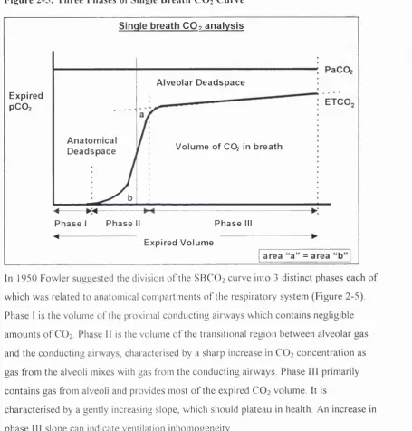
Figure 2-5: Three Phases o f Single Breath CO2 Curve... 57
Figure 2-6: Volum etric Capnography...58
Figure 3-1 : Variable flo w through an adult sensor with a 300mL syringe...66
Figure 3-2: Accuracy o f volume recordings w ith neonatal sensor...67
Figure 3-3: Accuracy o f volume recordings with paediatric sensor... 68
Figure 3-4: Accuracy o f volume recordings with adult sensor... 69
Figure 3-5: Volume accuracy o f lOOmL applied signal over range o f flows and measurement conditions... 70
Figure 3-6: Percentage error and CV o f measured pressure against manometer pressure... 71
Figure 3-7: Pressure-flow relationships o f the neonatal sensor w ith and w ithout 3.0mm tracheal tube attached... 72
Figure 3-8: Apparatus resistance-flow relationships in the neonatal flo w sensor.... 73
Figure 3-9: Pressure-flow relationships o f the paediatric flo w sensor w ith various tracheal tubes attached... 74
Figure 3-10: Resistance-flow relationships o f the paediatric flo w sensor w ith different tracheal tube attachments... 74
Figure 3-11: Pressure-flow relationships o f the adult flow sensor w ith different tracheal tube attachments... 75
Figure 3-13: Linear expression (from the equation o f m otion) o f Cm and Rrs from
several breaths in one individual...78
Figure 3-14: Example o f Crs measurements with lung model settings o f 0.5 and 1.0 m L/cm H iO during changes o f PIP and respiratory rate... 79
Figure 3-15: Influence o f PIP and respiratory rate on measured compliance, w ith lung model settings at 0.5, 1.0 and 3.0 m L/cm H 20... 80
Figure 4-1 : Four year old boy w ith “ CO2SMO Plus” flo w sensor attached between tracheal tube and ventilator tubing... 86
Figure 4-2: Physiotherapist performing manual hyperinflation w ith chest wall vibrations during expiration...90
Figure 5-1 : Examples o f trended data before and after treatment in a spontaneously breathing and a mechanically ventilated child... 97
Figure 5-2: Variability o f Vit. /kg and tracheal tube leak in 10 individuals during quiet clinical periods... 98
Figure 5-3: Comparison o f mean values fo r different measurement intervals against 45 minute mean... 99
Figure 5-4: Variability o f C,.s and R,s during quiet clinical periods... 99
Figure 5-5: R,s and C,s data fo r different time intervals compared w ith a 45 minute in te rv a l...100
Figure 5-6: Variability o f VCO2 and ETCO2 during quiet clinical perio ds 101 Figure 5-7: Variability o f CO2 parameters, comparing different data collection intervals to a 45 minute in te rv a l...101
Figure 5-8: The relationship between Vt and change in tracheal tube le a k ... 105
Figure 5-9: The relationship between tracheal tube leak and Vt e... 106
Figure 5-10 The relationship between tracheal tube leak and Crs and R rs ...106
Figure 5-11 : Flow-volume and pressur-volume loops before tracheal tube change in a 7 week old infant...107
Figure 5-12: Flow-volume and pressure-volume loops after tracheal tube change in a 7 week old infant...107
Figure 5-13: Influence o f tracheal tube change on Vte, in a 7 week old infant...108
Figure 5-14: Influence o f tracheal tube change on Crs and Rrs in a 7 week old infant 108
Figure 5-16: Difference (after - before intervention) in selected parameters in 10
patients after disconnection, physiotherapy or nursing suction...112
Figure 6-1 : Ages o f study patients...117
Figure 6-2: Body weight o f children in study...118
Figure 6-3: Diagnostic groups o f children in study... 119
Figure 6-4: Ventilation mode fo r different age groups and diagnoses... 119
Figure 6-5: Chart o f patients receiving physiotherapy treatm ents... 120
Figure 6-6: Chart o f patients receiving nursing suction... 120
Figure 6-7: Chart o f patients receiving both physiotherapy and nursing suction ...120
Figure 6-8: Magnitude o f tracheal tube leaks in study p o p u la tio n ... 121
Figure 6-9: Flow and volume range in the study population... 123
Figure 6-10: Duration o f physiotherapy and nursing treatments... 124
Figure 6-11 : Suction catheters used per treatment by physiotherapists and nurses. 125 Figure 6-12: Saline instilled per treatment by physiotherapists and nurses... 125
Figure 6-13: Number o f chest wall vibrations performed during physiotherapy treatment... 126
Figure 6-14: The difference between C,,s as a) absolute and b) weight corrected values against age...133
Figure 6-15: The influence o f a) age and b) weight on R,^ values in the study p o p u la tio n ... 134
Figure 6-16: Change in R,s relative to reported sputum yield after physiotherapy. 135 Figure 6-17: The influence o f age on a) VD;,i,^vay and b) VDaiv... 137
Figure 6-18: Significant individual changes in Vt e follow ing treatment... 140
Figure 6-19: Significant individual changes in R,s after treatm ent...141
Figure 6-20: Significant individual changes in C,^ after treatm ent...142
Figure 6-21: Significant individual changes in PeC02 after treatm ent...143
Figure 6-22: Relative changes in VDpi,ys...144
Figure 6-23: Individual changes in HCO?- after trea tm ent... 146
Figure 6-24: Individual changes in base excess after treatment...147
Figure 6-25: Individual changes in O2 saturation...147
Figure 7-2: SBCO2 before and after physiotherapy...167
List of Tables
Table 3-1: Rapp and Rtt through neonatal flo w sensor...73
Table 3-2: Rapp and Rtt through paediatric flo w sensor...75
Table 3-3: Rapp and Rtt through adult sensor at variable flo w s ... 76
Table 3-4: Mean C,^ calculations by the “ CO2SMO Plus” compared w ith set Crs on
neonatal lung simulator (variable ventilation settings)...81
Table 3-5: Accuracy o f CO2 measurements...82
Table 5-1: Effect o f disconnecting the flo w sensor compared w ith physiotherapy treatment and nursing suction...I l l
Table 5-2: Repeatability o f measurements in 33 individuals in the absence o f any
interventions... 114 Table 6-1 : Details o f tracheal tube leak during data collection in 11 patients
excluded from analysis... 122
Table 6-2: Differences between paired physiotherapy (p) and nursing suction (n) treatments...124
Table 6-3: Recordings o f ventilatory parameters during measurement periods 127
Table 6-4: Effect o f physiotherapy on respiratory fu n c tio n ...130 Table 6-5: Effect o f nursing suction on respiratory fu n c tio n ... 131
Table 6-6: Comparing nursing suction with physiotherapy (paired d a ta )... 138
Table 6-7: Demographic data on individuals w ith significant changes in Vte, C^ or
Rn,... 148
Table 6-8: Examples o f individuals w ith clinically significant improvements after
physiotherapy...149
Table 6-9: Examples o f patients with clinically significant deterioration follow ing
physiotherapy...150
Table 7-1 : Value o f F depends on power required to detect a difference w ith
specified precision (p < 0 .0 5 )... 194
Table 7-2: Numbers o f subjects needed to detect a 10% change (20% fo r R ^j at 5%
Table 7-3: Numbers o f subjects needed to detect 0.5 o f a standard deviation change
between nursing suction and physiotherapy at 5% significance w ith a power o f 80%,
90%, 95% and 99%... 195
Table 10-1: Accuracy o f volumes recorded by “ CO2SMO Plus” using the neonatal flo w sensor... 268
Table 10-2: Accuracy o f volumes recorded by “ CO2SMO Plus” using the paediatric flo w sensor... 269
Table 10-3: Accuracy o f volumes recorded by “ CO2SMO Plus” using the adult flo w sensor... 270
Table 10-4: Pressure recordings by “ CO2SMO Plus” compared to D ig itro n ...271
Table 10-5: W ithin subject variability o f parameters in 33 individuals over a 30 minute period... 272
List of Equations Equation 2 -1 ... 46
Equation 2 -2 ...50
Equation 2 -3 ...52
Equation 2 -4 :... 52
Equation 2 -5 :... 53
Equation 2 -6 :... 59
Equation 2 -7 :... 59
Equation 2 -8 :... 60
Equation 2 -9 :... 60
Equation 2 -1 0 :... 60
Equation 2-11 : ...60
Equation 2 -1 2 ... 63
Equation 2 -1 3 ... 63
Equation 3 -1 ... 77
Equation 3 -2 ... 77
Equation 5 -1 ...104
List Of Abbreviations and Definitions
Abbreviation Definitions and Units
abg ASD ARDS AVSD BE BPD BTPS CICU CO2 CPAP C Crs CV ds E ECMO ETCO2 F FEVi Fi02 FRO GLM
HCO3
-ICU IVH kg LA MAP N2 NS P Pa02 PaC02 PAO2 PCV
arterial blood gas atrial septal defect
acute respiratory distress syndrome atrio-ventricular septal defect base excess
bronchopulmonary dysplasia
body tem perature, barometric pressure and w ater vapour saturated conditions
cardiac intensive care unit carbon dioxide
continuous positive airways pressure, cmH20, kPa (1cmH20 = 0.098 kPa) compliance, m L/cmH20 or L/kPa, (1m L/cm H20 = 10.2 m UkPa)
respiratory system compliance: the distensibility of the respiratory system m L/cm H20 or L/kPa (may be expressed per kg body weight).
1mL/cmH20 = 10.2 mL/kPa
coefficient of variation, %, CV = (SD/mean) X 100 deadspace, mL/kg
elastance (the reciprocal of compliance) cm H2 0/m L
extra-corporeal membrane oxygenation end tidal CO2, kPa
gas flow
forced expiratory volume in one second fractional oxygen concentration of inspired gas functional residual capacity
general linear model
bicarbonate, abundant buffer in blood plasma, regulated by kidneys intensive care unit
intra-ventricular haemorrhage kilogram
limits of agreement (mean difference +/- 2 SD)
mean airway pressure, cmhi20 or kPa, (1cmH20 = 0.098 kPa) nitrogen gas
non significant pressure
partial pressure of O2 in arterial blood, kPa
partial pressure of CO2 in arterial blood, kPa
partial pressure of O2 in alveoli, kPa
Abbreviations and Definitions continued...
Abbreviation Definitions and Units
PeCOz PEEP PEF p H Physio PICU PIE PIP PRVC R Rapp
Resp. rate
Rtt Sa02 SBCO2 SD tE t |
t r s
Truncus A
Tube leak
V
V/Q ratio
VCO2
VCV
Voairway
Voalv
VDphys
VDphys/ Vt
VO2
Vt Vte
Vti
weight
mixed expired CO2. kPa
positive end expiratory pressure, cm H20 or kPa (1cm H20 = 0.098 kPa) peak expiratory flow, mL/s
measure of the balance between acids and bases in the blood plasma physiotherapy
paediatric intensive care unit pulmonary interstitial emphysema
peak inspiratory pressure, cmH20, kPa (1cmH20 = 0.098 kPa) pressure regulated volum e controlled
resistance
resistance due to apparatus, cm H20/L/s or kPa/L/s, (1cm H20/L/s = 0.098kPa/L/s)
respiratory rate, breaths/minute
total respiratory system resistance, cm H20/L/s or kPa/L/s, (1cmH20/l_/s 0.098kPa/L/s)
resistance due to tracheal tube, cm H20/L/s or kPa/L/s (1cmH20/L/s = 0.098kPa/L/s)
saturation of oxygen in arterial blood, % single breath CO2
standard deviation expiratory time, seconds inspiratory time, seconds
respiratory tim e constant, seconds truncus arteriosus
tracheal tube leak, %, tracheal tube leak = ( Vt i- Vt e) / Vti x 100
volume
ratio of pulmonary ventilation to pulmonary perfusion
volum e of CO2 expired in mL/min (expressed per kg of body weight)
volume controlled ventilation airway deadspace, mL alveolar deadspace, mL physiological deadspace, mL
ratio of physiological deadspace to tidal volume oxygen consumption
1. Introduction and literature review
1.1 Introduction
Chest physiotherapy and tracheal suction to clear pulmonary secretions from the
ventilated child are routine in many intensive care units. Historically, studies to assess
the clinical effectiveness o f such treatments have been limited by the lack o f objective,
non-invasive measures o f respiratory function. There is little firm evidence to
substantiate the use o f physiotherapy in ventilated children or to recommend it above the
suction techniques used by nursing staff. This study aimed to use a new portable non-
invasive respiratory m onitor (“ CO2SMO Plus” , Novametrix Medical Systems Inc.
version 3.0, CT, U S A) to measure respiratory function before and after physiotherapy
and nursing suction procedures. The principal aims o f the study were to assess the
effects o f respiratory physiotherapy treatments in ventilated children and to establish
whether such treatments offer a significant advantage over routine suction used by nursing staff in the intensive care unit. In addition, considerable efforts were made to
validate the “ CO2SMO Plus” respiratory m onitor and to evaluate its clinical usefulness
in objectively assessing therapeutic interventions in the intensive care unit. The first three chapters o f this thesis include a literature review, the theoretical
background pertaining to respiratory parameters o f interest in this project and how they
were measured or calculated by the m onitor and a description o f the validation w o rk
undertaken to evaluate the performance o f the “ CO2SMO Plus” .
The latter four chapters include a description o f the study design and methodology used
in the project, a report o f the in-vivo and pilot studies performed, results o f the study
and a discussion o f the findings with reference to other publications in relevant research
fields.
Finally tw o publications which issued directly or indirectly from the w o rk done in this
project are included preceding the Appendix and reference list.
1.2 Literature review
Mechanically ventilated patients are inefficient at clearing mucus from their own
airways. The tracheal tube covers normal ciliated tracheal tissue and prevents normal
movement o f mucus towards the epiglottis and oesophagus. In addition the “ foreign”
thereby stimulating further production o f mucus. M ost mechanically ventilated infants
are well sedated and may be pharmacologically paralysed, so that the ability to cough
effectively is either seriously compromised or absent. Accumulation o f mucus in the
airways because o f increased production or decreased clearance due to defects in the
ciliary clearance apparatus w ill contribute to the m orbidity o f airway diseases by predisposing patients to respiratory infections, airflow obstruction and discomfort.
There is a significant association between chronic production o f mucus and an increased
risk o f m ortality (Kim , 1997). The management o f mucus hypersecretion can be
undertaken either by improving clearance by physical methods or by pharmacological
methods (M arriott, 1981; Kim, 1997). Mucus hypersecretion may not be the primary problem in many ventilated children, but the presence o f the tracheal tube, as well as
respiratory pathology and sedation or muscle relaxation, w ill significantly reduce normal mucociliary clearance, thereby creating a vulnerability to further disease. Regular suction
(performed by nursing staff or physiotherapists) combined with a number o f other
modalities has become an important part o f reducing respiratory complications associated with mechanical ventilation.
Physiotherapy techniques in intensive care have developed over the last tw o decades but
there remains little conclusive evidence to support or guide airway clearance techniques
in ventilated children. Current practice is often based on locally established routines, experience, clinical intuition based on anatomical and physiological knowledge and some
wisdom from eclectic and often conflicting publications. There is also wide variability in
the role o f the physiotherapist in intensive care units depending on the country, training,
local traditions, staffing levels and expertise. There is little, i f any, evidence to
recommend dynamic physiotherapy airway clearance techniques above the use o f routine
airway clearance techniques employed by nursing staff in paediatric intensive care units
(Stiller, 2000).
The lack o f research evidence to support physiotherapy practice in intensive care has also been in part due to the difficulty in identifying appropriate and sensitive outcome
parameters. Until recently, equipment for measuring lung function in the intensive care
unit has been too cumbersome, invasive, inaccurate or expensive to use routinely.
continuous and non-invasive measures o f various respiratory parameters possible. Some o f these have the potential to be useful in evaluating the efficacy o f airway clearance
techniques.
1.2.1 Adverse effects of mechanical ventilation
The various cardio-respiratory complications associated w ith mechanical ventilation
have been extensively studied. They include atelectasis, barotrauma, pulmonary oedema,
tracheal tube obstruction, compromised cardiac output and pulmonary blood flow ,
hypoxia, hypo- or hyper-capnia and nosocomial pneumonia (Rivera and Tibballs, 1992; Clough et al. 1994; Meade et al. 1997; McGuire et al. 1997; Gannon et al. 1998;
Dreyfuss and Saumon, 1998; Stewart et al. 1998). Mechanical ventilation in preterm
infants has been associated with retinopathy o f prematurity, intraventricular
haemorrhage, and bronchopulmonary dysplasia (Greenough, 1999). It has also been
shown to reduce postnatal alveolar multiplication and increase the amount o f bronchial smooth muscle and submucosal glands (Greenough, 1992; Thompson et al. 1992). The
pathology that precipitated the need fo r assisted ventilation may be exacerbated by the high tidal volumes or airway pressures needed to obtain or maintain optimal gas
exchange (Parker et al. 1990; Dreyfuss and Saumon, 1993; Rosen et al. 1993).
There are few controlled paediatric studies comparing the various modes o f ventilation
in terms o f patient outcomes. Currently, the choice o f ventilator mode depends largely on the equipment available, the patient's disease state, and the clinician’ s preference
based on personal experience (MacDonald and Johnson, 1996). Survival o f small infants
has improved significantly in recent years and the focus is now on reducing the incidence
o f ventilation-induced lung injury and other complications. This involves improved
understanding o f conditions producing respiratory failure, the response o f the
respiratory system to various therapies and the effects o f changes in ventilator strategies
(Stenson et al. 1998).
The rationale fo r physiotherapy assumes that removal o f secretions from the airways w ill
improve delivery o f gas from the ventilator, help to resolve atelectasis and improve
from ventilation and reduce lung damage associated w ith mechanical ventilation. Preterm infants are particularly susceptible to lung damage from mechanical ventilation
and should be weaned as soon as possible (Hislop et al. 1987; H aw orth et al. 1989).
1.2.2 The general effects of physiotherapy or nursing suction
There are a large number o f publications which are relevant to the efficacy o f
physiotherapy or nursing suction procedures, but many o f them have significant flaws in
design or execution and results have to be interpreted w ith caution. Published reviews examining the efficacy o f respiratory physiotherapy have been frustrated by studies o f
poor quality or w ith small study numbers, multiple treatment modalities and inter and
intra-professional differences in performing techniques, multiple patient groups w ith
different diagnoses, and multiple outcomes (Hussey, 1992). The lack o f consensus and
conflicting results in the literature regarding optimal methods o f airway clearance diminish confidence in the interpretation o f research findings and extrapolation o f adult
studies or those in non-ventilated or selective paediatric populations to the general paediatric population. Despite quite extensive bibliographies, reviews on the efficacy o f
respiratory physiotherapy tend to arrive at similar conclusions; that the evidence is limited and that there is an urgent need for further research to be performed (Stiller,
2000). Sometimes airway clearance procedures are performed by nurses but referred to as "chest physiotherapy" (Dulock, 1991; Lewis et al. 1992). Unfortunately these
publications have the potential to reflect inaccurately upon the practices o f
physiotherapists (Higgins, 1974; Ciesla, 1996; Krause and Hoehn, 2000; Flenady and
Gray, 2000; van der Schans et al. 2000).
The rationale fo r chest physical therapy in intensive care units is to minimise pulmonary
secretion retention and to maximise oxygenation by re-expanding atelectatic lung
segments (Ciesla, 1996). Chest physiotherapy is regularly performed in ventilated
children w ith the justification that it helps prevent or resolve complications associated
w ith artificial respiration (retention o f secretions and atelectasis) and speeds up the
resolution o f primary respiratoi*y disorders. Prior to accessible measures o f respiratory
function in the intensive care unit, physiotherapists have had to rely on sub-optimal and
relatively subjective markers to evaluate the efficacy o f treatment. These markers have
Arterial blood gas measurements remain the gold standard fo r clinical assessment o f lung function, but are not always readily available and are not necessarily routinely taken
before and after chest physiotherapy.
1.2.2.1 Sputum clearance
The efficacy o f sputum clearance may be demonstrated directly by sputum weight or via
indirect measures o f pulmonary function such as radio-labelled aerosol clearance
(Bateman et al. 1981). Sputum weight has always been a controversial outcome measure in physiotherapy trials because o f intra- and inter-subject variability. Sputum yield varies
during the course o f the day in any individual and different pathophysiology w ill result in
different amounts o f sputum but not in a predictable or consistent fashion. Some studies dispute the correlation between the amount o f sputum and improved pulmonary function
in ventilated patients (Mackenzie et al. 1989; Hasani et al. 1994) while others suggest that sputum clearance is associated with an improvement in respiratory function
(Cochrane et al. 1977). Some authors suggest that the connection between sputum
production and pulmonary function is irrelevant as long as it can be demonstrated that physiotherapy treatments are advantageous in clearing secretions (Etches and Scott,
1978; M ay and M unt, 1979; Pavia, 1990; Gallon, 1991). Many studies to date which
used sputum weight as an outcome have been undertaken in non-ventilated patients, but a recent study found that total static respiratory system compliance (Crs) and sputum
clearance were improved by the addition o f manual hyperinflation to a physiotherapy
treatment o f positioning and suctioning in mechanically ventilated patients w ithout compromise o f cardiovascular stability or gas exchange (Hodgson et al. 2000).
1.2.2.2 Arterial blood gases
The reported effects o f physiotherapy treatments on arterial blood gases are conflicting.
Some publications report a significant fall in PaO] after physiotherapy techniques or
tracheal tube suction (Gormezano and Branthwaite, 1972) while others report no
changes in PaO] after physiotherapy despite treatment protocols w ithout pre
oxygenation (Mackenzie et al. 1978). Hypoxaemia has frequently been reported
follow ing tracheal tube suction in ventilated neonates (Holloway et al. 1966; Simbruner
et al. 1981 ; Fox et al. 1978; Durand et al. 1989; Hussey, 1992), although Finer and
Boyd showed a greater increase in PaO] in neonates follow ing physiotherapy and
Boyd, 1978). One study reported significant increases in Pa02 after percussion in premature babies, but the study design included increasing ventilation pressures by 25%.
There can be little doubt that the increase in peak inspiratory pressure is very likely to
have influenced the observed improvement in blood gases after suction (Tudehope and
Bagley, 1980).
Several studies in ventilated adults or infants have shown that pre-oxygenation,
delivered manually or by the ventilator, completely prevented post suction hypoxaemia
or produced an increase in PaOz immediately after treatment (Connors et al. 1980;
Goodnough, 1985; Preusser et al. 1988; Kerem et al. 1990; Stone et al. 1991). Some authors suggest that in the presence o f significant atelectasis, an increase in PaOz may be
seen after resolution o f atelectasis with physiotherapy, even i f pre-oxygenation is not incorporated (Holody and Goldberg, 1981; Mackenzie and Shin, 1985; Ciesla, 1996).
Atelectasis may be associated with more widespread disturbances o f gas exchange than
is generally realised, perhaps because o f distension o f adjacent lung regions (Fletcher and Larsson, 1985).
In summary, it appears that hypoxaemia may be associated w ith physiotherapy and / or
tracheal tube suction. Many publications have recommended pre-oxygenation or
hyperinflation breaths as effective ways o f avoiding or minimising the hazards o f suction and physiotherapy (Young, (a) 1984; Riegel and Forshee, 1985; Gunderson et al. 1986;
Kerem et al. 1990; Shah et al. 1992; Mancinelli-Van and Beck, 1992; Odell et al. 1993;
Wood, 1998). Alternatively, suction through closed ports to maintain PEEP and tidal
volumes during the procedure may reduce suction related hypoxaemia (Gunderson et al. 1986).
1.2.2.3 Respiratory mechanics (Crs snd Rrs)
The evidence fo r the effects o f physiotherapy on respiratory system compliance (C^) and
resistance (Rrs) in ventilated adults and children is also conflicting. This may be related
to institutional differences in treatment modalities, differences in patient populations or
differences in calculating respiratory mechanics. Suction has been associated w ith
significant deterioration in C,s in fully ventilated, muscle relaxed, new-born babies
reduction in Crs after treatment, while manual hyperinflation improved Crs by a maximum o f 16% which was sustained for more than an hour in patients w ith lung disease (Jones
et al. 1992). Another study found no significant difference in C , s between mechanically
ventilated adults who received pre-oxygenation and tracheal tube suction, w ith and
w ith out manual hyperinflation and chest wall vibrations after cardiac surgery (Eales et
al. 1995). B y contrast, Mackenzie et al, in tw o separate studies, found statistically significant (p<0.01) increases in Crs 2 hours after physiotherapy in mechanically
ventilated adults but no change in R,s(Mackenzie et al. 1980; Mackenzie and Shin,
1985). Other studies have also shown an increase in Crs after physiotherapy (W inning et
al. 1975). In animal studies, negative pressure applied to the trachea was shown to
decrease lung compliance and this was partly attributed to collapse o f alveoli and
increased venous admixture, possibly as a result o f the continued perfusion o f the collapsed alveoli (Velasquez and Farhi, 1964).
Very significant falls in both R , s and expiratory time constant ( t , s ) measurements have
been reported after tracheobronchial suction or lavage for infants whose pre-treatment
values o f resistance were elevated compared with reference data. N o changes in C^
values were noted (Prendiville et al. 1986). In the same study, severe but clinically inapparent mucus obstruction o f the airways was revealed in tw o infants by a
progressively rising R,-* during continuous monitoring. In addition, R^ has been shown to fall significantly (p<0.005) follow ing vibrations and suction in a neonatal population,
possibly as a result o f the removal o f secretions from the airways, but Rr$ returned to
baseline values within 2 hours o f suction, (Fox et al. 1978). In non-intubated adults,
significant reduction in airflow obstruction after chest physiotherapy has been reported
(Cochrane et al. 1977).
1.2.2A Acute lobar atelectasis and chest radiograph
Atelectasis at a macroscopic or microscopic level refers to the collapse o f lung regions
as gases are resorbed from underventilated alveoli. Atelectasis may be due to several
factors including regional airway obstruction by retained secretions. O f all the
pulmonary complications associated with ventilation (including pneumonia,
or chronic respiratory disease) acute lobar atelectasis has quite consistently been shown
to respond favourably to physiotherapy (Marini et al. 1979; Hammon and M artin, 1981;
Fourrier et al. 1994). However, w ith the exception o f one study (Galvis et al. 1994),
most o f these studies were performed on ventilated adult patients and results may not be
extrapolated to a paediatric population w ith confidence. The predominance o f upper
lobe collapse observed in paediatric intensive care patients contrasts w ith the high
incidence o f lower lobe collapse in their adult counterparts. M u ltiple factors are likely to be contributory and include the anatomical and physiological differences between adults
and children, the pathophysiology o f childhood respiratory disease and more critical
positioning o f tracheal tubes in younger patients and their movement w ith patient
positioning (Thomas et al. 1999).
Three randomised studies comparing the efficacy o f fiberoptic bronchoscopy to
respiratory therapy in resolving acute lobar atelectasis, found respiratory therapy alone to be superior or equivalent to fiberoptic bronchoscopy (M arini et al. 1979; Hammon
and M artin, 1981; Fourrier et al. 1994). A further study concluded that routine post
lobectomy bronchoscopy offered no advantage over physiotherapy in preventing the
development o f postoperative atelectasis (Jaworski et al. 1988). Ciesla et al, in tw o separate studies, found that physiotherapy assisted in the resolution o f atelectasis in
ventilated adults and improved PaO] w ithout the negative haemodynamic side effects o f
therapeutic bronchoscopy (Ciesla et al. 1981; Ciesla, 1996). Physiotherapy techniques in addition to hyperinflation and suction have been shown to enhance and hasten resolution
o f acute lobar atelectasis after a single treatment (Stiller et al. 1990; Stiller et al. 1996).
In a prospective study o f 47 ventilated adults, Mackenzie et al. found that chest
radiographs within 24 hours o f physiotherapy showed improvement in 68% o f patients.
Chest physiotherapy was most effective in the treatment o f unilobar densities and produced dramatic improvement in atelectasis o f acute onset (Mackenzie et al. 1978).
The single paediatric study on mechanically ventilated infants, found that physiotherapy was effective in the treatment o f various degrees o f lung collapse. Lung expansion,
1994). Zach et al reported improvement in atelectasis in unventilated children receiving
physiotherapy (Zach and Oberwaldner, 1987).
In contrast to the studies reported above. Reines et al. found that chest physiotherapy
was associated w ith significantly (p< 0.01) more frequent and severe atelectasis in
children after heart surgery than in patients who had not received physiotherapy (Reines
et al. 1982). However, this study was done almost 20 years ago, and relatively small subjects numbers (19 and 25 in each group), coupled w ith the fact that both
physiotherapy and ventilation techniques have evolved considerably since then, reduce
the confidence with which this evidence can be applied.
1.2.2.5 Prevention and treatment of pneumonia or common pulm onary
complications
T w o very early publications suggested that prophylactic chest physiotherapy could prevent post-operative pulmonary complications (including atelectasis and pneumonia) in adults follow ing abdominal surgery (Thoren, 1954; Palmer and Sellick, 1953). Since then however, there has been little consistency in the literature regarding the role o f
physiotherapy in prevention or treatment o f common pulmonary complications in
ventilated patients (w ith the exception o f acute lobar atelectasis). W hile some studies
suggest there is moderate evidence that routine prophylactic postoperative chest
physiotherapy in both adult and paediatric populations significantly reduced the frequency o f post operative complications (Bartlett et al. 1973; M orran et al. 1983;
Rockwell and Campbell, 1976; Rello et al. 1996), others have reported detrimental
effects or no effect in reducing the incidence or resolution o f nosocomial pneumonia
(Graham and Bradley, 1978; Reines et al. 1982; Ntoumenopoulos et al. 1998). One
randomised trial involving 171 patients concluded that chest physiotherapy was "at best
useless in patients w ith primary infectious pneumonia" (B ritto n et al. 1985).
Some authors have suggested that relatively recent policies o f encouraging early
mobilisation and ambulation follow ing many surgical procedures have reduced the need
fo r prophylactic physiotherapy treatment (Dull and Dull, 1983; Hallbook et al. 1984;
A recent Cochrane review based on three small trials found there was no evidence that
peri-extubation physiotherapy compared with non-active techniques (positioning and
suction alone) or no intervention prevented post-extubation lobar collapse, though there
appeared to be a reduction in re-intubation episodes when 1 to 2 hourly physiotherapy
treatments were performed prior to extubation (Flenady and Gray, 2000). The numbers o f babies studied were small and 2 o f the 3 studies were carried out over 10 and 20
years ago respectively. Since then, enormous changes have occurred both in the medical
and physiotherapy management o f ventilated patients. Extrapolation o f this information
to current practice must therefore be very guarded.
1.2.2.6 Deadspace and ventilation/ perfusion balance
Very few studies have reported deadspace or ventilation / perfusion matching as
outcomes fo r physiotherapy, although a major cause o f impaired gas exchange in ventilated patients is atelectasis, causing intrapulmonary shunt. The effect o f body position on ventilation / perfusion relationships has frequently been examined but this
has been particularly in relation to optimising positioning in acute respiratory distress syndrome (ARD S) or unilateral lung disease, rather than specifically w ith regard to
physiotherapy (Rattenborg and Holaday, 1967; Secker-Walker et al. 1975; M oran et al. 1977; Batra et al. 1981; Dean, 1985; McCarthy, 1987; M oriya et al. 1987; Cohen, 1997;
Walther et al. 1998). A single case study reported that alveolar deadspace (Voaiv) was
significantly reduced after resolution o f a partially atelectatic lung (Fletcher and Larsson,
1985).
Another study concluded that external mechanical vibration o f the chest was useful in
the management o f hypoxaemia in patients with atelectasis or pneumonia and that
improved ventilation / perfusion (V /Q ) balance was the reason fo r the observed
significant increase in Pa02 30 and 60 minutes after treatment (H olody and Goldberg, 1981). Yet another study showed a decrease in intrapulmonary shunt and significant
improvement in respiratory compliance 2 hours after physiotherapy in 19 mechanically
ventilated patients w ith post-traumatic respiratoi-y failure (Mackenzie and Shin, 1985).
1.2.2.7 Potentially adverse effects of multimodality treatments
The physiological and haemodynamic effects o f physiotherapy have been extensively
treatment modalities. Several authors have reported arrhythmia’ s during or follow ing suction or physiotherapy (Hammon et al. 1992), while others have found that patients
w ithout adequate sedation have had significant and sometimes dramatic increases in
heart rate, blood pressure, cardiac output, oxygen consumption (V O2), carbon dioxide
elimination (V C O2) and PaC02 during such treatments. Some o f these studies described
significant reduction in certain or all o f these effects w ith sedation or pharmacological
paralysis (Bodai, 1982; Weissman et al. 1984; Klein et al. 1988; Stone et al. 1991; Hammon et al. 1992; Weissman and Kemper, 1993; Harding et al. 1994; Weissman et al.
1994; Singer et al. 1994; Cohen et al. 1996; Horiuchi et al. 1997).
Physiotherapy and suction treatments have also been associated w ith significant
increases in intracranial pressure (ICP), although in most cases, the simultaneous
increase in blood pressure has resulted in adequate or only transiently inadequate cerebral perfusion pressures (GPP) (Parsons and Shogan, 1984; Gar rad d and Bullock,
1985; Durand et al. 1989; Singh et al. 1991; Crosby and Parsons, 1992; Paratz and Burns, 1993; Wang et al. 1993; Bru ci a, 1993; Bnjcia and Rudy, 1996; Imle et al. 1998).
Adequate sedation and pharmacological paralysis have been reported effective at
controlling these responses to tracheal suction (Parsons and Shogan, 1984; Ninan et al. 1986; Ninan et al. 1986; Fanconi and Duc, 1987; Ersson et al. 1990). Brim ioulle et al
claimed that passive mobilisation techniques were not associated w ith adverse effects in
patients w ith raised ICP (Brim ioulle et al. 1997).
In 1998, Harding et al proposed a connection between encephaloclastic porencephaly
and chest physiotherapy in extremely preterm infants (Harding et al. 1998). The study involved retrospective analysis o f 454 infants o f birth weight less than 1500gm delivered
between 24 to 27 weeks o f gestation. Patients with encephaloclastic porencephaly
received tw o to three times as many chest physiotherapy treatments in the second, third, and fourth weeks o f life as control infants (p < 0.001). Affected subjects also had more
prolonged and severe hypotension in the first week than control subjects (p < 0.01), and
were less likely to have had a cephalic presentation (p < 0.01). The brain lesion was
considered by the authors to be caused by impact o f the brain w ith the skull during
shaking movements that could occur during chest physiotherapy w ith percussion. Since
encephaloclastic porencephaly and physiotherapy and have also pointed out the significant methodological errors and risks o f type 1 error in this w o rk (Beeby et al.
1998; Gray et al. 1999; Vincon, 1999). Gray et al and Beeby et al reported that they had
not found any cases o f encephaloclastic porencephaly over the same 3-year period,
despite similar criteria for the initiation o f physiotherapy treatment. Gray noted that while Harding found a strong association (P < 0.001) between chest physiotherapy and
encephaloclastic porencephaly, severe hypotension and noncephalic presentation were also statistically significant and the “ model collapsed” when these variables were entered
into the multivariate analysis. It was thus likely that the lesion occurred only in the
sickest infants and the fact that they had more chest physiotherapy just reflected their degree o f illness.
1.2.3 The efficacy of individual components of physiotherapy treatments in ventilated patients.
M ost frequently physiotherapy treatments involve a combination o f treatment techniques. Research attempting to tease out the effects o f different components o f treatment rarely tests each component in isolation. Instead they involve designs where a
specific technique is included or not in a control group and then the differences between
the groups are attributed to the specific technique. There are several treatment
modalities commonly used in ventilated children which include those discussed below.
1.2.3.1 Positioning and postural drainage
Postural drainage refers to therapeutic drainage in bronchopulmonary diseases in which
there is copious mucus secretion, such as chronic bronchitis, bronchiectasis, pulmonary
abscess, or cystic fibrosis. The patient is placed so that gravity assisted drainage o f the
affected lobe may be facilitated. There are eleven described therapeutic positions which
have the aim o f allowing gravity to assist in accelerating drainage o f mucus from the
lung periphery to larger airways. The duration o f postural drainage may vary
considerably ( 1 5 - 6 0 minutes) depending on the age and tolerance o f the patient as well
as the clinical requirements o f the treatment (Bateman et al. 1981; Ciesla, 1996). Results
from some studies on non-ventilated adults claim that postural drainage adds benefit to
traditional chest physiotherapy (Sutton et al. 1982; Johnson et al. 1987). One study
found that postural drainage and manual hyperinflation in ventilated patients did not
studies reporting the efficacy o f these techniques in adult or paediatric intensive care have been found.
Chest physiotherapy including postural drainage has been implicated in causing or exacerbating gastro-oesophageal reflux in young patients w ith cystic fibrosis (Button et
al. 1997; Taylor and Threlfall, 1997; Heine et al. 1998; Button et al. 1998; Button,
1999), but this effect has been disputed by others (Phillips et al. 1998) and was not found in patients w ith chronic bronchitis and bronchiectasis (Chen et al. 1998).
Positioning refers to the more general aim o f optimising V /Q matching, and the
physiological rationale fo r it in the intensive care unit has been well described in some
disorders. Prone positioning has been shown to improve oxygenation in the short term in critically ill adults and children (Langer et al. 1988; Pappert et al. 1994; M izuno et al.
1995; Chatte et al. 1997; Mure et al. 1997; Numa et al. 1997; Jolliet et al. 1998; M izuno and Aizawa, 1999). Improvements in lung function have also been described fo r patients
w ith unilateral lung disease when they are positioned in side lying, although the optimal position is different in adult and paediatric patients because o f differences in regional
ventilation and perfusion (Zack et al. 1974; Ibanez et al. 1981; Prokocimer et al. 1983; Rivara et al. 1984; Gillespie and Rehder, 1987; Larsson et al. 1989; Lu mb and Nunn,
1991; Webster, 1999). In children and adults, the effect o f posture on the distribution o f
perfusion is similar, however, in children ventilation in response to gravity is the reverse
o f that seen in adults, being preferentially distributed to the upper lung whether diseased or normal (H eaf et al. 1983; Davies et al. 1985; Bhuyan et al. 1989). Positioning therapy
to assist resolution o f acute atelectasis has been recommended, but the evidence fo r this
is unclear (Dean, 1985; Dean and Ross, 1992; Dean, 1997).
1.2.3.2 M anual hyperinflation, hyperventilation or hyperoxygenation
Manual lung inflation involves disconnection o f the patient from mechanical ventilation to provide temporary manual ventilation. Specific techniques o f performing manual
inflation depend on whether the aim is to achieve hyperinflation, hyperoxygenation,
M anual hyperinflation commonly involves a slow, deep inspiration, inspiratory pause and fast unobstructed expiration, although the technique should be modified fo r patients
w ith reduced or compromised cardiac output. Specific aims o f this procedure are to
improve tidal volume and alveolar recruitment by re-inflating areas o f atelectasis,
thereby im proving compliance and ventilation/perfusion matching. Hyperinflation w ill
additionally often stimulate cough in self ventilating patients and thus assist in airway clearance (Clement and Hubsch, 1968; W indsor et al. 1972; B artlett et al. 1973; Stiller
et al. 1996). Hyperinflation manoeuvres have been shown to effectively re-expand
collapsed lung tissue and improve oxygenation in anaesthetised ventilated adults w ith
healthy lungs (Rothen et al. 1999).
Research examining the efficacy o f manual hyperinflation specifically in airway clearance
is conflicting or not comparable because o f significant differences in technique and methodology (Barker and Bales, 2000). Controversy exists regarding the safety and
effectiveness o f application o f manual lung hyperinflation in intubated patients. Tidal volumes, pressures and FiOz are not controlled and there are inherent dangers o f
barotrauma or hypoxaemia in the absence o f additional oxygen (Brandstater and Muallem, 1969; Fox et al. 1978; Gattinoni et al. 1993; M cKelvie, 1998; Clarke et al.
1999; Dorges et al. 2000). Whereas some studies suggest that the addition o f manual
hyperinflation in treatment does not offer any advantage (Novak et al. 1987; Bales et al. 1995), others have found that manual hyperinflation was associated w ith an
improvement in respiratory compliance and sputum clearance in mechanically ventilated
patients w ith out compromise o f cardiovascular stability or gas exchange (Jones et al.
1992; Hodgson et al. 2000).
H yperoxygenation involves delivering greater FiO], with the purpose o f improving oxygenation. It may be used in combination with manual hyperinflation during
physiotherapy treatments and is recommended in preference to hyperinflation alone in reducing suction-induced hypoxaemia, or facilitating recovery after suction (Chulay and
Graeber, 1988; Lookinland and Appel, 1991). Stone and others, comparing the efficacy o f ventilator versus manual hyperinflation in delivering hyperoxygenation or
hyperinflation breaths before, during, and/or after tracheal suctioning, found that
breaths in preventing suction-induced hypoxaemia, and that delivery o f manual
hyperinflation breaths resulted in increased airway pressure and increased
haemodynamic consequences (Stone, 1990; Grap et al. 1996). B y contrast, Goodnough
(1985) claimed that manual hyperinflation with FiOz o f 1 provided better protection than
ventilator delivered breaths o f the same FiOz, but recommended m onitoring o f
haemodynamic parameters because o f potential alterations in arterial blood pressure and
heart rate associated w ith manual hyperinflation (Goodnough, 1985).
M anual hyperventilation involves rapid delivery o f breaths usually w ith the aim o f
im proving oxygenation or blowing o ff CO2 It is rarely used in physiotherapy treatments,
but may be helpful in patients in whom hypercapnia should be avoided, fo r example
patients w ith head injuries in whom low PaCOi may be advantageous prior to treatment,
or patients in pulmonary hypertensive crisis who have extremely reduced C,s and may be difficult to oxygenate. The efficacy o f hyperventilation in physiotherapy treatments has
not been reported.
1.2.3.3 Chest percussion (clapping) and vibrations
Chest percussion or clapping in paediatric patients involves tapping the chest wall w ith
cupped hand, fingers or soft plastic c u ff to mobilise secretions. Vibrations refer to the manual technique o f shaking the chest wall during the expiratory phase o f respiration.
Chest percussion or vibrations are reported to enhance mucociliary clearance from central and peripheral aii*ways (May and M unt, 1979; Bateman et al. 1981; Mackenzie et
al. 1989; Ambrosino et al. 1995). The exact mechanisms by which percussion and
vibration are felt to achieve mucociliary clearance are unclear, but alteration o f airflow
and release o f pulmonary chemical mediators have been proposed (K in g et al. 1983;
Gallon, 1991). A t bronchoscopy, secretions have been seen to move into the upper
airways when vibrations are applied to the chest wall (Ciesla, 1996). Other authors have
suggested that chest wall vibrations and manual hyperinflation are associated w ith large
fluctuations in cardiothoracic pressure and hence are inadvisable (Laws and M cIntyre,
1969).
Some studies assessing the efficacy o f percussion and vibrations in ventilated adults and
(Finer and Boyd, 1978; Holody and Goldberg, 1981). Another study, however, found a
significant fall in PaC^ after chest percussion in ten patients who produced no sputum or
small amounts o f sputum, while there was no significant change in PaOz in 12 patients
who produced moderate to large amounts o f mucopurulent secretions (Connors et al.
1980). Some authors suggested that in non-ventilated patients postural drainage was the
most useful therapeutic component while percussion and vibrations had nothing to add
(K ir illo ff et al. 1985; Sutton et al. 1985; Pavia, 1990).
Bronchospasm, increased risk o f arrhythmia, decreased blood pressure, increased heart
rate and short term decline in FEV] (forced expiratory volume) have all been associated w ith percussion in both ventilated and non-ventilated patients (Campbell et al. 1975;
W ollm er et al. (b) 1985; Hammon et al. 1992; Ntoumenopoulos, 1994).
Pharmacological bronchodilation therapy has been proposed in cases where the benefits o f treatment are thought to outweigh the risks o f bronchospasm (Gallon, 1991).
1.2.3.4 Instillation of saline
Saline instillation into the tracheal tube o f ventilated patients is commonly used to stimulate cough or loosen thick, sticky secretions so that they may easily be removed w ith suction, but evidence for the practice is equivocal (Swartz et al. 1996; Schwenker
et al. 1998). Some authors suggest that at best saline instillation is not effective and at
w orst may be detrimental in terms o f O2 saturation (Bostick and Wendelgass, 1987;
Ackerman, 1993; Hagler and Traver, 1994; M cKelvie, 1998; Ackerman and M ick, 1998;
Blackwood, 1999; Kinloch, 1999), while others suggest it is well tolerated even in
infants and may be helpful in removing secretions adherent to the chest wall (Shorten et
al. 1991; Whitnack, 2000). Several reviews suggest normal saline instillation in
ventilated patients should be discontinued as a routine or standard practice until more
research has proved its efficacy (Raymond, 1995; Druding, 1997).
1.2.3.5 Tracheal suction
Tracheal suctioning is a necessary practice carried out in intensive care units. It involves
the removal o f pulmonary secretions from a patient w ith an artificial airway in place via
a catheter connected to a negative pressure source (Stone and Turner, 1989).
Sometimes suction is used to stimulate a cough in patients making spontaneous