University of South Carolina
Scholar Commons
Theses and Dissertations
6-30-2016
Enhancing The Efficacy Of Thymidylate Synthase
Inhibitors By Genetic Modulation Or
Pharmacological Inhibition Of Bone Marrow
Derived Cells In The Tumor Stroma
Nikeya LaShanna Tisdale
University of South Carolina
Follow this and additional works at:https://scholarcommons.sc.edu/etd Part of theBiology Commons
This Open Access Dissertation is brought to you by Scholar Commons. It has been accepted for inclusion in Theses and Dissertations by an authorized administrator of Scholar Commons. For more information, please [email protected].
Recommended Citation
E
NHANCING THEE
FFICACY OFT
HYMIDYLATES
YNTHASEI
NHIBITORS BYG
ENETICM
ODULATION ORP
HARMACOLOGICALI
NHIBITION OFB
ONEM
ARROWD
ERIVEDC
ELLS IN THET
UMORS
TROMA.
By
Nikeya LaShanna Tisdale
Bachelor of Science College of Charleston, 2006
Submitted in Partial Fulfillment of the Requirements
For the Degree of Doctor of Philosophy in
Biological Sciences
College of Arts and Sciences
University of South Carolina
2016
Accepted by:
Maria Marjorette O. Peña, Major Professor
Hexin Chen, Committee Member
Franklin G. Berger, Committee Member
Minsub Shim, Committee Member
Kim Creek, Committee Member
D
EDICATIONI must give all the honor and glory to God, my strength and endurance comes
from God’s grace and mercy. I am blessed for I have reached the finish line of
this journey, only to embark on another one provided that I trust in God’s
guidance. I will continue to look towards the heavens, searching for my purpose,
so that I may perform God’s work until the day I obtain God’s heavenly approval.
With much admiration, I dedicate my thesis to my parents, Kirk and Vivian
Tisdale for loving me unconditionally and for supporting all of my life adventures.
To my Daddy, a quiet man yet the rock of the family, who provided the foundation
on which I stand.
To my Mommy, a woman of many words and actions, who always knows how to
light my fire.
Thank you, for introducing me to God.
Thank you for disciplining and encouraging me.
Most importantly, thank you for loving me in spite of my flaws and many
A
CKNOWLEDGEMENTSFirstly, I want to acknowledge my parents Kirk and Vivian Tisdale, for
helping me to keep my eyes on the prize by reminding me to staying true to
myself with humility. To my brother, Jermaine Tisdale who taught me not to be
stressed, and to just let some problems work themselves out. To my little brother,
Brian Tisdale for sitting up late nights to offer listening ears and reassurance that
I can accomplish anything and everything. To Robert Macioce, my love and a
God sent blessing, who always knew how to lift my spirits with a tender kiss, a
clever joke, or getaway trips that allowed me to reset mentally. To Tiara Wilburn,
who has shown me by her actions that patience and understanding are obtained
by the very experiences that one must endure. To the little ones, my nephew
Zacchaeus Tisdale and my niece Nyara Tisdale, whose innocence brings joy and
laughter into my life. To Brittany Garvin, my happy go lucky friend, thanks for
always sharing a smile and laughs and always providing encouraging words. To
Mrs. Rachel Phelps, Mrs. Gloria Graham Boyd, Ms. Barbara Brown, and Ms. Ida
Tyler, God sent women, for taking the time to constructively criticize and edit my
writing assignments. Thank you, family and friends for supporting and
encouraging me through this program. The love from family and friends are the
To my major professor, Dr. Maria Peña who is a brilliant and beautiful
person, thank you for giving me the opportunity to perform research in your
laboratory. I want to thank my committee members Drs. Maria Peña, Franklin
Berger, Hexin Chen, Shim Minsub, and Kim Creek, who challenged me to think
outside of my comfort zone, so that my thirst for knowledge was not stunted by
my own limitations. I am grateful to Dr. Bert Ely, a mentor who provided words of
wisdom and encouragement during my graduate journey. Appreciation goes to
Celestia Davis, who trained me to handle mice and assisted me with all of my
mouse experiments. Thanks to Karen Barbour, Sapana Shah, and Yang Yang
Xing, for assisting with technical assays in the lab. One last shout, out to my lab
mates, Yu Zhang, Grishma Acharya, Daniel Hughes, and John Bonaparte, for
showing me that I was not the only one on a rollercoaster ride of emotions.
Thank you, mentors and colleagues for sharing your thrill for science and the
A
BSTRACTThe impact of tumor associated stromal cells in tumor formation,
progression, and response to therapy has led to a paradigm shift in cancer
therapy. For many years, tumors were considered as a mass consisting only of
actively proliferating cancer cells and therapies were design to target these cells.
However, research in the last two decades has shown that tumors are not only
comprised of a heterogeneous population of neoplastic cells, but they are also
comprised of and infiltrated by a heterogeneous population of non-tumor cells
that contribute to tumor progression and potentially affect the efficacy of
tumor-directed chemotherapies. Tumors are now considered as complex organ
consisting of both cancerous and noncancerous cells interacting with each other
within the tumor microenvironment to promote malignancy.
For decades, 5- fluorouracil (5-FU), an inhibitor of the enzyme thymidylate
synthase (TS), has been used in the clinical management of colorectal cancer.
Although it has been beneficial to some patients, its use has been severely
limited by cytotoxic side effects due to its lack of specificity, affecting both rapidly
proliferating cancer and healthy normal cells, and acquired resistance by cancer
cells over time. To circumvent these limitations, we propose that genetic
modulation of non-tumor or stromal cells in the microenvironment might enhance
Genetic modulation of stromal cells may be accomplished by utilizing
ribonucleic acid interference (RNAi) technologies. With RNAi, we were able to
suppress intracellular protein levels of TS prior to therapy and chemo-sensitized
cells to TS inhibitors. In vivo, we found that the use of constitutive H1 promoter
RNAi vector systems to sensitize hematopoietic stem cells in ApcMin/+ mice, was
problematic and toxic. However, using a mCMV constitutive RNAi vector which
was not toxic in an in vivo model system, we found that we can sensitize stromal
cells to the cytotoxic effects of FU. To specifically direct chemo-sensitivity to
5-FU to cells in the tumor microenvironment we utilized the promoter of
osteopontin, a gene that was only upregulated in hematopoietic cells in the tumor
microenvironment, to drive the expression of a TS silencing shRNA. This allowed
us to enhance chemo-sensitivity to 5-FU specifically within the tumor stroma.
In addition to enhancing the efficacy of TS inhibitors by modulating bone
marrow derived cells, we also targeted mast cells by incorporating a
pharmacological drug, Cromolyn that inhibits mast cells function, and combined
this with 5-FU therapy. We had previously found that mast cells are resistant to
5-FU and are recruited into the tumor microenvironment upon systemic treatment
of ApcMin/+ mice with 5-FU. We hypothesized that inhibition of mast cells in
combination with 5-FU treatment might enhance anti-tumor efficacy of 5-FU or
block the recurrence of tumors post-therapy. The results showed that
administration of Cromolyn after systemic treatment with 5-FU decreased
recurrence of tumor post therapy. These findings suggest that modulation of the
the potential to enhance the anti-tumor efficacy of TS inhibitors. It will be
necessary to assess the long-term effects and the mechanistic underpinnings of
T
ABLE OFC
ONTENTSDEDICATION ... iii
ACKNOWLEDGEMENTS ...iv
ABSTRACT ...vi
LIST OF FIGURES ... xii
LIST OF ABBREVIATIONS ...xv
CHAPTER 1 INTRODUCTION ... 1
1.1COLON CANCER ... 1
1.2HALLMARKS OF CANCER ... 4
1.3TUMOR MICROENVIRONMENT ... 6
1.4THE ROLE OF MAST CELLS IN TUMOR BIOLOGY ... 9
1.5THYMIDYLATE SYNTHASE AND ITS INHIBITOR 5-FU ... 13
1.6PARADIGM SHIFT IN ANTI-CANCER THERAPIES ... 16
1.7GENETIC MODULATION OF BONE MARROW DERIVED CELLS ... 17
1.8PHARMACOLOGICAL INHIBITION OF BMDCS ... 18
1.9EXPERIMENTAL MODELS ... 19
1.10REVIEW OF LITERATURE (GENETIC MODULATION OF BMDCS)... 21
1.11REVIEW OF LITERATURE (INHIBITION OF BMDCS) ... 24
1.12RESEARCH GOAL ... 27
CHAPTER 2DEVELOPING STRATEGIES TO INCREASE CHEMO-SENSITIVITY OF CELLS TO
THYMIDYLATE SYNTHASE INHIBITORS ... 29
2.1ABSTRACT... 30
2.2INTRODUCTION ... 30
2.3MATERIALS AND METHODS ... 33
2.4RESULTS ... 39
2.5DISCUSSION ... 46
CHAPTER 3 CHARACTERIZATION OF THE ABILITY OF THE MURINE OSTEOPONTIN PROMOTER TO DRIVE THE GENE EXPRESSION SPECIFICALLY IN BONE MARROW DERIVED CELLS IN THE TUMOR MICROENVIRONMENT ... 51
3.1ABSTRACT... 52
3.2INTRODUCTION ... 52
3.3MATERIALS AND METHODS ... 58
3.4RESULTS ... 63
3.5DISCUSSION ... 102
CHAPTER 4 THE EFFECTS OF MAST CELL INHIBITION ON TUMOR RESPONSE IN APCMIN/+ MICE ... 108
4.1ABSTRACT... 109
4.2INTRODUCTION ... 109
4.3MATERIALS AND METHODS ... 113
4.4RESULTS ... 115
CHAPTER 5SUMMARY AND FUTURE DIRECTIONS ... 125
L
IST OFF
IGURESFIGURE 1.1STAGES OF COLORECTAL CANCER ... 1
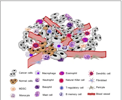
FIGURE 1.2TUMOR MICROENVIRONMENT... 7
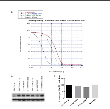
FIGURE 2.1DOWN REGULATION OF MUSTS PROTEIN WITH SIRNA ... 40
FIGURE 2.2GROWTH INHIBITION ASSAY WITH SIRNA ... 41
FIGURE 2.3GROWTH INHIBITION ASSAY WITH PSUPER CONSTITUTIVE VECTOR ... 43
FIGURE 2.4EXAMINING SENSITIVITY OF TRANSDUCED BMDCS EXPRESSING PSUPER VECTORS ... 44
FIGURE 2.5NON-RECOVERY OF MUSTS EXPRESSION IN BMDCS EXPRESSING PSUPER VECTORS ... 45
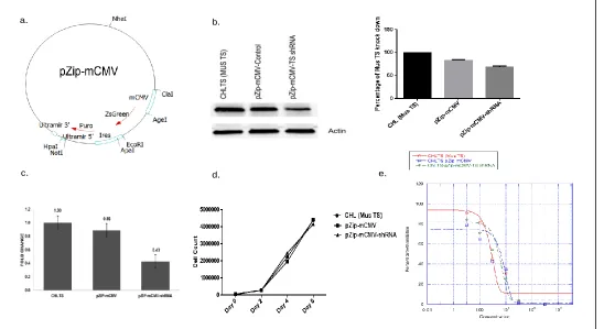
FIGURE 3.1EXAMINING THE ABILITY OF THE MCMVPROMOTER TO CONTROL AN RNAI SEQUENCE ... 66
FIGURE 3.2CONFIRMING INCORPORATION OF PZIP-MCMVLENTI-VIRAL VECTORS IN VIVO... 67
FIGURE 3.3INDUCIBLE CHARACTERISTIC OF THE OPN PROMOTER ... 69
FIGURE 3.4COLONY FORMATION OF MODULATE BMDCS EXPRESSING MSCV-OPN-MCHERRY VECTOR ... 71
FIGURE 3.5CONFIRMING INCORPORATION OF MSCV-OPN-MCHERRY VECTORS IN VIVO... 72
FIGURE 3.6FLOW ANALYSIS OF TRANSDUCED BMDCS EXPRESSING MSCV-OPN-MCHERRY VECTORS ... 74
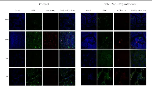
FIGURE 3.7CONFOCAL MICROSCOPY ANALYSIS OF THE OPN PROMOTER ACTIVITY IN VARIOUS TISSUES ... 75
FIGURE 3.9EXAMINING THE ABILITY OF THE OPNPROMOTER TO CONTROL AN
RNAI SEQUENCE ... 78
FIGURE 3.10EXAMINING ZSGREEN EXPRESSION ... 79
FIGURE 3.11APOPTOTIC INDICES OF TRANSDUCE CHLTS(MUSTS) CELLS ... 82
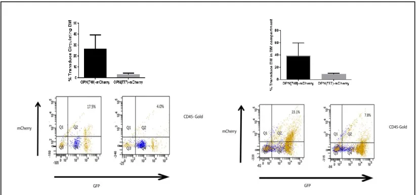
FIGURE 3.12CONFIRMING INCORPORATION OF PZIP-OPNLENTI-VIRAL VECTORS IN VIVO... 84
FIGURE 3.13FLOW ANALYSIS OF TUMOR BMDCS POPULATIONS ... 90
FIGURE 3.14APOPTOTIC AND PROLIFERATION INDICES FOR TUMOR MDSCS ... 91
FIGURE 3.15APOPTOTIC AND PROLIFERATION INDICES FOR TUMOR TREGS CELLS ... 92
FIGURE 3.16APOPTOTIC AND PROLIFERATION INDICES FOR TUMOR MAST CELLS ... 93
FIGURE 3.17APOPTOTIC AND PROLIFERATION INDICES FOR TUMOR TC CELLS ... 94
FIGURE 3.18APOPTOTIC AND PROLIFERATION INDICES FOR TUMOR CD45+ IMMUNE CELLS ... 95
FIGURE 3.19APOPTOSIS INDUCED IN GRANULOCYTES AT 5 DAYS AFTER 5-FU ADMINISTRATION ... 98
FIGURE 3.20APOPTOSIS INDUCED IN MONOCYTES AT 5 DAYS AFTER 5-FU ADMINISTRATION ... 99
FIGURE 3.21APOPTOSIS INDUCED IN MAST CELLS AT 5 DAYS AFTER 5-FU ADMINISTRATION ... 100
FIGURE 3.22THEORETICAL APOPTOTIC INDICES OF MODULATED BMDCS EXPRESSING ZSGREEN ... 101
FIGURE 4.1MAST CELL CHEMOTHERAPY REGIMEN ... 113
FIGURE 4.2TUMOR ANALYSIS FROM APCMin/+MICE SUBJECTED TO THERAPY WITH NO RECOVERY... 115
FIGURE 4.3 INFILTRATION OF MAST CELLS AND GRANULOCYTES IN INTESTINAL TISSUE FROM APCMin/+MICE SUBJECTED TO THERAPY WITH NO RECOVERY... 116
FIGURE 4.5INFILTRATION OF MAST CELLS AND GRANULOCYTES IN INTESTINAL TISSUE
FROM APCMin/+MICE SUBJECTED TO THERAPY WITH RECOVERY ... 119
L
IST OFA
BBREVIATIONS5-FU ... 5-Fluorouracil
APC ... Adenomatous polyposis coli
BMDCs ... Bone marrow derived cells
CAFs ... Cancer associated fibroblasts
CH2THF ... 5, 10- methylenetetrahydrofolate
CHL ... Chinese lung hamster cells
CPA3 ... Carboxypeptidase
CRC ... Colorectal Cancer
DAPI ... 4’, 6-diamididino-2-phenylindole
dTDP ... 2’ deoxythimdine – 5’diphosphate
dTMP ... 2’ deoxythymidine- 5’ monophosphate
dTTP ... 2’ deoxythmidine- 5’ triphosphate
dUMP ... 2’ deoxyuridine- 5’ monophosphate
dUTP ... Deoxyuridine triphosphate
ECM ... Extracellular matrix
EGF ... Epidermal growth factor
eGFP ... Enhanced green fluorescent protein
FAP ... Familiar adenomatous polyposis
FAT ... Folinic acid, aminopterin, thymidine
FBS ... Fetal Bovine Serum
FdUMP ... Fluorodeoxyuridine monophosphate
FGF ... Fibroblast growth factor
HAT ... Hypoxanthine, aminopterin, thymidine
HGF ... Hepatocyte growth factor
HGPRT ... Hypoxanthine-guanine phosphoribosyl transferase
HSCs ... Hematopoietic stem cells
hTS ... Human TS gene
i.p. ... Intraperitoneal
IgE ... Immunoglobulin E
IL-17 ... Interleukin 17
IL-1β ... Interleukin 1 beta
IL-3. ... Interleukin 3
IL-6 ... Interleukin 6
INF-γ ... Interferon gamma
LPS ... Lipopolysaccride
LV ... Leucovorin
MDSCs ... Myeloid derived suppressor cells
MMP9 ... Matrix metallopeptidase-9
MSCs ... Mesenchymal stem cells
MSCV ... Murine stem cell virus
MusTS ... Mus Musculus thymidylate synthase
OPN ... Osteopontin
OPTecTS ... Optimized Escherichia coli TS gene
RISC ... RNA-induced silencing complex
RNAi ... RNA interference
RTX ... Raltitrexed
SCF ... Stem cell factor
shRNA ... Short hairpin RNA
siRNA. ... Short interference RNA
TAMs ... Tumor associated macrophages
Tc ... Cytotoxic T cells
TGF-β ... Tumor growth factor beta
TME ... Tumor microenvironment
TNF-α ... Tumor necrosis factor
TP53 ... Tumor protein p53
Tregs ... T regulatory cells
TS... Thymidylate synthase
VEGF ... Vascular endothelial growth factor
CHAPTER
1
INTRODUCTION
1.1
COLON
CANCER
In 2014, the American Cancer Society estimated that approximately 136,830
individuals were diagnosed with colorectal cancer (CRC) and approximately
50,310 will die from this disease. CRC is the third most diagnosed and the
second cause of cancer related deaths in the United States. CRC begins with the
development of polyps in the colon or large intestines, with low incidents
occurring in the small intestines. Studies have shown that the formation of polyps
normally occur in a period of ten to twenty years, during which a non-cancerous
growth forms on the inner lining of the colon or the large intestine (Stryker, Wolff
et al. 1987; Winawer and Zauber 2002). These non-cancerous adenomatous
polyps are likely to become cancerous upon continuous growth, developing into
an adenocarcinoma, a malignant tumor of glandular cells (Figure 1.1). The
progression from adenomatous polyps to adenocarcinomas is the result of
mutations, which occur in tumor suppressor genes such as the adenomatous
polyposis coli (APC) and tumor protein p53 (TP53). Mutation of the APC gene
occurs in the early stage of CRC development, and is responsible for familial
adenomatous polyposis (FAP), an inherited form of CRC (Jasperson, Tuohy et
al. 2010). The APC gene encodes a multifunctional protein that regulates the
expression of β-catenin in the Wnt signaling pathway, cell adhesion, proliferation,
and apoptotic pathways (Coppede, Lopomo et al. 2014). Typically, the APC
protein forms a cytoplasmic complex with other proteins in the Wnt signaling
pathway. This complex targets β-catenin by ubiquitination for proteasome
degradation keeping its intracellular levels low. Conversely, loss of APC function
causes accumulation of β-catenin in the cytoplasm, and its translocation into the
nucleus where it assists in the activation of genes essential for cell proliferation,
resulting in abnormal growth and adenomatous polyps.
Mutations in TP53 typically occur after the APC mutation, as the polyp
continues to grow and become cancerous. TP53 is a nuclear protein that initiates
cell cycle arrest to initiate repair or apoptosis in response to DNA damage
(Vogelstein, Lane et al. 2000). Loss of TP53 function, which is critical for sensing
checkpoint and proceed with unregulated cell division to produce daughter cells
with mutated DNA. This mutation promotes the transition from an adenomatous
polyp to an adenocarcinoma.
Approximately 96% of colorectal cancers are adenocarcinomas (Stewart,
Wike et al. 2006). Common symptoms include bleeding from the rectum,
discomfort in the lower abdomen, constipation and diarrhea, and loss of appetite.
Fortunately, early screening via a colonoscopy allows visual examination of the
colon so that adenomas or adenocarcinomas can be excised from the colon and
large intestine. Although the advancement of colonoscopies has increased CRC
survival (Edwards, Ward et al. 2010), there is room for improvement to decrease
of CRC related deaths. To date, the 5-year survival rate is approximately 89.9%
for localized CRC, 70.5% when CRC spreads to regional lymph nodes, and
12.9% if CRC has metastasized to distant organs (Surveillance, Epidemiology,
and the End Results (SEER) Program). The majority of CRC related deaths
result from metastatic CRC cells entering into the lymph nodes and blood
vessels, and spreading to distant organs such as the liver and/or lungs. Current
treatments are not efficient in blocking this process. In addition, contributing to
the inadequacies of our anti-cancer treatments is the reoccurrence of the cancer
with more aggressive properties, or the development of resistance against
chemotherapy, such as 5-Fluorouracil (5-FU) that is commonly used to treat
CRC. Thus, in order to combat and eliminate this disease, it is important to
combination with novel therapies to enhance their efficacy while minimizing their
toxicities.
1.2
HALLMARKS
OF
CANCER
In order develop effective anti-cancer therapies and to resolve the
problematic side effects resulting from their lack of specificity for cancer cells, it is
important to understand the complexity of cancer development. Currently, eight
properties define the hallmark of cancer cells. These properties provide a
framework for the development of novel therapies. These hallmark properties
include: sustaining proliferative signaling, evading growth suppressors, resisting
cell death, enabling replicative immortality, inducing angiogenesis, activating
invasion and metastasis, reprogramming of energy metabolism, and evading
immune destruction (Hanahan and Weinberg 2011). The unregulated production
of growth signals provides cancer cells the ability to acquire the hallmark of
sustained proliferative signaling. This causes an imbalance in homeostasis,
wherein cancer cells uncontrollably produce growth factors for themselves.
However, it is not sufficient for cancer cells sustain proliferation because cellular
checkpoints within cell division can inhibit the continuous growth necessary to
develop a tumor mass. Thus, obtaining the ability to evade the regulation of
growth suppressors, through mechanisms such as TP53 is necessary for tumor
progression. Growth suppressors negatively regulate cell proliferation contingent
nucleotide, oxygen, and energy levels within or outside the cell. Mutations in
growth suppressors disrupt induced apoptosis.
Alternatively, the cancer cell’s quest for survival may also increase the
expression of anti-apoptotic regulators that contribute to resistance to apoptosis.
Under normal conditions, the balance of pro- and anti- apoptotic proteins
regulates programmed cell death. The abnormalities in cancer cells creates
imbalance in favor of inhibiting the trigger of death. Resisting cell death does not
guarantee cancer cells the ability for unlimited proliferation, but obtaining the
hallmark of immortality does. Immortality is achieved by up regulating the
expression of telomerase, an enzyme responsible for elongating telomeres,
resulting in extended protection of chromosomal DNA. With each cell division,
the telomere length decreases, until it reaches a point at which the cell goes into
a silencing phrase or program cell death. Hence, immortality ensures the tumors
capability to expand. However, as the tumor expands it must ensure the ability to
sustain a healthy environment to maintain its growth. Tumor progression requires
sources of energy, the import of nutrients, and the export of waste, which is
accomplished by increasing energy metabolism and inducing angiogenesis. The
newly discovered hallmark of reprogramming energy metabolism allows cancer
cells to survive by up regulating the transport of glucose into the cell where it is
broken down to generate energy and metabolites to compensate for the increase
in cell growth and division. Angiogenesis is the process of developing new blood
vessels from existing vessels that allow the import and export of the necessary
tumor in a localized region; however, the ability to travel to another organ
requires the activation of invasion and metastatic characteristics. Normally
aggressive cancers alter the expression of cell-to-cell adhesion molecules; giving
cancer cells the capacity to invade nearby blood vessels in an attempt to spread
to a distal organ. The accomplishment of metastasizing to a distal organ is
contingent on the cancer cell’s ability to detached from the primary tumor, survive
in circulation, extravagate and adapt to the microenvironment of the distal organ
to survive. Finally, the property of evading immune surveillance is a significant
hallmark that allows cancer cells to avoid destruction by the immune system,
which served to eliminate rogue cells. Delineation of these hallmark properties
has provided clarity to cancer development and its capability to resist therapies.
Cancer research has evolved from the perspective of exclusively targeting and
understanding the development of neoplastic cells, to the forefront of studying
the intricate relationship that develops between both neoplastic and
non-neoplastic cells derived from bone marrow to create the tumor microenvironment
(TME).
1.3
TUMOR
MICROENVIROMENT
Since the recognition of the TME, a paradigm shift in cancer therapy has
occurred, such that the tumor is perceived to be an organ or complex tissue
(Egeblad, Nakasone et al. 2010), composed of a heterogeneous populations of
both malignant and non-malignant cells and extracellular matrix, which function
hallmarks of cancer (Coussens and Werb 2002; Chantrain, Feron et al. 2008;
Whiteside 2008; Hanna, Quick et al. 2009; Peddareddigari, Wang et al. 2010;
Hanahan and Coussens 2012). This heterogeneous population of non-malignant
cells within a tumor includes, but is not limited to the following cells: tumor
associated macrophages (TAMs); myeloid derived suppressor cells (MDSCs),
mast cells, and cancer associate fibroblasts (CAFs), monocytes, endothelial
cells, and neutrophils (Figure 1.2).The TME has the potential to activate TAMs
into two subgroups: M1 (anti-tumor) or M2 (pro-tumor) macrophages (Gordon
2003; Mantovani, Sica et al. 2007). MDSCs have been shown to cooperate in the
non-responsiveness of T-cells to promote tumorigenesis (Nagaraj and
Gabrilovich 2008). Mast cells exhibit the ability to secrete many pro-tumorigenic
growth stimulatory factors, which can trigger the angiogenic switch to turn on
angiogenesis (Coussens, Raymond et al. 1999). In response to tumor growth,
CAFs are secrete a host of growth factors such as the epidermal growth factor
(EGF), transforming growth factor beta (TGF-β), and hepatocyte growth factor
(HGF) (Kalluri and Zeisberg 2006). These non-cancerous cells provide a
pro-inflammatory environment by secreting cytokines, chemokines, and growth
factors essential for the initiation and maintenance of tumor growth. In addition to
the infiltration of these heterogeneous population of cells within the tumor, the
composition of the extracellular matrix, for example, laminin, collagen,
fibronectin, and proteoglycans can promote tumor progression (Peddareddigari,
Wang et al. 2010) and provide resistance to drug therapies due to abnormal
tissue organization that provides a hindrance to drug delivery (Egeblad,
Nakasone et al. 2010). For example, type 1collagen plays a significant role in
regulating tumor sensitivity to various anti-neoplastic therapies (Loeffler, Kruger
et al. 2006).
These examples represent minuscule information on the intricate
relationship between cancer and non-cancerous cells. These studies confirm that
the neoplastic cells are not solely acting alone, but they collaborate and
co-evolve with various immune cells to orchestrate tumor growth and development.
morphological changes to occur within the TME to become supportive. Hence
advanced stages of cancer exhibit altered tumorigenic properties in comparison
to the early stages of cancer. The roles of these immune cells need intense
investigation, to have a better understanding of the supportive role of the TME at
the different stages of tumor development. In addition, this will contribute to the
knowledge of how the TME provides protection against chemotherapies,
providing insight into the development of more effective ways to target the tumor.
1.4
THE
ROLE
OF
MAST
CELLS
IN
TUMOR
BIOLOGY
As previously described, the TME consists of bone marrow derived cells
that contribute to tumor formation and potentially play a vital role in resistance to
current chemotherapies. In particular, there is a growing interest in the role of
mast cells in cancer development, and recent studies have identified functions of
these cells in tumor biology. Historically, mast cells are typically associated with
immunoglobulin E (IgE)-mediated response to an allergic reaction or an
asthmatic incident. The IgE mediated response causes the activation of mast cell
degranulation, which results in the crosslinking of antigens and IgE molecules
that are bound to the high affinity receptors, FC epsilon RI (FcεRI) located on the
cell surface of mast cells (Gould, Sutton et al. 2003). This immune response
provokes inflammation resulting in recruitment and activation of additional
immune cells to the local site. Thus, mast cells are effectors cells that exert
immune signals to regulate and recruit other immune cells. These granulocytes
differences between the subgroups is dependent the content of granules, which
occurs during maturation. Mast cell precursors are released from the bone
marrow as immature cells and migrate into various tissues within the body to
undergo maturation and differentiation. Maturation and differentiation influenced
by the infiltrated tissue environment resulting in a heterogeneous population of
mast cells throughout the body. Mast cell granules have three subgroups:
pre-formed granules, de novo synthesized granules, and cytokines and chemokines.
The pre-formed granules include histamine, heparin, and the proteases tryptase,
chymase, and carboxypeptidase A3 (CPA3). The second group is the de novo
synthesized lipid mediators, which includes: LTC4 and PGD2, and the third
category consists of the various cytokines and chemokines, such as but not
limited to tumor necrosis factor alpha (TNF-α), vascular endothelial growth factor
(VEGF), fibroblast growth factor (FGF), and matrix metallopeptidase-9 (MMP9)
(Theoharides, Kempuraj et al. 2007). Upon degranulation, the pre-formed
granule mediators are immediately release within minutes; while the de novo
synthesized mediators are released slowly within a period of three to twelve
hours later. In addition, the mechanism for releasing the mediators can occur by
either complete or selective degranulation (Dvorak and Kissell 1991;
Theoharides and Kalogeromitros 2006). The role of mast cells in tumor formation
and immunity is poorly understood, and accumulating evidence has shown
controversial roles in tumor biology. Mast cells can either display anti-tumor or
development (Groot Kormelink, Abudukelimu et al. 2009; Ribatti 2013; Oldford
and Marshall 2014).
Mast cells have been shown to mediate anti-tumor response to cancer in
in vivo model systems. In ApcMin/+mice, a model of colon carcinogenesis, genetic
ablation of mast cells resulted in larger and increased number of intestinal tumors
compared to wild type littermates, suggesting that mast cells can suppress
intestinal tumor development (Sinnamon, Carter et al. 2008). In addition, several
studies suggest that IgE mediated activation of mast cells within tumor results in
an anti-tumor immune response (Jensen-Jarolim, Achatz et al. 2008; Teo, Utz et
al. 2012; Singer and Jensen-Jarolim 2014). Mast cells can also regulate the
activity of immune cells. For example, mast cells can promote recruitment of
MDSCs into the tumor and activate their expression of IL-17, which in turn
mobilizes and suppresses the pro-tumorigenic activity of T-regulatory cells within
the tumor. Collectively, these studies suggest that mast cells induce anti-tumor
responses and play a role in promoting inflammation to recruit additional
leukocytes into the tumor region where they activate or regulate immune cell
activity and induce cytotoxic activity.
Alternatively, mast cells can display a pro-tumorigenic response. Studies
have shown that pro-inflammatory mediators from mast cells can promote tumor
proliferation and provide a suitable environment to sustain tumor growth and
development. Gounaris and colleagues showed that mast cells for polyp
development in ApcMin/+mice (Gounaris, Erdman et al. 2007). In addition poor
cancers such as melanoma, lung, gastric, colorectal, and prostate carcinomas
(Groot Kormelink, Abudukelimu et al. 2009). An in vitro study utilizing the human
mast cell line, LAD2 demonstrated the capacity of mast cells to regulate the
invasiveness of cervical cancer cells, yet cervical cancer cells were able to
stimulate the degranulation of mast cells, suggesting an intimate communication
between tumor and inflammatory immune cells (Rudolph, Boza et al. 2008).
Another study using a HPV16 transgenic mouse model of squamous cell
epithelial carcinoma showed that chymase released from tumor infiltrating mast
cells activated MMP9 resulting in extracellular remodeling (Coussens, Raymond
et al. 1999). This study also demonstrated that mast cells infiltrated tumors prior
to the angiogenic switch. To validate the role of mast cells in angiogenesis and
tumor remodeling, another study using a Myc-induced pancreatic islet tumor
model showed infiltration of mast cells within twenty-four hours of Myc activation.
The recruitment of mast cells resulted in the expansion of Myc-induced tumors
(Soucek, Lawlor et al. 2007). Together, these studies show that mast cells
perform pro-tumorigenic roles by contributing to tumor remodeling, angiogenesis,
and immune suppression.
The pro-tumorigenic roles of mast cells affect the efficacy of anti-cancer
therapies by contributing to tumor progression and assisting in tumor remodeling
and tumor sustainability. Currently, the efficacy of anti-cancer therapies is
dependent on its direct deliverable contact with cancer cells within the tumor
where its cytotoxic action can destroy or reduce tumor burden. However, tumor
important to decipher the pro-tumorigenic roles of mast cells to develop
strategies to enhance the efficacy of anti-cancer therapies.
1.5 THYMIDYLATE
SYNTHASE
AND
ITS
INHIBITOR
5-FU
Thymidylate synthase (TS) is an enzyme that catalyzes the reductive
methylation of 2’ deoxyuridine- 5’ monophosphate (dUMP) to 2’ deoxythymidine-
5’-monophosphate (dTMP) and dihydrofolate using
5,10-methylenetetrahydro-folate (CH2THF) as a methyl donor (Danenberg 1977; Carreras and Santi 1995).
Subsequently, dTMP is sequentially phosphorylated into 2’-deoxythimdine–
5’diphosphate (dTDP), and then into 2’ deoxythmidine- 5’ triphosphate (dTTP),
an essential component for DNA replication and repair in actively proliferating
cells. Importantly, this pathway provides the sole intracellular source of dTTP,
thus, TS has been an important target of chemotherapeutic agents. In addition to
its catalytic function, TS is an auto-regulatory protein that binds to its mRNA to
repress its translation and has been shown to interact with the mRNAs of TP53
and c-myc (Chu and Allegra 1996). TS inhibition results in depletion of dTMP and
accumulation of dUMP leading to increased levels of deoxyuridine triphosphate
(dUTP) that is mis-incorporated into DNA, resulting in DNA fragmentation and
ultimately cell apoptosis (Longley, Harkin et al. 2003; Garg, Henrich et al. 2010).
TS inhibitors are typically substrate analogs or analogs of the folate co-factor.
The most widely used TS inhibitor is the 5-FU, a fluoropyrimidine that has been
widely used in the clinical management of colon and head and neck cancers.
5-FU is structurally similar to uracil; however, a fluorine atom replaces a hydrogen
it is converted into the powerful TS inhibitor fluorodeoxyuridine monophosphate
(FdUMP). FdUMP has the same affinity to TS as dUMP: however, when bound to
TS, the fluorine atom at the 5’ position cannot be displaced, and it forms a stable
ternary complex with TS and 5,10-CH2THF resulting in TS inhibition. TS inhibitors
based on folate analogs include tomudex (raltitrexed), liposomal GW1843U89,
and capecitabine (Rose, Farrell et al. 2002).
Expression of TS after exposure to anti- therapy, such as 5-FU has shown to
increase proteins levels, a consequence of inhibiting TS activity, thus allowing
translation of the mRNA and production of the protein(Van der Wilt, Pinedo et al.
1992).
Although 5-FU has been used for many decades in the clinical management of
cancers, its use has been limited by drug induced toxicities and acquired
resistance by cancer cells. Cancer resistance to TS inhibitors can occur through
a number of mechanisms. TS can be over expressed in tumors due to gene
amplification or mutations that may cause resistance. TS auto-regulates its gene
expression, by binding to its mRNA. However, upon treatment with TS inhibitors,
bounded TS is released from its mRNA allowing its translation, which in turn
increases intracellular TS protein levels (Chu and Allegra 1996). The
effectiveness of 5-FU depends on the availability of 5,10-CH2THF. The active
metabolite, FdUMP forms an unstable binary complex with the enzyme, resulting
in poor inhibition. However, in the presence of 5,10-CH2THF stabilizes the
ternary complex inhibiting the catalytic function of the enzyme (Aherne,
resistant to 5-FU, however, addition of the drug Leucovorin (LV), increases
intracellular levels of 5,10-CH2THF resulting in enhanced sensitivity (Matherly,
Czajkowski et al. 1990). Another mechanism of resistance is the salvage
pathway which allows for the recovery of free bases, such as thymidine that are
byproducts of degrading DNA, to be recycled for nucleotide biosynthesis through
the action of the enzyme, thymidine kinase (Kinsella, Smith et al. 1997). The
salvage pathway can compensate for the lack of thymidylate production resulting
from TS inhibition. Acquired resistance cancan also result from mutations in
TP53 wherein damaged DNA resulting from mis-incorporation of FdUTP is
unrecognized allowing cells to escape apoptosis (Ahnen, Feigl et al. 1998). On
the other hand, cancer cells with wild type TP53 can still be resistant to 5-FU
therapy by altering the regulation of the cell cycle. In the presence of 5-FU
resistant cells had increased tendency to arrest in G1 and G2 phases, allowing
DNA repair prior to replication; unlike the parental cell line that had a tendency to
arrest in the S phase (De Angelis, Svendsrud et al. 2006). In addition to acquired
resistance, 5-FU therapy is limited by drug induced toxicities associated with its
lack of specificity, targeting both actively dividing cancer as well as healthy
normal cells. Some side effects from 5-FU include alopecia, myelosuppression,
and mucositis. Considerable attempts have been made to enhance the
effectiveness of 5-FU, while lowering its toxic side effects. These include the
combination of 5-FU chemotherapy with targeted therapies. The impact of 5-FU
on stromal cells is only beginning to be explored. Targeting both the tumor and
We hypothesize that stromal cells are also direct targets of 5-FU and that their
chemo-sensitivity to 5-FU might determine tumor sensitivity to the therapy.
Targeting the microenvironment is genetic modification or pharmacological
inhibition of pro-tumorigenic activities of bone marrow derived cells infiltrating the
tumor microenvironment, might be viable strategies to enhance anti-cancer the
anticancer efficacy of 5-FU.
1.6
PARADIGM
SHIFT
IN
ANTI-CANCER
THERAPIES
Targeting both the tumor as well as the tumor stroma is a paradigm shift in
our approach to enhance the efficacy of anticancer therapies while enhancing
selectivity and minimizing toxicity and relapse. Clinical trials targeting the
extracellular matrix (ECM), endothelial cells and pericytes, fibroblasts, and innate
immune cells have shown progress in combating cancer (Joyce 2005), however,
these strategies are only beginning to be developed. The recognition of tumors
as a complex collection of cancer cells interacting and recruiting normal
non-cancerous cells has altered our perception of cancer development, which has
also alter how we must combat this disease. Traditionally, anti-cancer therapies
only targeted actively proliferating cancer cell by surgical removal of the tumor
mass that may be combined with radiation or chemotherapy, or a combination of
both. These treatments have only been moderately effective. Since the
establishment of the critical role of the tumor microenvironment in cancer
initiation and progression, new anti-cancer therapies have emerged (Hanna,
existing chemotherapeutic drugs such as 5-FU. Furthermore, targeting stromal
cells might provide an opportunity to enhance the specificity of cancer
chemotherapies. Thus, combining genetic modulation or pharmacologically
inhibition of bone marrow derived cells (BMDCs) within the TME with 5-FU
therapy may be beneficial in reducing drug toxicity or the occurrence of relapse.
1.7 GENETIC MODULATION OF BONE MARROW DERIVED
CELLS
The plasticity of BMDCs has the potential to constitute a pro-tumorigenic
microenvironment for cancer cells. A review by Chantrain proposes three
mechanisms by which bone marrow cells support the initiation and progression of
cancer: first, through the recruitment of cancer cells to enrich the
microenvironment, second, by infiltration into the tumor stoma to suppress
immune response, and third, by promoting the development of the pre-metastatic
niche (Chantrain, Feron et al. 2008). Numerous studies have demonstrated an
array of pro-tumorigenic responses from BMDCs infiltrating the into primary
tumor microenvironment permitting tumor growth (Egeblad, Ewald et al. 2008;
Roorda, ter Elst et al. 2009; Fowler, Mundy et al. 2012; Kidd, Spaeth et al. 2012;
Srivastava, Andersson et al. 2012; Zou, Zheng et al. 2012). Tumors have
suppressed immune responses to recruit and activate MDSCs, which have been
associated with malignancy and poor prognosis (Srivastava, Andersson et al.
the tumor tissue, hampering the administration and reducing the effects of
anticancer drugs (Mizukami, Sasajima et al. 2012). The ability of BMDC to
circumvent immune response and reduce efficacy of anti-neoplastic drugs is the
subject of ongoing investigation. It is important to understand the role of BMDCs
in tumor stoma to determine if genetic modulation of these cells can enhance
efficacy of antitumor drugs. Transplantation of genetically modificed BMDCs may
be used to alter gene expression in stromal cells to enhance tumor response to
pre-existing therapies. For example, the knockdown of the Wnt signaling inhibitor
Dickkopf1(DKK1) in bone marrow derive mesenchymal stem cells (MSCs)
repressed myeloma development in mice (Fowler, Mundy et al. 2012), and
MSCs engineered to express lymphotoxin induced tumor regression (Zou, Zheng
et al. 2012). Genetic modification of BMDCs might be an effective tool to
enhance the efficacy of anti-neoplastic therapies since they can influence tumor
resonse to therapy by their ability to suppress immune responses.
1.8 PHARMACOLOGICAL INHIBITION OF BMDCs
An alternative to targeting the TME is by inhibiting the pro-tumorigenic
activities of BMDCs, such as mast cells, pharmacologically. Mast cells have been
shown to play critical roles in tumor initiation, progression, and response to
therapy. Thus, effective anti-cancer therapies may need to target mast cells in
addition to cancer cells. Several studies have used the pharmacological
stabilizer, sodium cromoglicate (Cromolyn), a drug known to inhibit mast cell
degranulation. Initially Cromolyn was used to treat acute inflammation which
shown a reduction in tumor burden mice after administration of Cromolyn
(Gounaris, Erdman et al. 2007; Soucek, Lawlor et al. 2007). Based on these
results, we have considered the possibility that Cromolyn might play a synergistic
role in anti-cancer therapy. However, the mechanism by which Cromolyn inhibits
mast cells is not fully understood. It is thought to stiffen the cellular membrane,
which indirectly inhibits the influx of chloride into the cell that in turn inhibits the
ability of the cell ability to trigger.
1.9 EXPERIMENTAL MODELS
A. In-vitro cell model
Mus musculus thymidylate synthase (MusTS) was stably transfected into
a TS-deficient Chinese hamster lung (CHL) cell line RJK88.13 (Nussbaum,
Walmsley et al. 1985). Stable transfectants were selected in media lacking
thymidine in the presence of the nucleoside transport inhibitor dipyridamole.
Stably transfected cells were grown in mass culture. The CHL(MusTS) cell line
was used to target TS specifically to assess the sensitizing effects of diminished
TS expression in response to 5-FU therapy.
B. Transgenic mouse model
The C57BL/6J-ApcMin/+ mouse has been used as an in vivo model for the
initiation and progression of intestinal adenomas. These mice are heterozygous
for a mutant allele of the APC tumor suppressor gene, which genetically
intestinal tract (Moser, Pitot et al. 1990). Tumors develop in these mice at an
early stage due to the loss of the remaining wild-type APC allele, which leads to
insufficient expression of APC (Powell, Zilz et al. 1992; Su, Kinzler et al. 1992).
The APC mutation is similar to that the germline mutation found in the human
hereditary form of colorectal cancer, familial adenomatous polyposis (FAP)
(Levy, Smith et al. 1994). Although FAP accounts for less than 2% of colorectal
cancer incidence, mutations in the APC gene have accounted for 80% of
sporadic colon tumors (Kinzler and Vogelstein 1996). Most of the adenomas that
develop in these mice are located in the small intestines with a few developing in
the colon, while in humans tumors develop exclusively in the colon.
Nevertheless, the ApcMin/+mouse model is useful for understanding the initiation
of tumor development and progression, and is a useful tool for developing or
examining the effects of therapeutic agents in vivo.
C. Syngeneic mouse model
The transplantable syngeneic CT26 tumor model in Balb/c mice is another
model to examine the role of microenvironment in tumor response to 5-FU
therapy. CT26 colon adenocarcinoma cells are injected into the flank of Balb/c
mice where tumors are allowed to develop prior to therapy. CT26 cells were
generated in Balb/c by treatment with the carcinogen
N-nitroso-N-methylurethane-(NNMU)where they were isolated and to grow in vitro (Brattain, Strobel-Stevens
et al. 1980). The tumorigenic property of this cell line was sustained when
rates. By using this model, it allows for rapid tumor growth, which can be excised
to quantify BMDC infiltration and expression of mRNA levels within the tumor.
Thus, this syngeneic mouse model is practical for studying the response of
BMDCs after exposure to different combinations of chemotherapeutic agents.
1.10 REVIEW OF LITERATURE (GENETIC MODULATION OF
BMDCs)
A. BMDCs infiltrate the intestinal tumor microenvironment in
Apc
Min/+mice.
In previous studies, Davis et al. showed that bone marrow can be
successfully transplanted and engrafted into ApcMin/+ mice. Prior to
transplantation, the recipient ApcMin/+ mice were lethally irradiated with 950 rads
to deplete existing hematopoietic system. Age matched donor marrow from
C57BL/6-UB1-eGFP mice expressing the enhanced green fluorescent protein
(eGFP) under the control of the human ubiquitin promoter was extracted from the
femur and tibia. The bone marrow is directly transplanted into recipient mice, or
grown in culture and fractionated to obtain hematopoietic stem cells (HSCs).
Transplantation of whole marrow or purified HSCs was performed by tail vein
injection. Full engraftment occurs within four to six weeks after transplantation.
Tissue sections were collected at eight weeks post transplantation to examine
the engraftment of donor eGFP bone marrow by confocal microscopy. The
results showed that donor derived eGFP marrow infiltrated both normal and
marrow was greater in tumor regions (75-80%) within the small intestines and
colon as compared to normal, non-tumor regions (10%) (Davis, Price et al.
2011). These results suggest that genetically modified bone marrow derived cells
transplanted into ApcMin/+ mice will successfully infiltrate the tumor regions of the
intestines and colon.
B. BMDCs expressing E. coli TS exhibits resistance to TS inhibitors
. Previous studies have demonstrated that the an Escherichia coli TS genewhose codon usage has been optimized for expression in mammalian cells
(OPTecTS) can confer high levels of resistance to TS inhibitors (Fantz, Shaw et
al. 2000; Shaw, Berger et al. 2001). Shaw et al. optimized the E. coli TS gene by
introducing mutations in the cDNA resulting in stabilization of the protein in
mammalian cells. OPTecTS was cloned into a Harvey murine sarcoma-based
retroviral vector and stably transfected into a TS-deficient CHL cell line
(Nussbaum, Walmsley et al. 1985); wild type ecTS (wtecTS) and humanTS (hTS)
were used as controls. Stably transfected cells expressing OPTecTS and the
controls were treated with various concentrations of the TS inhibitor, raltitrexed
(RTX) in the presence of dipyridamole to determine the concentration that
inhibited the growth of fifty percent of the treated cells (IC50). The results showed
that cells transfected with OPTecTs were 11-fold more resistant to RTX than
cells expressing hTS while cells expressing wtecTS were slightly more resistant
compared to cells expressing hTS. In addition, result showed greater than 75%
resistant to the TS inhibitors RTX and U89 respectively, at concentrations where
100% of non-transduced cells were inhibited.
C. The effects of chemo-resistant marrow on tumor response to TS
Inhibitors.
In previous studies
,
chemo-resistant marrow expressing the OPTecTSwas transplanted into ApcMin/+ mice. Retroviral vectors based on the murine stem
cell virus (MSCV) promoter were constructed to express OPTecTS, while the
MSCV empty vector was used as controls. Lethally irradiated ApcMin/+mice were
reconstituted with chemo-resistant (OPTecTS) or control BMDCs, and and then
subjected to systemic treatment of a combination therapy of 5-FU and RTX or
PBS as control treatment. After the treated mice were sacrificed, intestinal
tissues were isolated and the tumor burden was determined. The results showed
that in mice transplanted with mock transduced marrow, systemic treatment with
5-FU caused a statistically significant decrease in tumor burden by approximately
75% from 32±19 (PBS) to 8±5 (5-FU/RTX) (p=0.008). In contrast, in mice
transplanted with drug resistant marrow expressing OPTecTS, tumors were not
as responsive to the therapy exhibiting a statistically insignificant reduction in
tumor burden from 35±22 (PBS) to 19±23 (5-FU/RTX) (p=0.183) (Pena et al,
unpublished data). These results showed that genetically modified
chemo-resistant bone marrow does influence tumor response to chemotherapy. These
preliminary data provides the basis for our working hypothesis that (1) tumor
BMDCs and (2) it is possible that BMDCs can be genetically modified to chemo-
sensitize both BMDCs and tumors to chemotherapy.
1.11 REVIEW OF LITERATURE (INHIBITION OF BMDCs)
A. Effect of systemic 5-FU therapy on tumor burden in
Apc
Min/+mice
Tucker et. al. subjected ApcMin/+ mice to systemic treatment consisting of
three cycles of 40 mg/kg 5-FU administered intraperitoneal (i.p.) daily for five
consecutive days (Tucker, Davis et al. 2002). Mice were either immediately
sacrificed after the 5-FU regimen, or allowed to recover for 6-weeks post
therapy. At the end of the treatment regimens, intestinal tissue was removed
and tumor burden was assessed. The results showed that 5-FU reduced tumor
numbers by approximately 50% (p<0.001) compared to age matched control
groups treated with PBS. In addition, weekly blood samples were collected to
assess the effect of 5-FU on leukocytes. The results showed a reduction in
leukocyte counts after exposure to 5-FU, and a return to normal or higher levels
upon cessation of 5-FU therapy. When mice were allowed to recover for six
weeks post therapy, there was no significant difference in tumor numbers
between mice treated with PBS or 5-FU. This suggested that 5-FU therapy was
only cytostatic to the tumor cells and after removal of therapy; the tumors
relapsed to the same level as that in untreated mice. This study showed that
5-FU can inhibit tumor growth, but it also showed that it can affect the leukocyte
therapy, particularly in the tumor stroma may contribute to the growth of tumors
post therapy. Thus, it is important to identify and further investigate the full
impact of 5-FU not just on leukocytes but also on other BMDCs in the
microenvironment such as mast cells, to determine their effect on tumor
response to therapy and recurrence post therapy.
B. Examining the effects of 5-FU on mast cells and precursors
A previous study from our lab examines the effects of 5-FU on subsets of
BMDCs, including mast cells, T regulatory (Treg) cells, and MDSCs in the tumor
stroma in the ApcMin/+ mouse (Acharya, G., et.al, unpublished data). Briefly, 14-15
week old mice were injected i.p. with a single dose of 50 mg/kg 5-FU. The mice
sacrificed at 3,5,7,9 and 12 days post therapy. Tumors, spleen, and bone
marrow were collected, digested to create a single cell suspension and the
infiltrating BMDCs were quantified by flow cytometry. The results showed that
MDSCs were selectively sensitive to 5-FU while mast cells were resistant and
migrated to the tumor stroma in response to 5-FU while the Tregs were only
mildly sensitive to 5-FU. Analysis of apoptotic and proliferative indices of mast
cells and MDSCs showed that in mast cells, these indices were unaffected by
treatment with 5-FU. At 5 days post therapy, the number of mast cells infiltrating
the tumor stroma increased; this was accompanied by a corresponding decrease
in mast cell population in the spleen, suggesting that mast cells might migrate
from the spleen to the tumor stroma in response to 5-FU and molecular signals
most likely rendered them vulnerable to 5-FU. The number of MDSCs decreases
by three-fold within 5 days of 5-FU administration in tumor stroma, spleen, and
bone marrow. However, they returned to pre-treatment levels within 10 days.
In another study, the effect of 5-FU on mast cell precursors was
determined in 8-week old C57BL/6 mice (Ophir, Berenshtein et al. 1993). Mice
were injected intravenously once with 0–150 mg/kg 5-FU. BMDCs were collected
at 2 and 4 days after injection and cultured in enriched media for 14 days to
stimulate the proliferation of mast cell precursors. Toluidine and alcian blue
staining identified mast cells. In response to 50 mg/kg the sub lethal dose of 150
mg/kg 5-FU, BMDCs retrieved on day 2 after 5-FU injection yielded 12.2±0.9%
and 2.6±0.3% mast cell precursors, while day 4 yielded greater than 80% and
60% mast cell precursors, respectively. Normal levels of mast cells were
observed by day 8 for the sub lethal dose. Data from the sub lethal dose
indicated that mast precursors were continuously activated or cycling after 5-FU
injection. Thus, there was gradual replacement, instead of recovery of mast cells
in the bone marrow compartment. These preliminary data collectively provides
evidence which support our working hypothesis that (1) tumor response to
chemotherapy may be influenced by the sensitivity of tumor associated mast
cells, and may interfere with tumor development, (2) effect of 5-FU on mast cells
may play a synergistic role to enhance its anti-tumor efficacy, or (3) inhibition of
1.12
RESEARCH GOAL
The preliminary data showed that tumor response to TS inhibitors was
influenced by the sensitivity of tumor associated BMDCs to the therapy. Genetic
modification of BMDCs to make them resistant to TS inhibitors also made tumors
resistant to the therapy. The goal of this project is to develop a therapeutic
strategy to target both cancer cells and non-cancer tumor stromal cells to
enhance the efficacy of TS inhibitors. I will examine the ability of reducing
intracellular TS levels by utilizing small RNA interference technologies against TS
to enhance sensitivity to TS inhibitors. Genetically modified BMDCs that are
sensitized to TS inhibitors are predicted to enhance the cytotoxic effects of 5-FU
within the tumor upon infiltrating the TME while protecting normal non-cancerous
cells from drug-induced toxicity.
In addition, I will test the hypothesis that pharmacological inhibition of
mast cell activity will also inhibit their pro-tumorigenic role within the TME will act
synergistically with 5-FU as well as prolong tumor relapse post therapy. My
overall goal is to target the TME to enhance the cytotoxicity of 5-FU to cancer
cells and to reduce tumor recurrence post therapy.
1.13
RESEARCH INNOVATION
The realization that tumor stromal cells have profound influence on tumor
behavior and tumor response to anti-neoplastic therapies is leading to a
paradigm shift in cancer therapy. These findings suggest that effective therapies
possibility that altering the sensitivity of the cells in the microenvironment will also
alter tumor response to therapy. I will use the Osteopontin (OPN) promoter that
is primarily upregulated and highly activated within tumor stromal cells to drive
the expression of a TS shRNA sequence to sensitize the infiltrating BMDCs and
the tumor prior to chemotherapy. The use of a promoter that is only activated in
the tumor stroma, will not only enhance tumor sensitivity to the therapy but will
also specifically direct tumor sensitivity to tumor cells while reducing toxicity to
normal cells. This strategy to enhance the efficacy and the specificity of
anticancer therapies can be applied not just too intestinal tumors but to other
classes of therapeutic agents in different types of tumors.
In addition, to modifying BMDCs I will use a drug known to specifically inhibit
mast cell degranulation during and post post 5-FU therapy to determine if mast
cells inhibition can synergize with 5-FU anticancer efficacy or prolong tumor
recurrence of tumor after chemotherapy. The novelty of inhibiting mast cells is
the utilization of the drug Cromolyn that has been used in treating asthma and is
readily available on the market. Even though this drug has displayed success in
reduction of various tumors, the combination of Cromolyn and 5-FU needs further
CHAPTER 2
D
EVELOPINGS
TRATEGIES TOI
NCREASEC
HEMO-S
ENSITIVITY OFC
ELLS TOT
HYMIDYLATES
YNTHASEI
NHIBITORS1_______________________________
1 Developing Strategies to Increase Chemo-sensitivity of Cells to Thymidylate
2.1 ABSTRACT:
Thymidylate synthase (TS) is an essential enzyme necessary for DNA
replication and repair; it is responsible for the synthesis of thymidine. Typically,
cancer cells exponentially proliferate at higher rates, resulting in elevated TS
activity. Thus, using TS inhibitors has been promising in treating cancer.
Although, like any drug TS inhibitors do exhibit problematic pitfalls that include
harming normal non-cancerous cells, the occurrence of cancer resistance and
relapse to therapy. Attempting to circumvent these limitations, we used RNAi
vectors to suppress cellular levels of TS. Reduction of TS expression did
enhance sensitive to therapy, however the toxicity of the RNAi vector made it
impossible to examine the effects in vivo.
2.2 INTRODUCTION:
Thymidylate synthase (TS) is an enzyme that catalyzes the reductive
methylation of dUMP to produce dTMP and dihydrofolate (Danenberg 1977;
Carreras and Santi 1995). Subsequent, phosphorylation converts dTMP to dTTP,
an essential precursor for DNA synthesis. This catalytic activity provides the sole
intracellular de novo source of dTMP, thus TS is essential for actively dividing
cells. Due to this, TS is an optimal target for anti-cancer therapies(Wilson,
Danenberg et al. 2014).Examples of TS inhibitors are 5-fluorodeoxyuridnine
The most commonly TS inhibit is 5-FU which has been used in the clinical
management of many cancers. Inhibition occurs after 5-FU is transported into
cells and converted into several metabolites. One metabolite is FdUMP, a
powerful TS inhibitor. It disrupts DNA synthesis by forming a covalent ternary
complex with TS and the substrate 5,10-methylenetetrahydrofolate. Inhibition
results in the accumulation of dUMP, which is subsequently phosphorylated into
dUTP. The incorporation of dUTP into DNA, triggers DNA fragmentation and
ultimately cell death. Although, 5-FU is clinically used to treat cancer there are
some drawbacks to this therapy, such as acquired resistance and the lack of
specificity. Acquired resistance requires increase doses of 5-FU needed to kill
cancer cells; however, the dose administered is limited by its lack of specificity
resulting in toxicity to normal cells.
In addition, to its catalytic function, TS negatively auto-regulates its
intracellular protein level, by binding to its mRNA to block translation. To mitigate
the effects of 5-FU, TS protein bound to its mRNA are release to permit
translation leading to induction of TS protein, exhaustion of 5-FU, lowering cell
response and development of resistance to 5-FU. (Chu and Allegra 1996).
Another, disadvantage of 5-FU therapy is its lack of specificity, targeting both
actively dividing cancer and healthy non-cancerous cells. This leads to side
effects such as alopecia, myelosuppression, and mucositis, due to sensitivity of
non-cancerous cells in the hair follicles, the bone marrow compartment, and the
intestinal lining which are always cycling and are vulnerable to 5-FU. Thus, there
developing new therapies or technologies to be used in conjunction with 5-FU
(Natoli, Lupertz et al. 2013).
RNA interference (RNAi) is a novel technology that specifically lowers
cellular expression of a target gene of interest. It involves using short interfering
double stranded RNA (siRNA) or vectors driving the expression of short hairpin
sequences (shRNA) that are complementary to a target mRNA sequence.
Dependig on whether the siRNA is a perfect or imperfect match, binding to the
mRNA can cause its degradation or inhibit its translation, both resulting in
lowering the expression of the encoded protein (Hammond, Caudy et al. 2001;
Ku and McManus 2008). This technology has been beneficial in regulating gene
expression in cancers. A study targeting cyclin D1 with a silencing sequence
resulted in sensitivity to 5-FU (Seo, Jeong et al. 2013). More specifically a
number of studies have utilized RNAi against human thymidylate synthase in
cells to show that depletion of TS mRNA and protein levels can circumvent the
mechanism of resistance that occurs after exposure to TS inhibitors (Schmitz,
Chen et al. 2004; Yang, Cloud et al. 2006).
Although the use of RNAi against TS results to sensitize cells to TS
inhibitors is a promising strategy, these studies have only been performed in
cancer cell lines; the efficacy of this strategy and tumor response to this therapy
needs to be examined in mouse models in vivo. Targeting the TS siRNAs to cells
in the tumor strom to sensitize tumors to the therapy has not been explored and