·
BENEAR
APOLLO
L
OBANDA
,
A thesis su bmitted
in partial
fulfillment
of the
-c::»requirements
for the Degree of Master
of science
in Microbiology
of Kenyatta
L niversirv.
This thesis is my original work and has not been presented for a degree in any other university or any other award.
Mr. Benear Apollo Obanda. Sign~~ Date:
Ib/r/dS
We confirm that the candidate under our supervision carried out the work reported in this thesis.
Professor PaulO. Okemo.
Department of Biological Science. Kenyatta University.
Sign:~~'
II
D
E
DIC
ATI
O
N
ACKNOWLEDGEMENTS
The supervisors of the work were Prof Paul Okemo and Dr Ephantus Kabiru. I am grateful for their invaluable support, excellent guidance and enjoyed greatly the
innovative and challenging atmosphere created by them. I am deeply indebted to my supervisors without whom this thesis would have read like Edward Lear's "Complete Nonsense". They have devoted so much time and effort to teaching me both in this research and in writing it that my labours will never be able to match. I can only aspire to be like them.
Thanks also to Dr Noboru Inoue of Obihiro University, Japan. Though I have not troubled him, I know he had troubled himself helping me directly and indirectly, the extent of which I am not even certain. Dr Inoue made the topic of this thesis both "magnetic" and "stimulating". He also deserves my warmest thanks for many discussions and friendship. I thank all collaborators in Obihiro and staff of JICA.
I thank the, Kenya Medical Research Institute; Centre for Microbiology Research, Japan International Cooperation Agency and International Livestock Research Institute for financially supporting this project.
I thank the director, Kenya Medical Research Institute, Dr Davy Koech, for providing an enabling environment and study leave to undertake this research.
IV
research work at International Livestock Research Institute. I am grateful to Dr Edward Okoth for proofreading the version submitted to the university. I would like to thank other members of International Livestock Research Institute for their support.
I offer my gratitude and appreciation to Dr Pactric Orege, Monica and Brenda of. Kenya Medical Research Institute
DEFINITION OF OPERATIONAL TERMS
Heteroxenous: involving an invertebrate and a vertebrate host.
Monoxenous: development only ina single species (usually an arthropod). Hemoflagellates: development inthe blood or fixed tissues of vertebrates.
Kinetoplast: adisk-shaped, DNA containing body within the large mitochondrion. kDNA: can make up to 25% of total DNA. Maxi circles (when edited, coding for mitochondrial proteins), Minicycles (coding for guide RNA for RNA editing).
Kinetosome: centriole from which an axoneme arises.
Flagellar pocket: A depression on the parasite surface, where aflagellum arises, undulating membrane, surface coat (glycocalyx).
Salivaria: trypanosomes that develop in the anterior parts of digestive tract of the vector and are transmitted from the vector to the vertebrate host by injection during blood feeding.
Stercoraria: Parasites develop in the hindgut of the vector and transmission of the parasites from the vector to the vertebrate host is through fecal contamination.
VI
Epimastigote: kinetoplast and kinetosome are between the nucleus and anterior end, a short undulating membrane lies near the base of the flagellum
(Trypanosoma development intsetse midgut). Cryptogene: A gene whose transcript is edited.
Guide RNA or gRNA: A short 3'-uridylated RNA that can form a perfect duplex (except for the oligo [U] tail) with a stretch of mature edited mRNA. Anchor duplex: the RNA duplex formed by hybridization of the 5' end of the gRNA and
the mRNA sequence just downstream of the first editing site in an editing block.
Pre-edited region or sequence: Sequence that will be edited in the mature RNA. Unedited region or sequence: Sequence that is never edited.
Editing block: Edited mRNA sequence mediated by a single gRNA.
Editing domain: Edited mRNA sequence mediated by 2 or more overlapping gRNAs. Mature edited mRNA: A completely precisely edited mRNA.
Partially edited mRNA: An mRNA edited only inthe 3' region. Misedited sequence: Incorrectly edited sequence.
Pan-edited gene: extensively edited.
3' oligo [U] tail: The string of non-encoded U's at the 3' end of the gRNA.
gRNA-mRNA chimeric molecule: A molecule which consists of a gRNA 5' which is covalently linked atthe 3' end to an mRNA, usually at an editing site.
Post-transcriptional processing: Modification undergone by RNA during or after transcription.
Inducer: A substrate or chemical-related compound, which, along with the regulator protein, promotes the activity of a gene by aiding mRNA Polymerase.
Ligase: An enzyme that catalyes a condensation reaction that link two DNA molecules
via the formation ofa phosphodiester bond between the 3'hydroxyl and 5'
VIII
TABLE CONTENTS
DECLARATION ••••••••••••••••••••••••..••.••••••••••••••••.•..•••••••••••••••••.•.•.••••••••••••••••••••••••.••.•••••••••••••••••••••••••••••••1
DEDICATION ••••.•.•••.••••••••••••••••.•••••••••••••••••••••.••••••••••••••••••••.•••••••••••••••••••••••••••••••••••••••••••••••••11•••••••••••
ACKNOWLEDGEMENTS •••••••••••••••••••••••••••••••••••••••••••••••••••••••••••••••••••••••••••••••••••••..••••••••••••••••111••••••••••
DEFINITION OFOPERATIONAL TERMS ••••••••••••••••••••••••••••••••••••.••••••••••••••••••••••••••••••••••••••••••••••V•••••••
LIST OF FIGURES XI
LIST OF TABLES ••••••••••••••..••••••••••••••••...••..••••••••••••••••••..•••••••••••••••••••••••••••••••••••••••••••••••••••••••••••XIII ••••
LISTOF TABLES •••••••••••••••••••••••••••••••••••••••••••••..•••••••••••••••••••••••••••••••••••••••••••••••••••••••••••••••••X•II•••I •••••.
ABSTRACT ••••••••••••••••••••••••••••••••••••••.•••.•••..••••••••••••••••••••..•..•••••••••••••••••••••••••••••••••••••••••••••....XV...•.•••
CHAPTERONE: INTRODUCTION •.••••••••••••••••••••••••••.••••..••.••••••••••••••••••••••••••••••••••••••••..•.••..•...I•.•.••
1.1. Background I
1.2. Justification 3
1.3.Null hypothesis: 3
1.4. Main objectives 4
1.4.I.Specific objectives 4
CHAPTER TWO: LITERETURE REVIEW •••••••••••••••••••••••••••••••••..••••.••••.••••....••.•••••••••.••••....••••••••••••••5 ..•
2.1. Classification oftrypanosomes 5
2.2. Distribution of trypanosome 6
2.3. Morphology of genus Trypanosoma 7
2.3.1. Kinetoplastid DNA 10
2.4.Transmission oftrypanosomes 12
2.4.1. Vectors of trypanosomes 13
2.4.1.1. Tsetse-transm itted trypanosomes 13
2.4.1.2. Non-tsetse-transmitted trypanosomes 13
2.5. Life cycle of Trypanosoma brucei 14
2.6. Physiology oftrypanosomes 16
2.8.Clinical manifestations of Trypanosomiasis 17
2.8.I.Human trypanosomiasis 17
2.9. Diagnosis of Trypanosomiasis 20 2.9.1. Microscopic examination of trypanosome 20 2.9.2.Parasite concentration techniques 21
2.9.3.Microhaematocrit centrifugation technique 21
2.9.4.Serological tests 21
2.9.5. DNA amplification tests 23
2.9.6. In-vitro cultivation of salivarian trypanosome bloodstream forms 23
2.10. RNA Editing 24
2.11. RNA interference 27
2.11. Control of Trypanosomiasis 27
CHAPTER THREE: MATERIALS AND METHODS •••••••••••••••••••••••••••••••••••••••••••••••••••••••••••••••••••••••••••.33
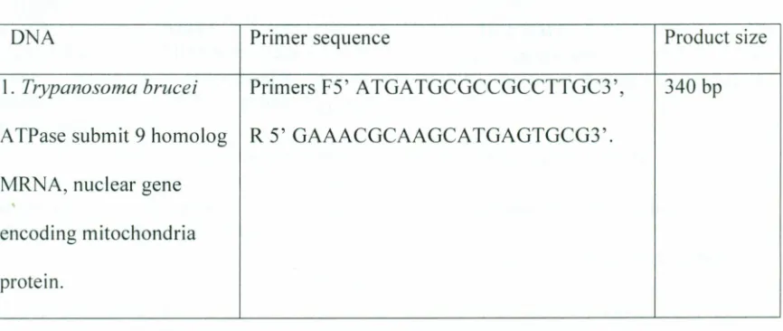
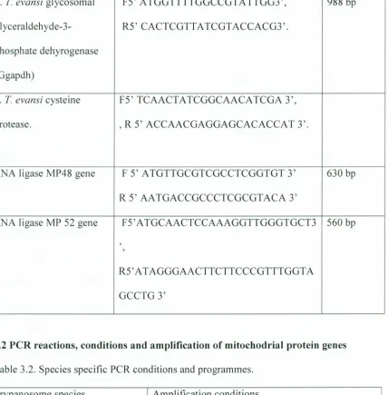
3.1. PCR of Tbrucei and Levansi specific Mitochondrial protein gene 33
3.2 PCR reactions, conditions and amplification ofmitochodrial protein genes 34
3.3 DNA sequencing 35
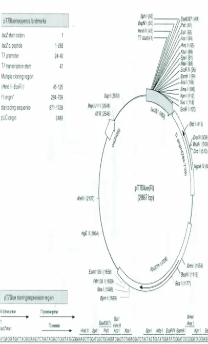
3.4. Cloning of MP 48 and MP 52 RNA editing ligase amplified gene of T evansi and
Tbrucei into pT7-Blue vector and p2T7Ti plasmid from LaCount and
Donelson 2000 36
3.5. Semi-large scale preparation of plasmid DNA using alkaline lysis method for
extraction of all plasm ids 40
3.6 Extraction and purification of plasmid by QIAGEN plasmid maxi protocol
commercial kit 40
3.7. DNA purified Gene Clean 41
3.8 Construction ofT7 Blue-T plasmid vector and p2T7Ti plasmid vector 42
3.9. p2T7Ti vector Plasmid construction 43
3.10 Generation of transgenic trypanosome cells for expression and phenotype
knockout analysis of MP 48 and MP 52 in Tevansi and T brucei 45 3.11. Construction pLEW82: GFP plasmid 46 3.13. Cell-line establishment of Bloodstream form Tbrucei Gutat 3.1 strain and T
evansi Tansui - 13 for RNA inteference analysis 49
3.14.PCR analysis 50
3.15. DNA Sequences analysis 50
3.16. Tievansi Green Fluorescent Protein analysis 50
3.17. Statistical analysis ofRNA Inference 50
CHAPTER FOUR: RESUL TS.••••••••••••••••.••••••••••••••••••••••••••••••••••••••••••••••••••••••••••••••••••••••.••••••••••••••••••52•• 4.0. PCR products from mitochodrial related genes 52
4.2 DNA Sequencing 54
4.3 Expression of Green Fluorescence Protein (GFP) in Tevansi 64 4.4. RNA interference of 48 and 52 RNA Editing ligase in Tevansi Tansui 13 and
T brucei Gutat 3.1 66
4.4.1 Effect of double stranded R A interference of 48 RNA Editing ligase in
'Levansi Tansui 13and Tbrucei Gutat 3.1. Within 36 hours 68 4.2.4 Effect of double stranded RNA interference of 52 RNA Editing ligase in
x
CHAPTER FIVE: DISCUSSION ••...•.•••...•.•...••...•...••...•••.•...•..•.••.•...•..•..•••••..•...•...••..•. 72 5.1 Relationship of T evansi and T brucei at genome level, with respect to
mitochodrial encoding genes 72
5.2 Presence of MP48 and MP52 RNA editing ligase in T evansi 75 5.3 Ability of T evansi to driveT7 RNA polymerase lead to the expression of Green
Fluorescent Protein gene with the help ofT7 promoter. 76 5.4 The generation of knockout phenotype due to the introduction of double stranded
RNA (ds RNA) MP 48 and MP52 RNA editing ligase genes into T evansi and
T brucei. CRNA interference) 77
CHAPTER SIX: CONCLUSION AND RECOMMENDATIONS 82
6.1. Conclusions 82
6.2. Recommendations 82
APPE 'DICES •••.•...•...••••...•••••..•••...••..•••..•••...•••..••.•••••.••••..•.•.••••••••••••.••.••...•...••... 93
APPENDIX 1:CLASSIFICATION OF PHyLUM 93
APPENDIX 2: GENOMIC DNA ISOLATION FROM BLOOD 94
APPENDIX 3: GROWTH OF BACTERIAL CULTURES 96
APPENDIX 4. TRANSFORMATION OF E. COLI BY ELECTROPORATION 98
APPENDIX 5. PREPARATION OF MINIPREP DNA 101
APPENDIX 6.TRANSFECTION OF T.EVANSITANSUI 13 AND T.BRUCEIGUTAT 3.1 ... 108
LIST OF FIGVRES
Figure2.1.Distribution of sleeping sickness foci in Africa 6
Figure2.2. Structures of T.brucei. 9
Figure2.3.Fine Structure of a Protozoan Parasite, T evansi, As Revealed By Transmission
Electron Microcopy Of Thin Sections 10
~igure2.4: (A.) T.evansi mini circle Kinetoplastid DNA. (B). T.brucei Maxi cycle and mini circle
Kinetoplastid DNA 11
figure2.5. Life Cycle Of Trypanosoma brucei 16
figure2.6. The enzyme cascade model for RNA editing. (I-IV) V-insertion editing. (lilA, IV A)
V-deletion editing 25
figure3.2. Map of pT7Ti-Insert MP 481Mp 52 39
figure3.3. Structure of plasmid pLEW82 47
[igure3.4. Structure of plasmid pLEWI3 48
igure4.1. PCR products for mitochodrial gene 52
igure4.2. PCR Specific primers of RNA ligase MP48 gene and RNA ligase MP52 were specific for subgenus Trypanozoon. The primers can detect and identify 1) T. evansi, 2) T.
brucei and 3) T. equiperdum in clinical specimen 53
'igure4.3.Aligned sequences MP 48 RNA ligase Gene, multiple alignments of T.evansi IL1934
(A), T.b.rhodesiense IL2343(B) and T.evansi IL1695 (C) 55
rigure.4.4.Aligned Sequences Mp52 RNA Ligase Gene, Multiple alignments of T.evansi IL1934
(A), T.b.rhodesiense IL2343 (B) and Tievansi IL1695 (C) 56
'igure4.5.Predicted amino acid sequence of MP 48 RNA editing ligase in T.brucei and T.evansi ... 57 'igure4.6. Predicted amino acid sequence of MP 52 RNA editing ligase gene in T.brucei and
T.evansi. 57
igure4.7. Multiple alignment of 630bp of T. evansi Mp48 ORF with the related genes 59 igure4.8.A.Multiple alignment of T.evansi MP52 ORF with those of related genes continues in
page 63 60
igure4.8.B.Multiple alignment of T. evansi MP52 ORF with those of related genes from page
62 61
igure4.9. Multiple alignment of Trypanosoma evansi MP 52 with related proteins 62 igure4.10.Multiple alignment of Trypanosoma evansi MP48 with related proteins 63
igure4.11.Green Fluorescent T.evansi. 65
igure4.12. Plasm ids are successful electropoarted by GFP expression in T.evansi Tansui 13 65 igure4.13.T.evansi IL1695 MP 48 and MP 52 RNA ligase gene PCR products cloned in p
T7-Blue 67
igure4."14.pT7-Blue vector/MP48 and MP52 gene recombinant plasmid cut at BamHI and Hind IIIsite resulted into two bands over 1000bp pT7-Blue and small band at
560bp for MP52 gene and 630bp for MP 48 gene 67
igure4.15. p2T7Ti was cut at BamHI and Hind IIIsite resulting in two bands, a large size DNA band 6000bp that is part of p2T7Ti and 1000bp. The large bands were used to ligate
mp48 and 52 from Figure 4.15 67
igure4.16. The bands as a result of ligation of large size plasmid DNA fragment with 6000bp Figure 4.15, which is part of p2T7Ti, and small DNA fragment band at 560bp
XII
Figure4.17. Knockout of T.brucei MP 48and 52 RNA Editing ligase and T.evansi MP 52 and 48
LIST OF TABLES
Table 3.1. Primer sequences and amplification product size 35 Table 3.2. Species specific peR conditions... 36 Table 4.3. Statistic analysis for RNAi using MP48 RNA editing ligase 71. Table 4.4. Total %of dead Tevansi Tansui 13 and Tbrucei Gutat 3.1 caused by RNAi
MP48 RNA ligase... 72 Table 4.5. Statistic analysis for RNAi using MP 52 RNA editing ligase 72 Table 4.6:Total %of dead Tevansi Tansui 13 and Tbrucei Gutat 3.1 caused by RNAi
MP 52 RNA editing ligase... 73
Table7.I.Electroporation 104
XIV
Amp ATPase
LIST OF ABBREVATION Ampicillin
ATPase Submit 9 Homolog mRNA, Nuclear Gene Encoding Mitochondria Protein.
Cysteine Protease Cerebrospinal Fluid Diethyltoluamide Difluoromethylornithine Deoxyribonucleic acid.
Drugs for neglected diseases initiative. Enzyme-Linked Immunosorbent Assay Green Fluorescent Protein.
Glycosomal Glyceraldehyde-3-Phosphate Dehyrogenase Guide Ribonucleic Acid.
Human African trypanosomiasis. Glycerol Kinase
Kinetoplastid DNA. Luria-Bertani medium. Mitochodrial Protein.
Messenger Ribonucleic acid.
non - tsetse transmitted animal trypanosomiasis. Polymerase Chain Reaction.
Red Blood Cells. RNA editing ligase.
RNA-Induced Silencing Complex. Ribonucleic acid.
RNA Interference
Sodium Dodecyl Sulfate. Tricarboxylic Acid. Transfer Ribonucleic acid.
ABSTRACT
Trypanosomiasis is a disease caused by parasitic protozoans of the genus Trypanosoma. The agents of the disease are obligate extracellular parasites that occur in blood,
cerebrospinal fluid and tissue fluids. Trypanosoma brucei causes sleeping sickness and agana insub-Saharan Africa. Trypanosoma evansi causing surra is endemic inAsia, Middle East northern Africa including Northern Eastern Kenya. Salivarian
trypanosomiasis isone of the most important and widespread diseases of domestic animals and man in the world. The causes of the re-emergence ofthis disease include widespread civil war, declining economies, reduced health financing and the dismantling of disease control programs. The current drugs in use are toxic and not effective because of drug resistance, hence, the need for developing new drugs. The study objective was to establish the evolutionary relationship between T brucei and Tevansi, with respect to cell differentiation life cycle specific antigens and phenotype knockout analysis. PCR was used to compare genes encoding mitochodrial protein of Tevansi IL1695, Tevansi IL1934 and Trypanosoma brucei rhodesiense IL2343. Plasmid construction, preparation of plasmid DNA was done using alkaline lysis method. Extraction and purification of plasmid was by QIAGENR plasmid protocol. Cell line of T evansi and T brucei for RNA interference experiments were established. Electroporation was by Gene-Pulse machine for generation of knockout phenotypes. Statistical analysis was by Student's t-test. Tevansi IL1695, Tevansi III934 and T b. rhodesiense IL2343 contain all the five genes for mitochodrial protein in their genomes. MP 48 and MP 52 RNA editing ligases genes were identified in Trypanosoma evansi. Specific RNA ligases MP 48 630 bp and MP 52 560bp primers were developed. These primers specifically identify T evansi, T brucei and Tequiperdum from other organisms. Alignment of MP 48 and MP 52 gene
sequences obtained in T evansi and T brucei show 100 % homology. Comparisons of MP 48 and MP 52 RNA ligase gene with data of closely related organisms available in Genbank® showed no significant homology with the RNA ligase sequences of
TcruziREL and L. majorREL2 sp. nor with the available sequences ofLt RNA ligase. Multiple alignment of T evansi MP52 and MP 48 with related proteins show aperfect -match with T brucei and near-perfect match of genes with data of closely related organisms available in Genbank. T evansi was able to use T7 promoter gene, to recognize bacteriophage T7 RNA polymerase and produce RNA polymerase that synthesize mRNA encoding Green fluorescent protein, that was observed as Green
CHAPTER
O
NE: INTRODUCTION
1.1. Background
Trypanosomiasis is a disease caused by a parasitic protozoan of the genus
Trypanosoma. Salivarian trypanosomiasis is one of the most important and widespread
diseases of domestic animals and man in the world. The causative agents of the disease
are obligate extra cellular parasites that occur in blood, cerebrospinal fluid and tissue
fluids. The salivarian trypanosomiasis is further divided into tsetse - transmitted
trypanosomiasis and non - tsetse transmitted animal trypanosomiasis (NTTA T).
Tsetse - transmitted trypanosomiasis, Trypanosoma brucei rhodesiense and
Trypanosoma brucei gambiense cause sleeping sickness. Nagana in domestic animals is
caused by T brucei brucei and transmitted by tsetse fly. The disease occur in scattered
foci throughout the sub-Saharan tsetse belts of Africa-an area of some 10 million sq km.
Sleeping sickness is closely related to a cattle infection known as nagana, which restricts cattle rearing in many areas of the continent (WHO, 1979/1986).
Non - tsetse transmitted animal trypanosomiasis (NTTA T); Trypanosoma evansi
causes a wasting disease of domestic livestock that is called surra. The parasite is transmitted mechanically by biting flies and it does not posses developmental stage in insect vector or a mitochondria (Aratama et al., 1992). Surra is prevalent in almost all
tropical and subtropical areas of the world, such as sub Saharan Africa, Asia, Middle East and South America.
Human African trypanosomiasis (HAT) or sleeping sickness, a disease thought to
problem over large swathes of sub-Saharan Africa (Welburn et al., 2001). It has been
estimated that 300,000-500,000 people are currently infected and 100,000 deaths are
caused each year by the disease (Cattand et al.,2001). However, less than 4 million
people are under surveillance and only about 40000 are diagnosed and treated, due to
difficulty of diagnosis and remoteness of some affected areas (WHO, 1998). These
figures are relatively small compared to other tropical diseases, but African
trypanosomiasis, without intervention, has the propensity to develop into epidemics,
making it a major public health problem (WHO, 1998). Furthermore, the case fatality rate
in untreated patients is 100% (WHO, 1998). In many countries the tsetse and
trypanosomiasis problem has been exacerbated by population pressure, which has forced
pastoralists deeper into high-risk areas with increasing risk of infestation. Partly as a
result of this incursion of man and his animals into tsetse tly habitats there are indications
that tly populations are increasing and that some tly belts are currently expanding (WHO,
1998).
The chemotherapy of African trypanosomiasis relies on a few drugs which have
adverse side effects and are unsatisfactory: pentamidine, suramin and melarsoprol.
Etlornithine is the only alternative registered drug for the treatment of Trypanosoma
brucei gambiense in sleeping sickness patients who do not respond to melarsoprol.
However, apart from other drawbacks, it costs $US 300-500 per patient (WHO, 2000).
The risk of spreading sleeping sickness is closely related to the movement of cattle
populations, and the screening and treatment of livestock to prevent disease spread has
3
needed. New molecular diagnostic tools are extremely useful for elucidation of sleeping sickness and animal trypanosomiasis epidemiology.
MP 52 and 48 RNA Editing ligases were identified as drug targets in Tevansi. Two new primers were designed from MP 48 and MP 52 RNA editing ligase DNA sequences for peR diagnosis of infections caused by T brucei and Tevansi.
1.2. Justification.
Attempts have been made to distinguish Tbrucei and Tevansi, since analyses of isoenzyme electrophoresis pattern are similar (Gibson et al., 1980) .The sequence
analyses of ribosomal RNA genes (rRNA) of these species have also been found to resemble (Urakawa, 1983). The peR amplification patterns of procyclic acidic repetitive protein (PARP) genes that encode amajor surface glycoprotein of procyclic forms of T
brucei (Mowatt et al., 1987) were found to be the same with T evansi. The we
ll-characterized difference between T evansi and T brucei was found in the Kinetoplast D A (kDNA) (Borst et al., 1979). peR was performed on various mitochodrial (Kinetoplast DNA) protein genes to determine whether T evansi is different from T brucei. This scientific research was an attempt, to develop Polymerase chain reaction diagnosis, to differentiate Tevansi from T brucei. These organisms have not been differentiated by any other scientific method with exception of electron microscopy of Kinetoplast DNA (kDNA). RNA interference was used to identifying new chemotherapy target for chemotherapy of diseases caused by T evansi and T brucei.
1.3. Null hypothesis:
1.4.Main objectives
To establish the evolutionary relationship between T brucei and Tevansi, with respect to cell differentiation life cycle specific antigens and phenotype knockout analysis.
1.4.1.Specific objectives
J) To find out the relationship of T evansi and T brucei at genome level, with respect to mitochodrial encoding genes.
2) To establish the presence of MP48 and MP52 RNA editing ligase in T evansi 3). To determine ifT7 RNA polymerase drives the expression of Green Fluorescent Protein gene with the help ofT7 promoter in T evansi.
4) To determine the effect of introducing double stranded RNA (ds RNA) MP 48 and MP52 ligase genes into T evansi and T brucei.
5
CHAPT
E
R TWO: LITERET
U
RE REVIEW
2.1.Classification of trypanosomes
The pathogens of the salivarian trypanosomiasis are members of the section
salivaria and belong to the genus Trypanosoma, (Hoare et al., 1972, Lun et al., 1993, Riou et al., 1979.) which are within the order Kinetoplastida and the class Mastigophora
(Appendix I). It is generally accepted that subgenus Trypanozoon isdivided into 3 species: Tbrucei, T evansi and T equiperdum, with T brucei further subdivided into 3 subspecies defined by pathogenicity, distribution and host range (Hoare et al., 1972).
Trypanosomes of subgenus Nannomonas are defined by their developmental
cycle in the tsetse fly, which involves the midgut and proboscis. As bloodstream forms
these trypanosomes are the smallest of the Salivaria, but there is considerable
morphological variation, both in dimensions (length and maximum width), and in
features such as body shape, prominence of the undulating membrane and presence ofa
free flagellum (Hoare etal., 1972). Coupled with variation in host range and
pathogenicity, Subgenus Nannomonas is split into 2 species: Trypanosoma congolense, which has a wide range of unregulated hosts, and T simiae, for which pigs are regarded as the most important hosts (Hoare etal., 1972). This simplifies clinical diagnosis: if you
find a small trypanosome in the blood of a sick ox, goat or sheep, it will be T congolense, while inpigs with acute trypanosomiasis itwill be T simiae. A number of hitherto
cryptic subgroups have been discovered by molecular characterization during the past 20
years or so. Only one of these has been described in sufficient detail to warrant
2.2. Distribution of trypanosome
The tsetse-transmitted trypanosomoses are prevalent in a vast region of Africa and
cover approximately 10million km", trypanosomoses distribution overlaps with that of
the tsetse fly (WHO 1979/1986). Thirty six countries are locate in the tsetse infested
areas (so- called tsetse belt) and approximately 50 million people, 50 million cattle, 30
million sheep and 40 million goats are at risk of infection (Figure 2.1) (WHO,
1979/1986). The non - tsetse transmitted trypanosomes are prevalent in almost all
tropical and subtropical areas of the world, such as sub Saharan Africa, Asia, Middle East
and South America. The area affected by non - tsetse transmitted trypanosomes NTTA T
is approximately 3 times greater than that of the tsetse transmitted trypanosomes (WHO,
1979/1986, Williams et al., 1986).
• \.. t> "<,Jz,..• ,., • (
.
.): ...•...~-- J
/
{;.pafpollj
Figure 2.1. Distribution ofsleeping sickness foci in Africa. WHO 1979/1986.
A. Trypanosoma brucei gambiense area of the foci dotted line in west and central Africa.
7
BDistribution of Glossina mositans and Glossina palpalis in Africa.
C. Distribution of Glossina palpalis inAfrica.
D.Distribution of Glossina tachinodes in Africa.
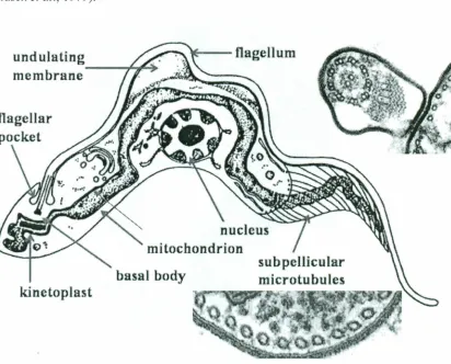
2.3. Morphology of genus Trypanosoma
Bloodstream form trypanosomes of the 3species are morphologically
indistinguishable, save for the occurrence of short-stumpy forms in T brucei.
Confusingly, the trait of pleomorphism can be lost in laboratory isolates of T brucei, and
they then become indistinguishable from the monomorphic species, T evansi (Figure 2.3)
and T equiperdum. On the other hand, in the monomorphic trypanosomes such as T
congolense, T vivax and T simiae, there are no distinguishable forms during the
infection in animals.
At the functional level pleomorphism reflects the ability ofT brucei to develop in
its vector, the tsetse fly, and this is in turn dependent on possession of acomplete and functional set of genes for mitochondrial operation. The mitochondrial genome is
contained in the maxicircle DNA of the kinetoplast of T brucei, together with the set of
minicircle-encoded genes necessary for editing the maxicircle transcripts so they can be
correctly translated. These features define T brucei, and their absence defines T evansi
and T equiperdum. T evansi lacks a mitochondrial genome and its kinetoplast contains
only a homogeneous set of mini circles. The few isolates of T equiperdum examined also
have missing kinetoplast DNA. One Chinese strain of T equiperdum had maxi circles
just over half the size of those of T brucei and homogeneous mini circles like T evansi
(Lun et al., 1993).
Two other laboratory strains of T equiperdum also had homogeneous mini
al., 1979) T brucei is a spindle shape, flagellated protozoan, which is about 8to 39 mm
in length. It has a centrally placed nucleus and a single unbranched mitochondrion, which
extends along the outer margin of the undulating membrane. The flagellum emerges from
the cell and there is a flagellar pocket. Th isis where most of the excretion and pinocytosis takes place. A Mitochondrial genome of the organism isknown as the
maxicircle kinetoplast, which is located beneath the flagellar pocket (Figure 2.2).
Trypanosoma brucei is a polymorphic trypanosome species. Two distinctly different
forms can be distinguished, i.e. a long slender form and a short stumpy form. Often,
intermediate forms, possessing characteristics of both the slender and stumpy forms, are
observed. The cytoplasm often contains basophilic granules in stained specimens.
Trypanosoma brucei (long slender form): 17-30 urn long and about 2.8 urn wide,
undulating membrane is prominet, free flagellum present at the anterior end, posterior
end pointed, kinetoplast small and subterminal. Trypanosoma brucei (short stumpy form): 17-22 urn long and about 3.5 urn wide, undulating membrane is conspicuous, free
flagellum absent, posterior end pointed, kinetoplast small and subterminal. Trypanosoma vivax: 20-27 urn long, undulating membrane is not obvious, free flagellum present at the
anterior end, posterior end rounded, and kinetoplast large and terminal. Trypanosoma congolense: 8-25 urn (small species), undulating membrane not obvious, free flagellum
absent, posterior end rounded, kinetoplast is medium sized and terminal, often laterally
positioned. Although T congolense is considered to be monomorphus, adegree of
morphological variation is sometimes observed. Trypanosoma theileri: 60-70 urn (large species), undulating membrane isconspicuous, long free flagellum present, posterior end
9
normally nonpathogenic, but its presence can confuse the parasitological diagnosis
(Frasch et al., 1979).
undulating
membrane
--...
,~
~
*"--
flagellum
nucleus
mitochondrion
kinetoplast
MlcroboC-l
/
Granular(lndOPlaGmlc relic"lum
Figure 2.3. Fine Structure of a Protozoan Parasite, T evansi, As Revealed By Transmission Electron Microcopy Of Thin Sections. (Adapted From Vickerman, 1969)
2.3.1. Kinetoplastid DNA
Kinetoplastid DNA (ktDNA) is relatively abundant and consists of mini circles and
maxi circles. The two types of ktDNA occur in a concatenated mass within the
mitochondria (mt) (Figure 2.4). Maxi-circles encode several mitochondrial genes and are more-or-Iess equivalent to the mtDNA (Borst et al., 1979). Mini circles are
I I
which need to be edited. The guide R As assist in RNA editing that takes place in
the mitochondria of kinetoplast ids. The editing of these 'cryptogenes' is believed to occur
in a hypothetical 'editsome' particle. The extent of editing seems to correlate with
different parasite life cycle stages and the corresponding changes in metabolism (i.e.,
aerobic vs. anaerobic) that isassociated with the different life cycle stages. Mini circle
DNA is also used for parasite detection and distinguishing different isolate. (Lun et al., 1992, Riou et ai., 1979, Frasch eta!., 1979).
Mini-cycle DNA
A B
2.4.Transmission of trypanosomes
on-tsetse-transmitted trypanosomes (NTTA T) are caused by T evansi that is
mechanically transmitted by haematophagous flies, but ingestion of meat from infected
carcasses by carnivores can result in infections (Luckins et aI., 1977). The most
important vectors in the field are Tabanus spp. Stomoxys spp may be more important in
spreading infection in stables due to the presence of large numbers of these flies, whereas
in open conditions, tabanids are more important. Although the mode of mechanical
transmission is well established, its dynamics are not understood and it is still necessary
to define the relationship between the host species, the duration of infection and the level
of parasitaemia, the period between feeds and the relative efficiency of different vector
species. Other blood-sucking flies such as Tabanus and Stomoxys also transmit
Trypanosoma vivax mechanically (Luckins et aI., 1977).
Dourine is a chronic trypanosomal disease of Equidae caused by T equiperdum.
The disease istransmitted almost exclusively by coitus and is characterized by edematous
lesions of the genitalia, nervous system involvement, and progressive emaciation
(Williamson et al., 1986).
Both T brucei gambiense and T'brucei rhodesiense are biologically transmitted
bytsetse flies, and are the pathogens of chronic and acute sleeping sickness in man. T
congolense, T.brucei brucei, T vivax and T simiae are also tsetse-transmitted
13
2.4.1. Vectors oftrypanosomes
2.4.1.1. Tsetse-transmitted trypanosomes
The tsetse-transmitted trypanosomes are biologically transmitted by tsetse flies
(Glossina sp). There are about 30 known species and subspecies of tsetse flies belonging
to the genus Glossina. They can be divided into three distinct groups or subgenera:
Austenia (G.fusca group), Nemorhina (G.palpalis group) and Glossina (G. morsitans
group). Only nine species and subspecies, belonging to either the G.palpalis or the G.
morsitans group known to transmit sleeping sickness. Most species fall into one of two
major groups represented by G.palpalis and G. morsitans. The palpalis group is
associated with riverine ecologies and is frequently found near streams, rivers, and lakes
inwest and central Africa (Ford et al., 1970). The morsitans group is most frequently
associated with savannah woodlands and dry bush country in East Africa. The palpalis
and morsitans groups are associated with the transmission of T'brucei gambiense and
Tbrucei rhodesiense, respectively. The differences in the ecologies and interactions with
reservoirs of these two types of tsetse contribute the different manifestations of diseases
caused by the two African trypanosome species (Minter et al., 1987).
2.4.1.2. Non-tsetse-transmitted trypanosomes
Tabanids are relatively large stout flies belonging to the sub-order Brachycera.
They have acquired various colloquial names including horseflies mainly for Tabanus
spp, clegs mainly for Haematopota spp, and deerflies mainly for Chrysops (Burger et al,
1981) Body length ranges from 5-25mm and the compound eyes are well developed. As
with most haematophagous diptera it is only the females that take blood in addition to
their hosts is sight and so the large eyes serve this function well. There is also evidence that CO2 acts as an Oduor source especially in some Chrysops spp (Burger et al., 1986),
At present there are over 3,000 known species of Tabanids. The three main genera of
economic importance are the Chrysops, Tabanus, and Haematopota. The three genera
differ in their distributions although there is considerable overlap (Abbassian-Lintzen et
al. 1964, Chainey et al. 1994), Tabanus has a cosmopolitan distribution with Chrysops
being predominantly holoartic and oriental. Haematopota is found in the palaeartic, the orient and afrotropical regions, Tabanus and Haematopota both show similar
morphological features in having reduced ocelli and aproboscis, which is shorter than the
head, Chrysops has a longer proboscis although not longer than the head and the ocelli
are functional. Tabanids are major mechanical transmitters of T evansi, causing "surra"
in horses, camels, and dogs, It also affects cattle and other mammals but this isto alesser extent.
Other trypanosomes transmitted include T vivax viennei, in cattle and sheep, T
simiae in pigs, T theileri of cattle (Krinsky et al., 1976),
2.5. Life cycle of Trypanosoma brucei
The trypanosome life cycle involves complex development of morphological
distinct forms, These forms are present within the mammalian host and the insect vector.
The major morphological difference involves changes in the cell structure and
repositioning of the mitochondrial DNA that makes up the kinetoplast. During the progression of their life cycle, repositioning of the kinetoplast from the anterior to the
posterior end of the trypanosome body occur, the flagellum begins anteriorly, passing to
the posterior end and forming the end of the undulating membrane, These events were
15
discovered to be fundamental, after transfer of the parasite from the host blood to the tsetse midgut (Matthews et al., 1995).
The first prerequisite for successful transmission is that bloodstream forms must differentiate into procylic form in the tsetse fly mid gut. The infection becomes
established, proliferate and parasites migrate to the tsetse fly salivary glands. Parasite differentiates further into epimastigote forms and subsequently into mature metacyclic forms capable of initiating a fresh infection when transmitted to a new mammalian host through a tsetse fly bite. T. brucei life cycle is between mammals and the tsetse fly; they
alternately express two types of surface coats. Bloodstream forms are covered by Variant Surface Glycoproteins VSG (Matthews et al., 1995). that shield underlying membrane proteins, preventing lysis of the parasites by serum components. The antigenic variation
of the blood forms and their consequent evasion of the host immune responses, are caused by the consecutive expression of potentially as many as 1000 different VSG. When bloodstream forms differentiate to insect procyclic forms, the parasite surface is completely remodeled, VSG is repressed and the coat is shed, as it is progressively
replaced by a new invariant coat composed of procyclins also known as procyclic acidic repetitive protein (Mowatt et al., 1987). The insect form has fully developed
mitochondria (Figure 2.5). In most species of trypanosome multipl ication is active in the
Eplmasttgotes multiply
0
T:e~~~: ::15 ~In sallvaJY gland. They I' ted t r
transform mto rnetacvcnc (Injects n>elacychctrypomas eeesj nJec me acycIC
,,-"--IIrj.~[4J--=.
..
-
.
...--- ...
-
.
.
.
.
trypomastJgotes transform trypornastigoles. ,. ~.e
into bloodstreamo
-=c~ trypomasbqotes. which, are cameo to other srtes
e
Tilypomastigotes multiply by binary rlSsionin various body fluids. eg..blood.7
"
'",,,
'
'"'
,
----
-
--~
OTrypomastigotes inbloodA
Tsetse fly StagesA.
o
~
Procyclic trvpomasuaotes leavethe midgut and transform
.ftTsetse fly takes V a blood meal
(bloodS ~M .ryp(N'l'l;!$~1901s
reIngeS!ed)
Bloodstream tryPOmaSI~-- •
~f\
transform into procyciicL
l\
~
trypornasnqotes intsetse fly'smidgut Procycbc tryposmatiqotes
mulbply by binary fission
A
:
:
Infective StageA
::
Diagnostic StageHuman Stages
Figure 2.5. Life Cycle Of Trypanosoma bruce; (Adapted From Vickerman)
2.6. Physiology of trypanosomes
The nutrition of most protozoa isholozoic; that is,they require organic materials,
which may be particulate or in solution. Many trypanosomes have a permanent mouth,
the cytosome or micropore, through which ingested food passes to become enclosed in
food vacuoles. Pinocytosis is a method of ingesting nutrient materials where by fluid is
drawn through small, temporary openings in the body wall. The ingested material
becomes enclosed within a membrane to form a food vacuole (Englund et al., 1998).
Trypanosome has metabolic pathways similar to those of higher animals and
17
advances have been made in devising chemically defined media for the cultivation of
parasitic trypanosomes. The resulting organisms are free of various substances that are
present inorganisms grown in complex media or isolated from a host and that can
interfere with immunologic or biochemical studies. Competition for nutrients is not
usually an important factor in pathogenesis because the amounts utilized by parasitic
trypanosomes are relatively small. Extracellular or intracellular parasites that destroy cells while feeding can lead to organ dysfunction and serious or life-threatening
consequences (Neva et al., 1994).
All living organisms make A TP as an energy carrier. This is produced mainly by
the oxidation of carbohydrates using glycolysis and the Tricarboxylic Acid (TCA) cycle.
Because free-living organisms do not have an abundance of food, they rely on the much
more efficient TCA cycle for most of ATP production. Trypanosoma brucei meets very
different environments at different stages of its life cycle. When this organism lives inthe
mammalian bloodstream, it depends completely on glycolysis for its supply of A TP. It
possesses neither a functional Krebs cycle nor oxidative phosphorylation, nor does it
store any carbohydrate. In the mammalian bloodstream forms there is an abundance of
glucose. The opposite istrue in the insect gut or hemolymph (Bakker et al.; Kaeser et al.,
1973; Heinrich et al., 1974;Westerhoff etal., 1987). The insect forms of T brucei in the
insect gut have mitochondria with full complement ofTCA and glycolysis enzymes.
2.8.Clinical manifestations of Trypanosomiasis
2.8.1.Human trypanosomiasis
After an infected fly bites aperson, ared painful swelling develops at the site of the fly
stream, causing episodes of fever, headaches, sweating, and generalized enlargement of
the lymph nodes. Parasites then invade the central nervous system (Early with
rhodesiense and later with gambiense) where they produce the symptoms typical of
sleeping sickness (Greenwood etal., 1980). Ultimately the parasites invade the brain,
first causing behavioral changes such as fear and mood swings, followed by headache,
fever, and weakness. Simultaneously, the patient may develop myocarditis (Greenwood
etaI., 1980).
Without treatment, death may occur within six months from cardiac failure from
Tbrucei rhodesiense infection. T brucei gambiense infection may require up to two
years before symptoms of infection in the central nervous system appear. Gambiense-infected people develop drowsiness during the day, but insomnia at night. Sleep becomes uncontrollable as the disease progresses until the patient becomes comatose (Dumas et
al., 1999).
2.8.2.Animal trypanosomiasis
2.8.2.1.Nagana
The clinical signs of the animal trypanosomiasis are parasitemia intermittent
fever, anemia, weight loss, progressive weakness and infertility in breeding animals. Host
susceptibility and virulence of parasite may vary, if left untreated hosts die of anemia
(Losos et al., 1972).
Initial replication oftrypanosomes is at the site of inoculation in the skin; this
causes a swelling and a sore (chancre). Trypanosomes then spread to the lymph nodes
and blood and continue to replicate. Trypanosoma congolense localizes in the endothelial
19
tissues. Antibody developed to the glycoprotein coat of the trypanosome kills the
trypanosome and results in the development of immune complexes. Antibody, however,
does not clear the infection, for the trypanosome has genes that can code for many
different surface-coat glycoproteins and change its surface glycoprotein to evade the
antibody. Thus, there is a persistent infection that results in acontinuing cycle of
trypanosome replication, antibody production, immune complex development, and
changing surface-coat glycoproteins. T brucei brucei has a relatively short incubation period and causes severe to fatal infection in horses, camels, dogs, and cats (Losos et al.,
1972). It usually causes mild, chronic, or subclinical disease in cattle, sheep, goats, and
pigs. A febrile response occurs in the horse 4-14 days after infection. This is followed by
recurrent febrile reactions. The heartbeat and respiration may be accelerated and
weakness are seen, whereas the appetite remains good (Losos etal., 1972).
Progressive anemia and icterus, and edema of the ventral regions, especially the
male genitalia, are characteristic. The organisms are not always easily perceived in blood
smears and are best demonstrated in tissue smears or sections, (e.g., lymph nodes).
Infected animals die in a few weeks or several months, depending on the virulence of the
strain of T brucei bruce i(Losos etal., 1972).
2.8.2.2.Surra.
An infected bite by Tabanus and Stomoxys, leads to Tevansi entering the blood stream of the host. Thereafter it multiplies to a sizeable population and causes the
appearance of a number of symptoms like intermittent fever, progressive anemia,
weakness, loss of condition and terminal nervous signs, including paralysis and
though incamels it may run for a few years the working capacity is severely impaired
(Lun et al., 1993).
2.9.Diagnosis of Trypanosomiasis
2.9.1. Microscopic examination of trypanosome
Several diagnostic techniques are available for the disease. The most primitive but
reliable technique isdirect detection oftrypanosomes by microscopic examination of
blood and lor Cerebrospinal Fluid (CSF).
The simplest techniques are examination of wet, thick or thin films of fresh blood,
usually obtained from the ear vein, jugular vein or the tail and stained by Giemsa staining
technique.
These are made by placing a drop of blood on a clean microscope slide and
covering with a cover-slip (22 x 22 mm). The blood is examined microscopically or by
phase-contrast. Approximately 50-100 fields are examined. Trypanosomes are recognized by their movement in red blood cells (RBCs). The method is simple, inexpensive and
gives immediate results. Depending on the trypanosome size and movements a
presumptive diagnosis can be made of the trypanosome species. Final confirmation of the
species ismade by the examination of the stained preparation. The diagnostic sensitivity
of the method is generally low but depends on the examiner's experience and the level of
parasitaemia. Sensitivity can be improved significantly by lysing the RBCs before
examination using a haemolytic agent such as sodium dodecyl sulfate (SDS) (Ndao et al.,
21
2.9.2.Parasite concentration techniques
The probability of detecting trypanosomes ina sample from an infected animal depends largely on the amount of blood examined and the level of parasitaemia. The amount of blood examined with direct examination techniques is low and parasites are often very scanty in the blood of an infected animal. Both of these factors contribute to the low sensitivity of direct examination techniques. Sensitivity can be improved by
increasing the volume of blood to be examined and by concentrating the trypanosomes (Woo et al., 1970).
2.9.3.Microhaematocrit centrifugation technique
The microhaematocrit centrifugation technique, or the Woo method (Woo et al.,
1970), is widely used for the diagnosis of animal trypanosomiasis. It is based on the separation of the different components of the blood sample depending on their specific gravity. The microhaematocrit centrifugation technique ismore sensitive than the direct examination techniques (Woo et al., 1970).
2.9.4.Serological tests
2.9.4.1.Indirect fluorescent antibody test
The original method for this test (Wilson et al., 1969) has been replaced by a new
technique for the preparation of trypanosoma Iantigens (Katende et al., 1987), which
involves fixation of live trypanosomes using a mixture of 80% cold acetone and 0.25%
formalin in normal saline (Platt et al., 1976, Well et al., 1982).
2.9.4.2. Card agglutination test
This test was originally developed for the diagnosis of human sleeping sickness
caused by T. brucei gambiense (Magnus et al., 1978) and has been further adapted for the
detection of antibodies against T.evansi (Pathak et al., 1997).
2.9.4.3. Enzyme-linked immunosorbent assay ELISA
The original ELISA (Luckins et al., 1977) has recently been further developed for
use in large-scale surveys of bovine trypanosomiasis (Hopkins et al., 1998). Both
antibody-detection tests have high sensitivity and specificity. Their species specificity is
generally low. They detect immune responses to current and past infections and can,
therefore, only provide a presumptive diagnosis of active infection. This
immunodiagnosis needs expensive, sophisticated equipment and expertise, which isnot
always available. It has to be performed inspecialized laboratories and there is a
substantial delay between the actual sampling and the availability of the results.
Nevertheless, the ELISA diagnosis lends itself to a high degree of automation and
standardization. Sample collection and storage is made easy through the use offilter
papers. All of these factors make the antibody ELISA a very useful test for large-scale
23
2.9.5. DNA amplification tests
A polymerase chain reaction (PCR) method has been developed as a tool for the
diagnosis of infections with African trypanosomes in humans and animals, as well as
tsetse flies. Specific repetitive nuclear DNA sequences can be amplified for T vivax and
each of the five T congolense subgroups (Desquesnes et al., 1997). A common primer set
is available for detection of the three T brucei subspecies. The primer sets available for
different trypanosome species, subspecies and subgroups are referred to as follows:
Tbrucei subspecies: TBRI and TBR2 Tcongolense (Savannah subgroup); TCN I and
TCN2 T congolense (Forest subgroup); TCFI and TCF2 Tcongolense (Kenya Coast
subgroup); TCK I and TCK2 vivax: TVW I and TVW2. Standard PCR amplifications are
carried out ina reaction mixture containing Tris-HCI, MgCI2, KCI, each of the four
deoxyribonucleotide triphosphates, primers, DNA template and Taq DNA polymerase.
Samples are incubated during several cycles at varying temperatures. The PCR products
are electrophoresed through agarose. Gels are stained with ethidium bromide. However,
at the moment, the cost ofPCR analyses is prohibitive for the routine use of the test
(Katakura et al., 1997).
2.9.6. In-vitro cultivation of salivarian trypanosome bloodstream forms
Serum-free medium for the cultivation of Tbrucei and T evansi BSFs was
achieved by Hirumi (Hirumi etal., 1997) .The serum-free culture isthe ultimate in vitro
system for studying growth-promoting factors, drug-sensitivity test, mode of trypanocidal
activity, gene-selection, cloning, cell division cycle and gene expression that have been
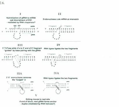
2.10. RNA Editing
The RNA modification phenomenon ofuridine (U) insertion/deletion RNA
editing was discovered in mitochondria of trypanosomatid protists more than IS years
ago (Benne et al. 1986). These phenomena can be grouped into two basic c1
asses-insertion/deletion editing and substitution editing. The term RNA editing (Benne et al.
1986) has been historically limited to certain RNA nucleotide modifications discovered
after 1986, such as C to U editing of the mammalian apoB mRNA, plant mitochondria
and chloroplast mRNAs, A to [editing of glutamate receptors mRNAs (Gott et al., 2000),
C-insertion editing of Physarum mitochondrial mRNAs. Until very recently, however,
little was known in detail about the proteins and enzymes involved in this process and
their interactions, but this israpidly changing due to the availability of Leishmania major
and T brucei genomic sequences and rapid gene identification techniques such as mass
spectroscopy.
In kinetoplastid protozoa, RNA editing is a post-transcriptional RNA-processing
event that occurs inthe mitochondrion and results in the addition and deletion of uridine
residues at specific sites in mRNA transcripts. Editing produces the sequence information
necessary for functional mRNAs by correcting frame shifts, creating start codons, or in
some cases, forming complete reading frames (Stuart et al., 1991). The genetic
information necessary to direct the insertion or deletion of uridine residues in these
mRNAs ispresent in small, SS-70-nucleotide primary transcripts called guide RNAs
(gRNAs). The gRNAs are complementary to the edited mRNAs and contain a
nonencoded poly (U) tail of 5-1S nucleotides (Blum et a!., 1990). The uridine tail may
25
gRNA/mRNA molecule consisting of the gRNA covalently linked via the poly (U) tail to
the 3' segment of the mRNA (Blum et al., 1991). The gRNA also contains a short 7-10
-nucleotide anchor region at its 5'-end, which is complementary to the pre-edited
sequences immediately 3' to the editing site. The anchor region recognizes and base pairs
with unedited mRNA and directs the initiation of the editing process. Uridine addition
and deletion continues until the mRNA is fully complementary to the gRNA as in Figure
2.6.
I
Hybridization ofgRNA 10mRHA just down5tr~m ofES~ •mCldi;rtod by RNAcru,perono?
eu Ell; ..rofA
5' Ci" Ale; A Q C U G IJ U UA "
• •• I I I I I I I I
]' ·11 IJ tJ 0 t) 0 Ce A.e A " AU. a
-c' •
u 9 <jOINA
o •
••• u
II
Endonuclease culS mRNAat mismatch
~' ..13" A CAe 0 0 U11UA I II t I I I I UVVtlUU CGACJ.Aa.U
c •
U 9
U •
• CO
III
3'rUTa.S4IaddsU'·8.10 3'end of 5'fTilgment
"gulded" bytJ.a·slI p.alrlng with the gRNA
~Ira nt \),'oil; 1"'-g""""" C)'AC.a.V .cc:ucuOUa. • •. .• I I, I I I I I
lJ 0 U U UU .... vc CAe a. A AU'
c t! u \v u & oiL C.tr
iliA J'·5' exonuclee settltnOvU
the"bulged~ U
.
-\1" •.CAQC u4l1Uu" J
••.• '.~.IIIII
I1UUqVUQ~c._,-. ~ c c lllIiX&
u •
rv
RtA lig~.$elIg;l't05thotwo fragmoni.
!I·'''fItJW"l I"'f;:;t,. :}tr
•••.•+.l'ii~~nnn
uuouu A9.CGACAA~U C u u '0
u •
lVA
RNAligno IIgal85 thetwo f"'l/mant"
,..
.
QlA"C.a.GC UQ"~OA .' ••••• ~' III I U09PUOtt 1::1..•.4U."·I < <
,
..
u • ••:;1: ••••
EdrtJngmoyersto noxt .it,
·If end or block,no·.t IIRNAfOrma ~nchor
•duplu{mc>di~1 d byRNA~Iie;l.$o?)
Figure 2.6. The enzyme cascade model for RNA editing. (I-IV) V-insertion editing. (IliA, IV A) If-deletion editing:( Adapted from Simpson,2003)
The second model proposes multiple rounds of cleavage-ligation and draws
chimera is formed by endonuclease and RNA ligase activities (Harris et al., 1992). The
pre-edited mRNA is cleaved by an endonuclease at the editing site and RNA ligase joins
the gRNA to the 3'-fragment of the mRNA producing the chimeric molecule. A second
round of cleavage and ligation resolves the chimera.
Support for the cleavage-ligation mechanism comes from the identification of
mitochondrial RNPs that are thought to be involved in RNA editing (Pollard etal., 1992).
These were shown by glycerol gradient sedimentation of mitochondrial extract to consist
of two ribonucleoprotein particles which sedimented as 19 Sand 35-40 S complexes
containing RNA ligase, chimera formation, and terminal uridylyl-transferase activities.
Uridylyl-transferase presumably is required for addition of the nonencoded poly (U) tail
to the gRNA. These results led to examine the role of the RNA ligase in chimera
formation (Simpson etal., 2003).
The mechanism ofRNA ligation has been studied for ligases from wheat germ
(Konarska et al., 1982), yeast (Greer et al., 1983), and T4-infected Escherichia coli cells
(Uhlenbeck et al., 1982). Even though the enzymes differ in the structures of their
substrates and products, their overall mechanisms are quite similar. The T4 enzyme, for
example, joins RNAs with a 5'-phosphate to 3'-hydroxyl termini in three distinct and
reversible steps:
I. E +A TP &lrhar2; E-AMP +PP
2. E-AMP
+
pN- &lrhar2; E+
AppN-3. -N +AppN- &lrhar2; -NpN- +AMP
The first step involves the adenylylation of the enzyme (E), by the transfer of
27
second step, AMP from the adenylylated enzyme is transferred to the 5'-phosphate of the R A molecule forming an activated R A molecule with as', 5'-phosphoanhydride
bond. In the third and final step, the 3'-hydroxyl of the same or different R A molecule attacks the activated R A forming aphosphodiester (Sabatini et al., 1995, Panigrahi et al., 200 I).
2.11. RNA interference
Regulation of gene expression, deciding how much of what proteins are produced in the cell, is controlled by a myriad of different molecules. One type of naturally
occurring regulatory molecule is small interfering R A (siR A), which selectively disrupts the production of a protein it is programmed to recognize, a process called R A interference (Hutva gner et al., 2004). These short stretches of nucleotides combine with other cellular proteins to form an R A-induced silencing complex, called RISe, which locates and destroys atargeted messenger R A, the molecule that carries a protein recipe from the nucleus to the site of production in the cytoplasm. RNA interference has been widely exploited tool to knock out gene expression and infer the function of missing proteins, very little is known about the mechanisms behind this regulatory process (Hutvagner et al., 2004). Trypanosoma brucei cells containing gene-specific double-stranded R As (dsR As) leads to specific degradation of the homologous messenger R A leading to formation of a knockout phenotype (Ngo et al., 1998)
2.11. Control of Trypanosomiasis
The control of trypanosomiasis is by vector control, curative and prophylactic drugs.
insecticides were developed, the large-scale insecticides 'spraying was carried out in
West Africa where over 200,000 km2ofland was cleared of tsetse by the Insecticides
spraying. Thus the large-scale insecticides spraying were very effective method for the
vector control. However, the insecticides spraying have become widely unacceptable in
terms of the protection of environment.
The sterile insect technique was one of the method for the vector control, the
method was not only expensive but also some of the sterile males of some species have
been shown to be efficient vectors of pathogenic Trypanosoma species and would
potentially increase the risk of trypanosomiasis (Moloo et al., 1982, 1988).
It is rare to change a small local environment to eliminate the breeding grounds of
these flies, such as small ponds. This method however is not effective in most cases and
rarely are ponds removed to at control a horsefly problem that mechanically transmits
Tievansi. This is due mainly to the close locality of another site, which cannot be removed, and the flight and dispersal of the adult flies (WHO, 1989).
The main attempts at present are the use of traps, pour on and spot on insecticides,
and for humans the use of repellents. These traps can be effective in localized areas and
involve killing horseflies without the need for insecticides in traps, which can easily be
home made(WHO, 1989)..
The use of insecticides as spot and pour one isused mainly to treat against other
livestock flies such as blowflies and warble flies. There is evidence that insecticides such
as the pyrethroids are effective when first applied but the effects are short lived in
relation to horsefly control. The use of self-application methods such as face bags or back
29
hornflies and faceflies, they are not very effective for horseflies. As far as humans are
concerned the methods employed can be either repellents or traps or a combination of
both. Traps are most effective when placed around the periphery of an area such as a
garden to create a horsefly free zone. The positioning and amount of traps however need
to be determined by trial and error as the population density and species will differ in
different localities. The use of repellents involves either topical application onto the body
with chemical such as diethyltoluamide (DEET), citronella oil or eucalyptus oil. DEET
can cause certain plastics to degrade, spectacle frames, watch faces etc, and no repellents
should contact the eyes or lips (WHO, 1989) ..
The use of these repellents is fine for the occasional trip out but become both
expensive and laborious to apply when wanting to go out into the garden. In these
situations it is probably better to use an impregnated patch or article of clothing. A light
cotton jacket or a stick on patch can be effective at repelling horseflies. A garment can be
soaked in DEET diluted in water to give effective protection. A cotton jacket (DEET
affects some artificial fibers) weighing 120 grams can be impregnated by pouring 30ml
of DEET into 250ml of water and immersing the garment into the solution. When not in
use the jacket can be sealed inaplastic bag to retain the life of the DEET. A garment so
soaked can be effective for several weeks (WHO, 1989).
While there are no drugs suitable for preventing sleeping sickness, there are drugs
that can be used to treat it. Treatment of infections with African trypanosomes in humans
is limited to chemotherapy, with diamidines (pentamidine), suramin,
melaminophenylarsenicals (melarsoprol"), and dl-o::-difluoromethylornithine (DFMOR)
(Fries et al., 2003). The latter two are effective against late-stage sleeping sickness (Pepin
etal., 1994), with melarsoprol being the drug of choice against the acute form of the
disease caused by T brucei rhodesiense (Bacchi et al., 1990). Pentamidine isothionate
and suramin are the drugs of choice to treat the early haemolymphatic stage of West and
East African trypanosomiasis, respectively. Melarsoprol" is the drug of choice for late
-stage disease where there is central nervous system involvement (Atouguia et al., 1995).
Recently, eflornithine has been licensed for use to treat late-stage sleeping sickness.
Eflornithine provides an alternative drug for the treatment of Tbrucei gambiense, the
form of sleeping sickness that occurs in West and Central Africa. For the Tbrucei
rhodesiense form that occurs in East and southern Africa, there is no alternative treatment
(Sunkara et al., 1987)
Insevere cases of the disease where the trypanosomes appear in the cerebrospinal fluid,
Mel BR is the drug of choice for the reasons of its ability to penetrate into cerebrospinal
fluid. However, the Mel BR, which is organic arsenic, must be used with utmost care
because of fatal side effects (WHO, 1989).
Trypanocidal drugs for use incattle are limited to the salts ofthree-compounds:
-Isometamidium chlorides, Homidium bromide and humdrum chloride and Diminazene
aceturate. Diminazene aceturate is the most widely used chemotherapeutic drug. It has
virtually no prophylactic activity (Leach et al., 1981). The homidium salts are used
mainly for chemotherapy, but they do have some prophylactic activity. Isometamidium
chloride ismainly used as a chemo prophylactic drug, but it too has some
chemotherapeutic activity.
Strategies for control have relied heavily on the use of chemotherapy to treat