_____________________________________________________________________________________________________
*Corresponding author: E-mail: [email protected];
(Past name: British Journal of Medicine and Medical Research, Past ISSN: 2231-0614, NLM ID: 101570965)
Comparative Study of Two Different Rapid
Diagnostic Tests with Microscopy Method for
Malaria Parasite Detection
Stella U. Ken-Ezihuo
1*, Sigmund S. Itatat
1and Ebirien-Agana S. Bartimaeus
11
Department of Medical Laboratory Science, Rivers State University, Nkpolu-Oroworukwo, Port Harcourt, Nigeria.
Authors’ contributions
This work was carried out in collaboration among all authors. Authors SUK and ESB designed the study, performed the statistical analysis, wrote the protocol, and wrote the first draft of the manuscript. Authors SUK and SST managed the analyses of the study. Author ESB managed the literature searches. All authors read and approved the final manuscript.
Article Information
DOI: 10.9734/JAMMR/2019/v30i930238
Editor(s):
(1)Dr. Kalpy Julien Coulibaly, Department of Environment and Health, Institut Pasteur of Ivory Coast, Félix Houphouet-Boigny University, Ivory Coast.
Reviewers:
(1)Oluboyo Bernard Oluwapelumi, Afe Babalola University, Nigeria. (2)Nyasha Chin’ombe, University of Zimbabwe, Zimbabwe. Complete Peer review History:https://sdiarticle4.com/review-history/51505
Received 09 July 2019 Accepted 15 September 2019 Published 22 October 2019
ABSTRACT
Introduction: Accurate rapid diagnosis is one of the most important steps in the effort to reduce morbidity and mortality of malaria. Blood-specific malaria rapid diagnostic tests (RDTs) are currently in use but reports on their sensitivity and specificity and comparison with the established blood film microscopy methods are dearth. The aim of the present study was to evaluate the performance characteristics of Nova and SD bioline RTDs and compare with microscopic method as a reference standard to detect the presence of malaria parasites in the blood.
Methods: A total of 100 subjects were conveniently selected from consented subjects attending out-patient Department of Rivers State University Teaching Hospital, Port Harcourt, and the samples were analyzed using blood film examined with Giemsa staining technique and Nova and SD bioline kits.
Results: Of the 100 samples examined, 57(57%) were positive for malaria parasite by light microscopy, 37(37%) were positive by Nova and 22 (22%) were positive by SD bioline. The
sensitivity of the two RDTs used were found to be 54% for Nova and 30% for SD bioline, the specificity were 86% for Nova and 88% for SD bioline, the PPV for Nova was 83% and 77% for SD bioline while the NPV for Nova was 59% and 49% for SD bioline. Percentage positivity of 50.9% and 49.1% for males and females respectively means that both sexes are equally susceptible to malaria parasites. There was a slight increase in parasitaemia in females (1931±2801) than males (1139±2415) but these results were not statistically significant (P >.27).
Conclusion: The SD bioline showed a very poor sensitivity in contrast to Nova and light microscopy. Inspite of the relative advantages of RDTs, microscopy remains the best method of detecting malaria parasite and Nova should be preferred to other RDTs.
Keywords: Malaria; parasites; sensitivity; specificity; microscopy; blood film.
1. INTRODUCTION
Malaria is one of the most widespread human parasitic diseases ranking first in terms of its socioeconomic and public health importance in tropical and sub tropical region of the world, especially in sub-Saharan African and South east Asian countries [1-3]. Various estimates have been made to measure the global burden of malaria. In 2010, the World Health Organization estimates more than 216 million cases of malaria and 655.000 deaths occur every year worldwide, with 106 countries at risk of malaria infection, among which 91% of deaths occurred in sub-Saharan Africa, 6% in South east Asia, and 3% in Eastern Mediterranean Region [1,4]. Reports show that Nigeria and Republic of Congo are two major African Countries that contributes to the high malaria burden, as 36% of the malaria cases worldwide occurred in these two countries [5].
Malaria is also a major public health problem and obstacle to socioeconomic development and also remain the leading cause of death in Nigeria with approximately 227,645 deaths in 1990 and 192,284 deaths recorded in 2015 [6]. Malaria is a major public health problem in Nigeria where it accounts for more cases and deaths than any other country in the world. Malaria is a major risk for Nigeria’s population because 97% of Nigerians live in malaria prone areas while the remaining 3% of the population live in the malaria free highlands. The estimated cases of malaria in Nigeria is about 100 million with over 300,000 deaths per year in Nigeria [7]. This data is comparable with 215,000 deaths per year in Nigeria that results from HIV/AIDS. Malaria also contributes to an estimated 11% of maternal mortality. In Nigeria 60% of outpatient visits are attributable to malaria alone and 30% of
hospitalization cases in most hospitals
nationwide among children under five years of age in Nigeria are reportedly due to malaria. Malaria has the greatest prevalence, close to
50%, in children age 6-59 months in the South West, North Central, and North West regions. Malaria has the least prevalence, 27.6 percent, in children age 6 to 59 months in the South East region. However, there is dearth of data on malaria prevalence in children in the South South region of Nigeria [7]. The two major African Countries contributing to the high malaria burden are Nigeria and Republic of Congo as 36% of malaria cases malaria cases worldwide occurred in these two countries [5].
It has been reported that in malaria endemic areas where there is increasing population of malaria infected individuals, rapid and efficient diagnostic methods are needed for rational therapy especially when it is realised that several tropical infectious diseases have many signs and symptoms that are similar to malaria [8]. Although examination of a thick blood smear after Giemsa staining remains the preferred standard method for malaria diagnosis, it is labour-intensive and time consuming. Moreover, as reliable as this technique is, it is possible to miss some few cases. Rapid diagnostic test is a device that detects malaria antigen in a small amount of blood, usually 5-15 μL, by immune
chromatographic assay with monoclonal
antibodies directed against the target parasite antigen and impregnated on a test strip [8]. The low capital involvement, non-requirement of
electricity, easy operation and result
interpretation are its advantages over
microscopy. Despite these advantages of RTDs, blood slide microscopy makes it possible to count the number of parasites and is more useful than rapid diagnostic tests for monitoring the effectiveness of malaria treatment [9]. The adoption of RDT by community health workers could facilitate diagnosis in local malaria-endemic areas with limited health personnel and facilities [10]. However, the accuracy of a clinical
diagnosis is dependent on the disease
discrepancies observed in RDT sensitivities in several observational studies [13-15].
Microscopy has low sensitivity when performed by poorly trained personnel in endemic areas, especially in primary and secondary healthcare facilities. This may result to the over- or under-diagnosis of malaria, with excessive use of anti-malarial drugs or negligent treatment, which invariably contributes to malaria morbidity and the development of resistance [16].Therefore, in the absence of well-prepared technicians for microscopic diagnosis in many areas of sub-Saharan Africa, the WHO recommends RDTs as a good alternative method for malaria diagnosis [16,17]. In remote parts of sub-Saharan Africa, RDTs have become the primary tool for the parasitological diagnosis or confirmation of malaria [18].
Continuous influx of imported RDT kits into the Nigerian market with little or no regulation is a cause for concern and has the subtle tendency to compromise malaria management and control. Similarly, the implementation of RDTs is also faced with many difficulties such as logistics, transport and continuous supply, limited shelf life and need for proper storage rooms. RDTs are quickly affected by humidity and extreme temperature. Comparative investigation on RTDs used in Port Harcourt Metropolis and microscopy is lacking in literature. This study was therefore aimed at undertaking a comparative performance of two popular brands of RDT kits found in Port
Harcourt markets alongside standard
microscopy.
2. MATERIALS AND METHODS
2.1 Study Design and Study Area
The study was a random cross sectional and comparative study.The study was carried out in Braithwaite Memorial specialist Hospital (BMSH) Port Harcourt, also known as Rivers State University Teaching Hospital (RSUTH) Port Harcourt. Port Harcourt is the capital city of Rivers State, Nigeria. It lies along the Bonny Rivers (an Eastern distributary of the Niger Rivers) 41miles (66 km upstream from the Gulf of Guinea. It has a land mass of 360 Km2 (140 sqm).
2.2 Study Subjects
A total of 100 subjects within the ages of 1-65 years attending the outpatient department of Rivers State University Teaching Hospital
(RSUTH) Port Harcourt, were selected using convenience method of sampling and blood samples were collected from them and analyzed at the Medical Laboratory Department, RSUTH.
2.3 Ethical Approval
Oral informed consent was obtained from the patients. Ethical approval was obtained from the Rivers State Ministry of Health Research Ethics Committee. Subjects were informed about their right to voluntary participation and assured of confidentiality of their test results. They were also allowed to withdraw at any time without hesitation as stipulated by the principle of Helsinki Declaration [19].
2.4 Eligibility Criteria
Subjects who were not within this age bracket were not eligible and those who took antimalaria drug recently were also not eligible.
2.5 Sample Collection
Three (3) mls of venous blood was collected aseptically into Ethylene diamine tetra Acetic acid (EDTA) bottles and well mixed to prevent clothing.
2.6 Laboratory Procedures
2.6.1 Microscopy
2.6.1.1 Preparation of blood films
Thin and think films were made on the same
slide and Stained with Giemsa staining
techniques. Giemsa is a type of Romanowsky stain containing methylene blue (a basic dye), eosin (an acidic dye) and polychrome methylene azure. The methylene blue stains the acidic part of the cell, and thus appearing bluish purple colour while the eosin stains the basic part of the cell and thus appearing pinkish red colour. The chromatin of the parasite appears dark red. Cytoplasm of parasite appear blue, red cells appears grey to pale mauve, while the reticulocytes appears grey blue [20].
2.6.2 Rapid diagnostic test (RDT)
Blood samples collected from the subjects were also tested for malaria parasite using carestat
and SD bioline RDTs. RDTs immune
medium that detects specific parasite antigens in the blood, mainly histidine rich protein 2 (HRP2) or Plasmodium lactate dehydrogenase (PLDH). In this study, the RDTs used were based on HRP-2 and detects only P. falciparium malaria. It involves the capture of dye labelled antibodies to produce a visible band on a strip of nitro-cellulose. The dye labelled-antibody binds to a corresponding parasite antigen and a resultant antigen-labelled antibody complex is formed and shown on the strip by the presence of a visible line [20].
2.7 Interpretation of the Result
The presence of two (2) colour bands, as control band and test band indicates a positive result. The presence of only one colour band indicate a negative result while if no colour band appears, the result is invalid.
2.8 Calculations
The sensitivity, specificity and predictive values (positive and negative) of each test method were calculated by comparing a composite reference gold standard generated from the two methods. The composite Reference method was defined by Okita and Amuta (2017) as a method that is positive for malaria parasites by the two methods (microscopy and RDTs) and also negative for malaria parasites by the two methods [21]. This gives the method 100% hypothetical sensitivity, specificity and positive and negative predictive values (PPV and NPV). The sensitivity, specificity, and predictive values of each of the methods were then calculated using the following the methods of Okita andAmuta [21].
2.9 Parasite Density Estimation
A portion of the thick film where white cells were evenly distributed and parasites well stained was selected for examination. 200 White blood cells (WBCs) were systematically counted. Asexual forms of the parasites in the field covered were
estimated concurrently. The number of parasites per μl of blood was calculated using the WHO formula [22]:
A slide was declared negative when there was no parasite detected/counted within 200 WBCs.
Two laboratory scientists independently
examined the slides; the third reading of discordant results by another scientist was taken as final. Sensitivity was defined as the probability that a truly infected individual will test positive and specificity as the probability that a truly uninfected individual will test negative [23].
2.10 Statistical Analysis
The results obtained from the blood samples were analyzed using GraphPad Prism Software Version 6.0, produced by GraphPad Software Inc, U.S.A. Data are presented as means and
standard deviation and as percentages.
Comparison between two means was done using the students’ t-test analysis.
3. RESULTS
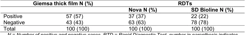
The relative malaria positivity by rapid diagnostic test kits used and microscopy using Giemsa thick film is shown in Table 1. The result shows that the Giemsa thick film had 57 (57%) positive results and 43 (43%) negative result, while Nova had 37(37%) positive results and 63 (63%) negative results and SD Bioline had 22 (22%) positive results and 78 (78%) negative result.
The comparisons of the sensitivity, specificity, positive predictive value and negative predictive value of the rapid diagnostics test kits used is shown in Table 2. The result showed that Nova had sensitivity of 54%, specificity of 86%, positive predictive value of 83% andnegative
Table 1. Relative malaria positivity and negativity by RDT kits and microscopy
Giemsa thick film N (%) RDTs
Nova N (%) SD Bioline N (%)
Positive 57 (57) 37 (37) 22 (22)
Negative 43 (43) 63 (63) 78 (78)
Total 100 (100) 100 (100) 100 (100)
predictive value of 59% whereas SD Bioline had sensitivity of 30%, specificity of 88%, positive predictive value of 77% and negative predictive value of 49%.
Table 2. Diagnostic performance of rapid diagnostic tests used
RDTs
Nova (%) SD bioline (%)
Sensitivity 54 (54) 30 (30)
Specificity 86 (86) 86 (86)
Positive predictive value (PPV)
83 (83) 77 (77)
Negative predictive value (NPV)
59 (59) 49 (49)
RDT= Rapid Diagnostic Tests
Malaria positivity and mean ± SD parasite density according to gender (sex) is shown in Table 3. The total number of subjects were 100 of which 54 subjects were males and 46 were females. Twenty nine (29) males were positive and 28 females were positive for malaria while 25 males and 18 females were negative. When compared the percentage positivity of males and females were 29% and 28% respectively, while the percentage negativity for male and female subjects were 25% and 18% respectively. The mean ± SD parasitaemia of the male subjects was 1139±2415 per µL of blood while that of the females was 1931±280 per µL of blood. No significant difference (p=0.2651) was seen in
malaria parasitaemia between the male and female subjects.
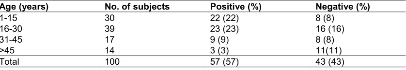
The comparison of age group with malaria infection is shown in Table 4. In this study, age group 16–30 years had the highest malaria infection, a total number of subjects examined within this age bracket were 39 (39%), and 23 (23%) were positive and 16 (16) were negative, this was closely followed by age 1–15 years, where 30 (30%) subjects were examined, 22 (22%) were positive and 8 (8%) were negative, followed by age 31–45 with 17 (17%) subjects examined, 9 (9%) were positive, and 8 (8%) were negative and age > 46 years with the lowest malaria infection, 14 subjects were examined, 3 (3%) were positive and 11 (11%) were negative.
4. DISCUSSION
There are four principal methods for diagnosing malaria. These are symptomatic, microscopy,
antigen test and molecular methods.
Symptomatic diagnosis, also known as
presumptive diagnosis is the most common in Nigeria especially in the rural communities where majority of the people are poor although it is known that many other diseases present similar symptoms to malaria, thus, malaria diagnosis by symptoms can be misleading and dangerous [24]. Presumptive diagnosis of malaria is based on nonspecific signs and symptoms like
headaches, fever, weakness, dizziness,
vomiting, abdominal pains, myalgia, chills, and pruritus [25]. The World Health Organization [5] reiterated that early diagnosis and treatment
Table 3. Malaria positivity and negativity and parasite density (mean ± SD) according to gender
Gender Total Number positive
(%)
Number negative (%)
Mean±SD parasitaemia (µL) of blood
Male 54 29 (29) 25 (25) 1139 ± 2415
Female 46 28 (28) 18 (18) 1931 ± 2701
Total 100 57 (57) 43 (43)
P- value .27
Table 4. Comparison of age with malaria infection
Age (years) No. of subjects Positive (%) Negative (%)
1-15 30 22 (22) 8 (8)
16-30 39 23 (23) 16 (16)
31-45 17 9 (9) 8 (8)
>45 14 3 (3) 11(11)
of malaria, especially in rural areas or at primary health care facilities level is important to fighting the disease in endemic region. This study, therefore, compares the diagnostic performance of two different rapid diagnostic test (RDT) kits namely Nova and SD Bioline with Giemsa stain microscopy as the reference standard. When malaria positivity for 100 subjects were examined using two different rapid diagnostic test kits along side with microscopy method, microscopy was able to detect the highest number of positive malaria cases 57 (57%). The best of the RDT kits (Nova) had a malaria positivity of 37 (37%) of the total samples diagnosed whereas SD bioline had malaria positivity 22 (22%) of the total sample diagnosed. Result obtained from this study confirmed that microscopy remains the reference standard and a better diagnostic tool for malaria parasite detection than the RDTs [26]. The use of microscopy in diagnosis of malarial infection in adult population that could comprise pregnant women is very challenging because of placental sequestration of parasites thus reducing the sensitivity of microscopy. However, the detection of peripheral blood HRP-2 genes (the principle through which the RDT works) is possible with malaria parasites RDT therefore, making RDT very useful in such condition despite its low percentage positivity when compared with microscopy [27]. Furthermore, the ability to detect placental infection by antigen detection when microscopy does not identify parasitaemia could have a significant impact on maternal and fetal health care [28]. This option makes RDT a utilizable alternative in community-base malaria care, however, it is difficult to sustain due to
problems of low cost effectiveness and
availability. In line with this argument, it was a major limitation in this study that we could not obtain and compare data on malarial RDT diagnostic performance in pregnant women in the present study.
In the study, Nova had sensitivity of 54%, specificity of 86%, positive predictive value of 83% and negative predictive value of 59% while SD bioline had sensitivity of 30%, specificity of 88%, positive predictive value of 77% and negative predictive value of 49%. By definition, sensitivity in this study is the probability that the test result will be positive when malaria is present and specificity is the probability that the test result will be negative when malaria is not present. Thus, the high specificity of 86% and 88% for Nova and SD bioline and the high positive predictive value of 83% and 77% for Nova and SD bioline respectively means that subjects stand the chance of being correctly
diagnosed as positive for malaria. The moderate negative predictive value of 59% and 49% for Nova and SD bioline respectively also means that the RDTs were not strongly reliable in ruling out the chance of malaria. This result is similar to the findings of Xiaodong et al. [29]. However, similar study in a rural community in Zaria, Northern Nigeria showed that some RDTs are capable of showing higher performance in sensitivity and specificity that the result obtained in this study [30].
Malaria positivity and mean parasite density ±SD among gender was not statistically significant (P > .27). Malaria parasitaemia is slightly higher in females than males going by both RDTs and microscopy findings are similar to results presented by Kalu et al. [31]. The higher infection rates recorded among females may be due to physiological differences between females and males (ovulation, pregnancy and childbirth) which tend to lower the female immunity, hence making them vulnerable to malaria infection and other diseases [21].
In the current study, out of the 100 subjects that participated in the study, 63 subjects were captured by Nova RDT as negative and 78 subjects were negative using SD Bioline. Of these confirmed negatives by the RDTs, field microscopy detected only 43 negative subjects (25 males and 18 females). The recorded parasitic density of ≥1000 parasites/ L was seen for both the male and female subjects. Studies have reported that patients with high levels of parasitaemia that give false negative RDT results are due to the deletion of Pfhrp2 antigens or genetic variability in the Pfhrp2 gene in certain Plasmodium falciparum parasites [32,33].
The result shows high prevalence of malaria (23% and 22%) among age group 16 –30 years and 1–15 years respectively, and 3% among age group 46 years and above. The result is similar to reports by other researchers [20]. The high prevalence among these individuals may be due to lack of protection against mosquito bites or lack of knowledge of malaria transmission or both [21]. It was also observed from the study that the intensity of the colour produces by the RDTs is proportional to the degree of parasitaemia.
5. CONCLUSION
Giemsa staining technique has been reconfirmed in this study to be a better reference routine
method for malaria diagnosis. We also
demonstrated that Nova RDT kit shows higher sensitivity, specificity, positive and negative predictive value than SD Bioline. This makes it valuable where facilities for microscopy are not available. Although Nova has shown a proven performance in the detection of malaria parasites in this study, it should serve as preliminary adjunct to microscopy especially since it is an antigen-based method. We thus recommend that only the antigen based method RDTs should be imported in Nigeria and perhaps other parts of the tropics within malaria endemicity.
CONSENT
Written informed consent was obtained from the patients.
ETHICAL APPROVAL
Ethical approval was obtained from the Rivers State Ministry of Health Research Ethics Committee.
ACKNOWLEDGEMENTS
We acknowledge the technical assistance of the laboratory scientists of Rivers State University Teaching Hospital, Port Harcourt, Rivers State, Nigeria.
COMPETING INTERESTS
Authors have declared that no competing interests exist.
REFERENCES
1. World Health Organization. World Malaria Report 2011. World Health Organization, Geneva, Switzerland; 2010.
2. World Health Organization. Malaria and
HIV interactions and their implications for public health policy, Geneva, Switzerland; 2004.
3. Hochman S, Kim K. The impact of HIV and malaria coinfection: what is known and suggested venues for further study, Interdiscip Perspect Infect Dis. 2009;1-8. Article ID 617954
4. Ramos JM, Reyes F, Tesfamariam, A.
Change in epidemiology of malaria
infections in a rural area in Ethiopia, J Travel Med. 2005;12(3):155–6.
5. World Malaria Report. World Health
Organization, Geneva, Licence: CC BY-NC-SA 3.0 IGO; 2016.
6. Khanam S. Prevalence and epidemiology
of malaria in Nigeria: a review, Inter J Res Pharm Biosci. 2017;4(8):10-12.
7. World Malaria Report. World Health
Organization, Geneva, Licence: CC BY-NC-SA 3.0 IGO; 2016.
8. Wongsrichanalai C, Barcus MJ, Muth S,
Sutamihardja A, Wernsdorfer WH. A
review of malaria diagnostic tools:
microscopy and rapid diagnostic test (RDT) anthones inhibitors of α-glucosidase and glycation from Garcinia nobilis. Amer J Trop Med Hyg. 2007;77:119-27.
9. Global Health, Division of Parasitic
Diseases and Malaria; 2018.
10. Harvey SA, Jennings L, Chinyama M,
Masaninga F, Mulholland MK, Bell
DR. Improving community health worker use of malaria rapid diagnostic tests in Zambia: Package instructions, job aid and job aid-plus-training. Malaria J. 2008; 7(160):1-12.
DOI: 10.1186/1475-2875-7-160
11. Dicko A, Mantel C, Kouriba B, Sagara I, Thera, MA. Season, fever prevalence and pyrogenic threshold for malaria disease definition in an endemic area of Mali. Trop Med Inter Health. 2005;10:550-6.
12. Mwangi TW, Mohammed M, Dayo H,
Snow RW, Marsh K. Clinical algorithms for malaria diagnosis lack utility among people of different age groups. Trop Med Inter Health. 2005;10:530-6.
13. Grobusch MP, Hanscheid T, Gobels K,
Slevogt H, Zoller T, Rogler G, Teichmann D. Comparison of three antigen detection tests for diagnosis and follow-up of falciparum malaria in travellers returning to Berlin, Germany. Parasitol Res. 2003;89: 354-7.
14. Iqbal J, Muneer A, Khalid N, Ahmed,
MA. Performance of the OptiMAL test for
malaria diagnosis among suspected
malaria patients at the rural health centers. Amer J Trop Med Hyg. 2003;68:624-8.
15. Fernando SD, Karunaweera ND, Fernando
WP. Evaluation of a rapid whole blood immunochromatographic assay for the
diagnosis of Plasmodium falciparum
and Plasmodium vivax malaria. Ceylon
Med J. 2004;49:7-11.
16. Ugah UI, Alo MN, Owolabi JO,
17. Kim SH, Nam MH, Roh KH, Park HC, Nam DH. Evaluation of a rapid diagnostic test specific for Plasmodium vivax. Trop Med Inter Health. 2008;13:1495–500.
18. Kozycki CT, Umulisa N, Rulisa S,
Mwikarago I, Musabyimana JP. False negative malaria rapid diagnostic tests in Rwanda: impact of Plasmodium falciparum isolates lacking hrp2 and declining malaria transmission. Malaria J. 2017;16:123. 19. World Medical Association. World Medical
Association Declaration of Helsinki. Ethical principles for medical research involving human subjects. Bull World Health Organ. 2001;79:373-4.
20. Mbata CA, Nwagu C, Adegoke OA.
Comparative analysis of Microscopy and rapid diagnostic test (RDT) for the laboratory diagnosis of Malaria among pregnant women attending Braithwaite Memorial Special hospital, Port Harcourt. J Med Sci Clin Res. 2015;3(1):3913-3920. 21. Okita FO, Amuta EO. Comparative use of
rapid diagnostic test and thick film microscopy in the diagnosis of malaria in sub-urban settlement in Makurdi, Nigeria. Inter J Infect Dis Ther. 2017;2(2):25-34. 22. Nicastri E, Bevilacqua N, Schepisi MS,
Paglia MG, Meschi S, Ame SM et al.
Accuracy of malaria diagnosis by
microscopy, rapid diagnostic test and PCR methods and evidence of anti malarial over prescription in non-severe febrile patients in two Tanzanian hospitals. Amer J Trop Med Hyg. 2009;80:712-17.
DOI: 10.4269/ajtmh.2009.80.712
23. Indrayan A. Sensitivity, specificity,
bayes’rule and predictivities. J Med Biostat. 2012;3(2):240-300.
24. Azikiwe CCA, Ifezulike CC, Siminialayi M, Amazu LU, Enye JC, Nwakwunite OE. Asian Pacific J Trop Biomed. 2012;2(4): 307-10.
25. Ojurongbe O, Adegbosin OO, Taiwo SS,
Alli OAT, Olowe OA, Ojuronbe TA, Bolaji OS, Adeyeba OA. Assessment of clinical diagnosis, microscopy, rapid diagnostic tests, and polymerase chain reaction in the
diagnosis of Plasmodium falciparum in Nigeria, Malaria Res Treat. 2013;1-5. Available:http://dx.doi.org/10.1155/2013/30 8069
Article ID 308069
26. Ilesanmi RI, Olalubi OA, Adetunde OT, Ilesanmi AO, Effedua H, Amoo AO. Comparative assessment of malaria rapid diagnostic test (RDT) in Ibadan Nigeria. Malaria World J. 2017;8(17):1-7.
27. Leke RFG, Djokam RR, Mbu R, Leke RJ, Fogako J. Detection of the Plasmodium falciparum antigen histidine-rich protein 2 in blood of pregnant women: implications for diagnosing placental malaria. J Clin Microbio. 1999;37:2992-6.
28. Murray CK, Gasser Jr. RA, Magill AJ, Miller RS. Update on rapid diagnostic testing for malaria. Clin Microbiol Rev. 2008;21:97-110.
29. Xiaodong S, Tambo E, Chun W, Zhibin C,
Yan D, Jian W, Jiazhi W, Xiaonong Z. Diagnostic performance of CareStart™ malaria HRP2/pLDH (Pf/pan) combo test versus standard microscopy on falciparum
and vivax malaria between
China-Myanmar Endemic Borders malaria J. 2013;12:6.
30. Sheyin Z, Bigwan IE. Comparison of care start HRP2 rapid malaria test with light microscopy for guiding patient's treatment off ever in Nigeria endemic areas. J Med Med Sci. 2013;4(9):353-6.
31. Kalu KM, Obasis NA, Nduka FLO,
Otuchristian G. A comparative study of the prevalence of malaria in Abia and Umuahia urban areas in Abia Sate, Nigeria. Res J Parasitol. 2012;7(1):17-24.
32. Mouatcho JC, Goldring JPD. Malaria rapid
diagnostic tests: Challenges and
prospects. J Med Microbiol. 2013;62(10): 1491–1505.
33. Wilson ML. Laboratory diagnosis of
malaria: Conventional and rapid diagnostic methods. Arch Path Lab Med. 2013; 137(6):805-811.
Available:https://doi.org/10.5858/arpa.2011 -0602-RA
_________________________________________________________________________________ © 2019 Ken-Ezihuo et al.; This is an Open Access article distributed under the terms of the Creative Commons Attribution License (http://creativecommons.org/licenses/by/4.0), which permits unrestricted use, distribution, and reproduction in any medium, provided the original work is properly cited.
Peer-review history: