Type of the Paper: Article 1
2 3
Reproductive toxicity of 4-octylphenol induced
4mitochondria-mediated apoptosis in male
mouse-5specific niche cells
67
Mingtian Zhang1,†, Hyun Jung Park1,†, Kwonho Hong1,, Chankyu Park1,, Hyuk Song 1,* 8
9 10
1 Department of Stem Cell and Regenerative Biology, Konkuk University, 1 Hwayang-dong, Gwangjin-gu, Seoul 11
05029, Republic of Korea
12 13 14 15
† These authors contributed equally to this work
16
*Correspondence: Prof. Hyuk Song, Department of Stem Cell and Regenerative Technology, Konkuk
17
University, 1 Hwayang-dong, Gwangjin-gu, Seoul 05029, Republic of Korea; Tel: +82-2-450-0562; e-mail:
18
19 20
Abstract 21
The toxic effects of 4-octylphenol (4-OP) have been studied in species such as mouse and fish; however,
22
the toxic effects of 4-OP in male specific niche cells has not been researched. In this study, we
23
investigated the molecular mechanism of toxicity of 4-OP in mouse TM4 Sertoli cells. TM4 cells were
24
treated with four concentrations (0, 10, 30, and 50 µM/mL) of 4-OP at time points 24, 48, and 72 h. Cell
25
viability and apoptosis assay was conducted following exposure. 4-OP significantly decreased cell
26
viability in a concentration- and time-dependent manner, and increased apoptosis. Quantitative PCR
27
analysis showed that Bad, Bax, and Bak mRNA expression levels were higher in exposed cells than in
28
the control, but Bcl-2 expression was decreased. Western blotting revealed that 4-OP induced activities
29
of caspase-3 and phosphorylation of Bad in a concentration- and time-dependent manner. Additionally,
30
cytochrome C protein did not colocalize with mitochondria marker dye by 24 h. Cytochrome c protein
31
expression increased in a time-dependent manner with 50 µM/mL. These results suggest that 4-OP
32
induces mitochondria-mediated apoptosis by regulation of Bcl-2 family proteins and caspase-3
33
activation in male Sertoli cells.
34 35
Keywords: 4-octylphenol, Male Sertoli cells, Reproductive toxicity, Apoptosis, Mitochondria 36
37
2 1. Introduction
38
4-Octylphenol (4-OP), one of many long-chain alkylphenols (APs), has been reported to cause
39
environmental contamination through its use by industries worldwide to optimize the manufacturing
40
of common products such as lubricants, plastics, cosmetics, and detergents [1]. APs are accumulated in
41
the human body via ingestion, inhalation, and dermal absorption, and have even been found in
42
maternal blood plasma, amniotic fluid [2], and breast milk [3].
43
Several studies have reported that 4-OP is a typical endocrine disruptor with estrogenic action, and
44
environmental exposure to endocrine disrupting chemicals (EDCs) has adverse effects on the human
45
reproductive system [4]. It has been reported that long-term 4-tert-octylphenol (OP) exposure in bank
46
voles resulted from disturbed androgen and estrogen synthesis and action (20850518). In addition, a
47
study of mice leydig cells exposed to 4-OP showed a decrease in the secretion of
48
dehydropiandrosterone, androstenedione, and testosterone, and reactive oxygen species
49
overproduction [5]. Similarly, juvenile mouse exposure to OP inhibited steroidogenesis by decreasing
50
the expression of steroidogenic enzymes in the testes [6]. Subcutaneous injections of 80 mg OP in an oil
51
vehicle 3 times weekly decreased sperm production in adult male rats [7], and the number of mitotic
52
germ cells and pre-spermatogonia was reduced in human fetal gonads during a 3-week culture period
53
with 4-OP treatment [8]. In females, the proliferation of uterine luminal, glandular, and stromal cells,
54
and vaginal epithelial cells were increased in adult ovariectomized rats following subcutaneous
55
injection of OP [9].
56
Fetal exposure to the weak estrogenicity of OP enhanced the induction of mammary carcinomas in
57
rats [10]. Early neonatal exposure to 4-OP (50 mg/kg) by oral gavage caused delayed sexual maturation
58
and deceased ventral prostate weight [11].
59
Apoptosis is a common form of programmed cell death that causes morphological changes including
60
cell shrinkage, nuclear fragmentation, and chromatin condensation [12]. Cellular apoptosis is regulated
61
by two typical activation mechanisms: the intrinsic pathway and the extrinsic pathway [13].
62
Apoptosis via the intrinsic pathway can be induced by the release of cytochrome c from mitochondria
63
and a change in the level of pro-apoptotic Bcl-2 family proteins such as Bax [14]. Among APs,
4-64
nonylphenol induced thymocyte apoptosis via the intrinsic pathway, including caspase-3 activation
65
and mitochondrial depolarization [15]. It is well-known that Sertoli cells are necessary for the
66
progression of germ cells into sperm in male testes. The damage of Sertoli cells caused by
67
environmental toxicants can negatively affect spermatogenesis.
68
Although many studies have reported the toxic effects of OP in reproductive organs and various cells,
69
the molecular mechanism of 4-OP in the niche of male germ cells has not been studied in detail.
70
Therefore, the present study examined the molecular mechanism underlying 4-OP mediated toxic
71
effects in TM4 sertoli cells.
72 73
2. Results 74
2.1. Effect of 4-OP on viability of TM4 cells 75
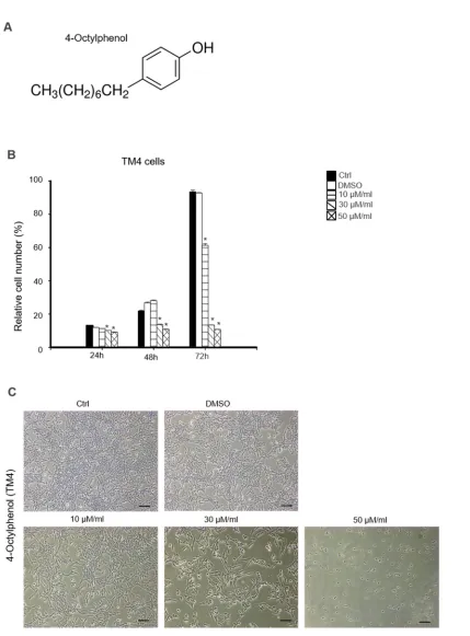
Figure 1A illustrates the chemical structure of 4-OP. We determined the cytotoxic effects of 4-OP on
76
the cell proliferation of TM4 sertoli cells. MTT assay was done at 24, 48, and 72 h. TM4 cells were treated
77
with the concentrations of 4-OP (10, 30, 50, and 100 µM/mL). As shown in Figure 1B, cell viability
78
significantly decreased when cells were exposed to the lowest concentration of 4-OP (10 µM/mL) for 72
79
h, although cell viability was only slightly affected when the cells were exposed to the same amount for
80
24 and 48 h. Notably, marked decrease in cell viability was observed following incubation with 4-OP at
81
concentrations of 30 and 50 µM/mL at 24, 48, and 72 h (Fig. 1B). The morphological change observed 72
82
h after treatment was dose dependent. 4-OP induced dose-dependent apoptotic cell death on TM4 cells.
83
Cell shrinkage and cytoplasm condensation appeared in 30 and 50 µM/mL of 4-OP treatment (Fig. 1C).
3 86
87
Figure 1. Cytotoxic effects of 4-OP on mouse TM4 Sertoli cells. (A) Chemical structure of 4-OP after 88
treated with increasing doses of 4-OP (0, 10, 30, and 50 µM/mL) at different time points (24, 48, and 72
89
h), (B) TM4 cell viability was measured by MTT assay. Results are represented as mean ± SD from 3
90
determinations per condition repeated 4 times. P<0.05, compared with the control. (C) Morphological
91
changes of TM4 cells were photographed under an inverted microscope after treatment with indicated
92
concentration of 4-OP (0, 10, 30, and 50 µM/mL) for 72 h. Scale bars = 100 µm.
4 2.2. Effects of 4- OP on apoptosis of TM4 cells 94
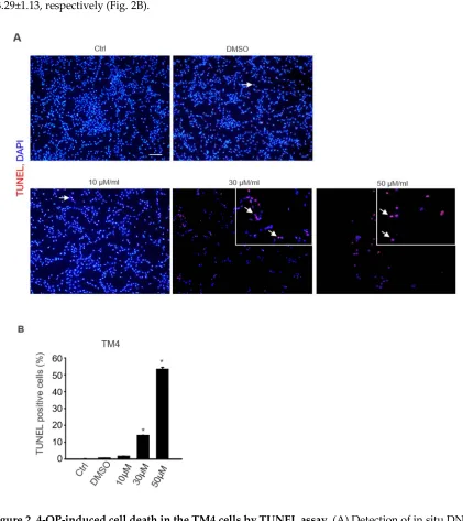
Cell apoptosis was examined using a TUNEL assay to quantify cellular death. As shown in Figure
95
3, 4-OP significantly increased the number of TUNEL-positive cells in a dose-dependent manner (0, 30,
96
and 50 µM/mL). In particular, 30 and 50 µM/mL of 4-OP markedly increased TUNEL-positive TM4 cells
97
(Fig. 2A). The data indicated that TUNEL-positive rates of TM4 and control were 13.95±0.14 and
98
53.29±1.13, respectively (Fig. 2B).
99 100
101 102
Figure 2. 4-OP-induced cell death in the TM4 cells by TUNEL assay. (A) Detection of in situ DNA 103
breaks by TUNEL assay. Cells were exposed to 0, 10, 30, and 50 µM/mL 4-OP for 24 h, the TUNEL
104
positive cells (arrow) increased dose-dependently in the 4-OP-treated TM4 cells. Bar represents
105
100µm. (B) Graph representing average number of TUNEL-positive cells in each group. The
106
percentage of TUNEL positive cells in each case were counted and the cumulative data from 3
107
independent experiments in shown here as mean ±SD (n =3, *p < 0.05 significantly different from
108
control).
5
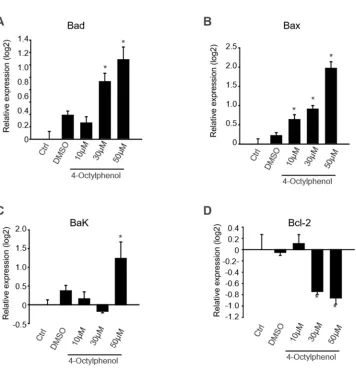
2.3. 4-OP induces the expression of pro-apoptotic gene and protein TM4 cells 112
To understand the apoptotic effect of 4-OP signaling in detail, we further analyzed several
113
apoptosis-associated molecules. The expression levels of Bax, Bad, Bcl-2, and Bak, mRNAs that play
114
essential roles in modulating apoptosis, were analyzed. Bax, Bad, and Bak mRNA levels in TM4 ells
115
treated with 4-OP increased in dose-dependent manner (Fig. 3). Bad and
116
Bax mRNA levels in TM4 cells treated with 4-OP increased in a dose-dependent manner (Fig. 3A and
117
B). In addition, the expression level of Bak significantly increased in TM4 cells treated with 50 µM/mL
118
of 4-OP, when compared with the control, and decreased Bcl-2 levels were observed in TM4 cells treated
119
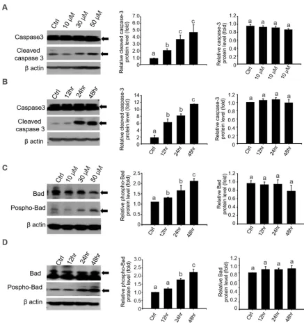
with 30 and 50 µM/mL of 4-OP (Fig. 3C and D). Since caspase-3 activation is considered a hallmark of
120
the apoptotic process, activation of caspase-3 by 4-OP was confirmed by western blot analysis to
121
confirm whether pro-apoptotic protein is involved in this apoptosis induction (Fig. 4). Caspase-3
122
activity was significantly increased in 4-OP exposed TM4 cells in a dose-dependent manner (Fig. 4A).
123
Consistently, 50 µM of 4-OP treatment increased the level of cleaved caspase-3, the active form of
124
caspase-3, in TM4 cells 24 h after treatment, but total caspase-3 was not elevated at all time points when
125
compared with the 0 h control (Fig. 4B). Based on our QPCR result, we examined the effect of 4-OP on
126
the phosphorylation of Bad and found remarkable detection of Bad phosphorylation in TM4 cells after
127
exposure to 4-OP in a dose-dependent manner (Fig. 4C). We found that 50 µM/mL 4-OP also induced
128
Bad phosphorylation of TM4 cells at 24 and 48 h, when compared with the control (Fig. 4D). For
129
normalization, total Bad protein levels were determined with Bad antibody, which reacted with both
130
the phosphorylated and non-phosphorylated Bad.
6 160
161
Figure 3. mRNA expression of anti-and pro-apoptotic genes in 4-OP-exposed TM4 cells. The mRNA 162
levels of Bad (A), Bax (B), Bak (C), and Bcl-2 (D) were examined by QPCR on 4-OP exposed TM4 cells
163
in a dose dependent manner (0, 10, 30, and 50 µM/mL 4-OP). Data shows significant difference of 4-OP
164
exposed cells compared to the control group. Results are represented as mean ± SD from 3
165
determinations per condition repeated 4 times. (n=4, *p < 0.05).
7 183
184
Figure 4. The expression of pro-apoptotic protein in 4-OP-exposed TM4 cells. 4-OP increased the 185
expression of cleaved caspase-3 in 4-OP-exposed TM4 cells. (A) After treatment with different
186
concentrations of 4-OP (0, 10, 30, and 50 µM/mL) for 24 h. (B) TM4 cells were treated with 50 µM/mL
187
4-OP, respectively, at different time points (0, 24, and 48 h). The expression levels of cleaved caspase-3,
188
caspase-3 and β-actin were examined by western blot. (C) After treatment with different concentrations
189
(0, 10, 30, and 50 µM/mL) for 24 h and (D) different time points (0, 24, and 48 h) of 4-OP. The expression
190
level of phospho-Bad, Bad, and β-actin were examined by western blot. Bar graphs represent the
191
relative density of each band normalized to β-actin or the no active form of each protein. Values
192
represent the mean ± SD of 3 independent experiments (n=3, *p < 0.05 compared with the controls).
8
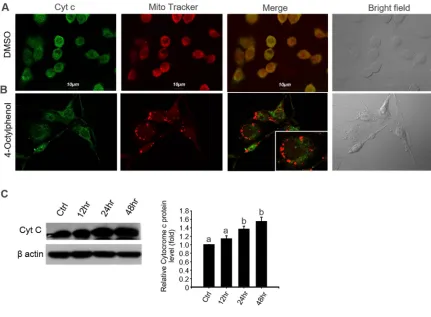
2.4. 4- OP induces cytochrome c release from mitochondria in TM4 cells 197
We determined whether the mitochondria pathway is involved in TM4 apoptosis induced by 4-OP.
198
Generally, cytocrome c release from mitochondria has been proposed to be a critical event that initiates
199
apoptosis in mammals [16]. We examine whether 4-OP induced the release of cytochrome c from
200
mitochondria to the cytosol. The cellular localization of cytochrome c protein was examined by confocal
201
immunofluoresence microscopy in TM4 cells. Labeling was done with MitoTracker, a red fluorescent
202
dye that targets mitochondria. The results showed an apparent difference in the levels of cytochrome c
203
between the treatment and control groups at 24 h post treatment. The pattern of cytochrome c
204
immunofluorescence and mitochondrial dye showed complete colocalization in the untreated controls
205
as overlapping red and green pixels seen as yellow in the TM4 cells (Fig. 5A). In contrast, the pattern of
206
staining observed after treatment with 4-OP revealed that mitochondria were stained with red
207
MitoTracker dye but were no longer colocalized with cytochrome c in the TM4 cells (Fig. 5B). In addition,
208
western blot analysis indicated that 4-OP significantly increased the expression level of cytochrome c
209
(Fig. 5C).
9 246
247 248
Figure 5. 4-OP induces cytochrome c release in TM4 cells in a time-dependent manner. (A) Effect of 249
4-OP on cytochrome c expression in TM4 cells. After treatment with 50 µM/mL 4-OP for 24 h, cells were
250
stained with Mito Tracker red (Mito Red) dye followed by immunostaining for cytochrome c and
251
observed under a confocal microscope. Scale bars = 10 µm. (B) Cytochrome c was examined by western
252
blot on 50 µM/mL 4-OP-exposed TM4 cells in time-dependent manner (0, 10, 30, and 50 µM/mL). Bar
253
graphs represent the relative density of each band normalized to β-actin. Values represent the mean ±
254
SD of 3 independent experiments (n=3, *p < 0.05 compared with the controls).
10 3. Discussion
272
Our results suggested that 4-OP was toxic to TM4 cells. In addition, we found that 4-OP could induce
273
TM4 cell apoptosis, which was verified by TUNEL analysis. Qian et al similarly reported that OP
274
decreased viability and increased apoptosis in concentration- and time-dependent manner in cultured
275
Sertoli cells derived from Sprague-Dawley rats; however, 30 µM/mL OP did not affect the proliferation
276
of Sertoli cells, in contrast to our TM4 cell viability results [17].Another study reported that OP had a
277
toxic effect on spermatogenic cells or Sertoli cells in rats [18]. 278
4-OP induces apoptosis through various molecular mechanisms, such as the induction of apoptotic
279
proteins and gene expression. There is much research showing that the Bcl-2 family plays an important
280
role in the regulation of both pro- and anti-apoptotic signals in healthy and stressed cells.
Anti-281
apoptosis proteins in the Bcl-2 family include Bcl-XL, BCL-W, and Bcl-2,and these proteins appear to 282
directly or indirectly preserve the integrity of the outer mitochondrial membrane, thereby preventing
283
cytochrome c release and cell death initiation. In contrast, Bax, Bak, and Bad are pro-apoptotic proteins
284
that mediate mitochondrial outer membrane permeabilization (MOMP) during apoptosis [19, 20].
285
In this study, Bad, Bax, and Bak gene expression was markedly higher in 4-OP-exposed TM4 cells
286
than in the control group. Results also showed that 4-OP induced activities of caspase-3 and
287
phosphorylation of Bad in TM4 cells. These findings not only help us understand the anti-proliferation
288
and apoptosis effect of 4-OP, but also improve our understanding of apoptotic signaling pathways.
289
According to previous research, caspases are crucial mediators of apoptosis. In particular, caspase-3
290
has been extensively studied as a protease that activates cell death, catalyzing the specific cleavage of
291
many proteins, and is dependent on mitochondria cytochrome c release function [20]. Caspase-3 is
292
indispensable for apoptotic chromatin condensation and DNA fragmentation in all cell types
293
investigated. In addition, a previous study showed Bcl-2, Bax, and caspase-3 activation were involved
294
in the regulation of the PO-induced apoptotic process in cultured rat Sertoli cells, supporting our results
295
[16]. 296
Generally, mitochondria play a key role in mediating apoptosis induction by diverse stimuli. The
297
release of cytochrome c from mitochondria and downstream caspase activation are important in
298
regulating apoptosis [21, 22]. Bcl-2 family proteins regulate MOMP and it has been reported that Bax
299
and Bak are also essential for MOMP and both Bax and Bak deficient cells are resistant to cytochrome
300
c release and apoptosis [23].We found thatBax and Bak expression was increased in TM4 cells with
4-301
OP treatment; thus, we analyzed the cytochrome c in these cells and found that cytochrome c protein
302
does not colocalize with mitochondria marker dye in exposed cells 24 h after treatment. In addition,
303
cytochrome c protein increased in a time-dependent manner in 50 µM/mL 4-OP-exposed cells.
304
4-OP is one of many alkylphenol compounds and is a known an environmental pollutant. Moreover,
305
it is a known EDC with estrogenic effects [24].Multiple studies have shown the impact of 4-OP in the
306
reproductive systems of different species such as frog[25],swine[26],fish[27, 28], and rodent [5, 29].
307
Even though Gregory et al. demonstrated that OP treatment of adult rats does not appear to have a
308
major effect on the male reproductive system at a relevant environmental exposure dose [30], many
309
studies have reported the negative effects of 4-OP in testes or testicular cells. In rodent testes, OP
310
appears to inhibit cAMP formation and steroidogenesis in mLTC-1 leydig tumor cells[31]. In addition,
311
4-OP induced reproductive abnormalities including small testes weight and decreased daily sperm
312
production[32]. Another study reported that administration of 80 mg/kg OP to adult male rats caused
313
shrinkage of the testes and accessory sex organs, and disrupted spermatogenesis. In humans, 4-OP
314
significantly reduced the mitotic index and the number of pre-spermatogonia in cultured fetal gonads
315
[8].
316
The seminiferous tubules in testes are composed of two major cell types, spermatogenic cells and
317
Sertoli cells. It is well-known that Sertoli cells normally control germ cell apoptosis and
318
spermatogenesis, and facilitate the progression of germ cells to spermatozoa through direct contact in
319
11
of di-(2-ethylhexyl) phthalate, targets Sertoli cells and makes them dysfunctional, resulting in the rapid 321
induction of testicular germ cell apoptosis [34]. In addition, several models of Sertoli cell injury showed
322
that spermatogenesis is vulnerable to disruption and that targeting critical Sertoli cell functions can 323
lead to rapid and massive germ cell death [35].Therefore, the apoptosis of Sertoli cells may result from
324
the abnormal effect of 4-OP. Maternal exposure to OP suppressed gonadotropin secretion with decrease
325
in testis size and Sertoli cell number during the fetal life of lamb but there was no effect to development
326
of the reproductive tract in male or female rats, although body weights were significantly decreased
327
[10]. Toxicity of OP has been extensively investigated in other organs as well. For example, OP induced
328
splenocyte apoptosis in rats and mice through Ca2+-dependence [36] and OP also had toxic effects on
329
liver in male rats [37].Therefore, toxicity evaluation of 4-OP in TM4 Sertoli cells is needed to ensure normal 330
sperm production in humans and animals. 331
In summary, our data suggest that 4-OP-induced TM4 cell apoptosis occurs in a time- and
dose-332
dependent manner directly through the mitochondrial apoptotic pathway. Of particular importance is
333
that high dose of 4-OP induced TM4 apoptosis via the down-regulation of Bcl-2; in contrast, Bak, Bad,
334
Bax expression was upregulated with subsequent activation of the caspase-3 pathway. These findings
335
can contribute to understanding the mechanism of 4-OP on the male reproductive system through its
336
action on the TM4 Sertoli cells.
337 338
4. Materials and Methods 339
340
4.1. Cell culture and treatment 341
Mouse TM4 Sertoli (TM4) cells were purchased from the Korean cell line bank (KCLB 21715, South
342
Korea). The cells were cultured in Dulbecco’s Modified Eagle’s medium, 10% fetal bovine serum (FBS),
343
and 1% penicillin streptomycin solution, in a humidified atmosphere of 5% CO2 at 37 ℃. 4-OP was 344
purchased from Sigma Aldrich (Sigma-aldrich, St. Louis, MO, USA), and dissolved in dimethyl
345
sulfoxide (DMSO) to make a stock solution. The stock solution was diluted into the cell culture media
346
prior to treatment to prepare the desired concentration.
347 348
4.2. Cell viability and morphologic analysis 349
Cell viability was determined using the EZ-Cytox Viability assay kit (Daeil Lab Services Co, Seoul,
350
Korea, #EZ1000) following the manufacturer's instructions. TM4 cells were seeded in 96-well plates at
351
a density of 5 × 10³ per well in culture medium and incubated for 24 h at 37 ℃. After 24 h, medium was 352
replaced with fresh medium containing different concentrations of 4-OP (10, 30, 50, or 100 µM/mL).
353
Cell viability assay was performed at multiple time points (24, 48, and 72 h). Assay reagent was added
354
(10 µL per well) and incubated for 30 min. The incubated plate was read on a spectrophotometer
355
(SunriseTM, TECAN) at a wavelength of 490 nm. Cell images were collected for each dose of 4-OP after 356
72 h.
357 358
4.3. Apoptosis detection with TUNEL assay 359
To evaluate cell death by apoptosis, an in situ cell death detection kit, TMR red (Roche, Germany),
360
was used to quantify DNA and chromatin morphogenic features. The procedures were followed
361
according to the manufacture’s guidelines. Cells were cultured on glass slides for 24 h, then exposed to
362
4-OP (10, 30, 50, or 100 µM/mL) for 48 h. Cells grown on coverslips were washed twice with PBS
(Sigma-363
Aldrich) and fixed with 4% paraformaldehyde in PBS for 60 min at 24℃. Following washing with PBS,
364
these cells were incubated in permeabilization solution (0.1% Trition X-100 in 0.1% sodium citrate) for
365
2 min on ice. Samples were incubated in 50 µL TUNEL reaction mixture (Roche, Mannheim, Germany)
366
for 60 min at 37 °C in a humidified chamber and in the dark. An in situ cell death detection kit provided
367
the negative control (label solution without terminal transferase, Roche) for the assay; and
368
12
BSA for 10 min at room temperature to artificially induce DNA strand breaks served as positive control.
370
Samples were incubated with or without 1 µg/mL 6-diamidino-2-pheylindole (DAPI) in PBS for 10 min
371
and coverslips were applied with mounting solution (Dako, Carpinteria, CA, USA; S3025) and analyzed
372
under fluorescence microscopy (Nikon, Tokyo, Japan).
373 374
4.4. Isolation of RNA and quantitative PCR 375
Total RNA was extracted from TM4 cells using a RNeasy Mini Kit (Qiagen, Hilden, Germany) with
376
on-column DNase treatment (Qiagen). Complementary DNA was synthesized from 1 µg of total RNA
377
using SuperScript™ III Reverse Transcriptase (Invitrogen, Carlsbad, CA, USA) with the Oligo(dT)30
378
primer according to the manufacturer’s instructions. Target gene PCR amplification was carried out for
379
30 cycles of 30 s at 95 °C, 10 s at 57 °C, and 20 s at 72 °C. Primers were designed using Primer3
380
(http://frodo.wi.mit.edu). The QPCR was achieved using a total volume of 20 µL, containing 10 ng of
381
cDNA and 1 pM of each primer, in a reaction buffer containing iQ SYBR Green Supermix (170–8880;
382
Bio-Rad Laboratories). The cycle threshold values were normalized against GAPDH gene expression,
383
a denaturation and polymerase activation step at 94 ℃ for 1 min and then 40 cycles consisting of 94 ℃ 384
for 10 s, 57 ℃ for 10 s, and 72 ℃ for 20 s. The primers used to detect porcine transcripts are listed in
385
Table 1.
386 387
4.5. Western blot analysis 388
Whole cell lysates were prepared using RIPA buffer (Thermo Fisher Scientific #89900) supplemented
389
with protease inhibitor cocktail (Roche, #1836153). Protein samples containing equal quantities of
390
protein were subjected to 4% to 20% Mini-TGX (Bio-Rad, Hercules, CA, USA; #456–1096) gel
391
electrophoresis and transferred onto polyvinylidene difluoride membranes. Membrane nonspecific
392
binding was blocked by incubation of the membranes in blocking solution (1% bovine serum albumin
393
(BSA) in tris buffered saline (TBS)) for 1 h at 22 °C, and then membranes were incubated overnight at
394
4 °C with a primary antibody diluted in TBST (20 mM Tris-HCl with pH 7.5, 150 mM NaCl, and 0.1%
395
Tween-20). The following primary antibodies were used: phospho-Bad (1:1000 dilution; Cell signaling,
396
#5284T), Bad (1:1000 dilution; Cell Signaling, #9239T), caspase-3 (1:1000 dilution; Cell Signaling, #9665T),
397
cleaved caspase-3 (1:1000 dilution; Cell Signaling, #9661T), cytocrome c (1:1000 dilution; Abcam,
398
#ab76107) and β-actin (1:1000 dilution; Santa Cruz Biotechnology, #sc47778). Membranes were washed
399
in TBST and incubated for 1 h with anti-rabbit and anti-mouse IgG and HRP-linked antibody (1:10000
400
dilutions; Jackson Immuno-Research Laboratories) in TBST. Blots were visualized using Pierce ECL
401
western blotting substrate and HyBlot CL autoradiography film (Denville Scientific, Metuchen, NJ,
402
USA; # E3018).
403 404
4.6. Immunofluorescence 405
TM4 cells were seeded on 12 mm glass coverslips (BD Biosciences, Franklin Lakes, NJ) at a density
406
of 2 × 10⁵ cells per coverslip and allowed to attach for 1 d prior to treatment with 10, 30, 50, or 100 407
µM/mL of 4-OP for 24 h. After washing once with cold PBS, cells were fixed with 4% paraformaldehyde
408
and blocked with 1% BSA in PBS containing 0.2% Triton X-100. Samples were subsequently incubated
409
with mouse anti-cytocrome c antibody (Santa Cruz Biotechnology, sc-13156, 1:200) diluted in blocking
410
solution overnight at 4 °C. After washing 3 times, the samples were incubated with secondary antibody
411
(Alexa Fluor 488 anti-mouse IgG; 1:1000) diluted in blocking buffer (1% BSA in PBS) for 1 h. Then
412
samples were washed once with warm PBS and incubated for 30 min with 100 nM of MitoTracker® red
413
CMXRos (M7512, Life Technologies, Carlsbad, CA, U S A) and washed 3 times. Nuclei were
414
counterstained with TO-PRO-3 (Life Technologies) and DAPI (Sigma-Aldrich). Samples were mounted
415
with mounting medium (Sigma-Aldrich) and images were taken under a confocal microscope (Carl
416
Zeis, Oberkochen, Germany; LSM 700).
13 4.7. Statistical analysis
419
The SPSS statistical package, version 15.0 for Windows (IBM Corp, Somers, NY, USA) was used for
420
data analysis. All the data were expressed as mean ± standard error. The differences between controls
421
and experimental samples were evaluated by one-way ANOVA, followed by Tukey`s
honestly-422
significant difference test. Significance levels of 0.05 and 0.01 were applied during data analysis using
423
Student`s t-test, and different significance levels have been indicated ( * P < 0.05). A significance levels
424
of 0.05 was applied during data analysis using ANOVA.
425 426
Competing Interests 427
The authors declare that they have no competing interests.
428 429
Acknowledgements 430
This work was supported by the Science Research Center (2015R1A5A1009701) from the National
431
Research Foundation of Korea, Republic of Korea.
432 433
Reference 434
435
1. Ying, G.G. Fate, behavior and effects of surfactants and their degradation products in the
436
environment. Environ. Int. 2006, 32, 417-31.
437
2. Shekhar, S.; Sood, S.; Showkat, S.; Lite, C.; Chandrasekhar, A.; Vairamani, M.; Barathi, S.; Santosh,
438
W. Detection of phenolic endocrine disrupting chemicals (EDCs) from maternal blood plasma and
439
amniotic fluid in Indian population. Gen. Comp. Endocrinol. 2017, 241, 100-107.
440
3. Ademollo, N.; Ferrara, F.; Delise, M.; Fabietti, F.; Funari, E. Nonylphenol and octylphenol in human
441
breast milk. Environ. Int. 2008, 34, 984-987.
442
4. Mendis-Handagama, S.M.; Ariyaratne, H.B. Differentiation of the adult Leydig cell population in
443
the postnatal testis. Biol. Reprod. 2001, 65, 660-671.
444
5. Jambor, T.; Greifova, H.; Kovacik, A.; Kovacikova, E.; Tvrda, E.; Forgacs, Z.; Massanyi, P.; Lukac, N.
445
Parallel. Effect of 4-octylphenol and cyclic adenosine monophosphate (cAMP) alters
446
steroidogenesis, cell viability and ROS production in mice Leydig cells. Chemosphere. 2018, 199,
447
747-754.
448
6. Kim, S.K.; Kim, J.H.; Lee, H.J.; Yoon, Y.D. Octylphenol reduces the expressions of steroidogenic
449
enzymes and testosterone production in mouse testis. Environ Toxicol. 2007, 22, 449-458.
450
7. Boockfor, F.R.; Blake, C.A. Chronic administration of 4-tert-octylphenol to adult male rats causes
451
shrinkage of the testes and male accessory sex organs, disrupts spermatogenesis, and increases the
452
incidence of sperm deformities. Biol. Reprod. 1997, 57, 267-277.
453
8. Bendsen, E.; Laursen, S.; Olesen, C.; Westergaard, L.; Andersen, C.; Byskov, A. Effect of
4-454
octylphenol on germ cell number in cultured human fetal gonads. Hum Reprod. 2001, 16, 236-243.
455
9. Katsuda, S.; Yoshida, M.; Isagawa, S.; Asagawa.; Kuroda, H.; Watanabe, T.; Ando, J.; Takahashi, M.;
456
Maekawa, A. Dose- and treatment duration-related effects of p-tert-octylphenol on female rats.
457
Reprod Toxicol. 2000, 14, 119-126.
458
10. Kawaguchi, H.; Miyoshi, N.; Miyamoto, Y.; Souda, M.; Umekita, Y.; Yasuda, N.; Yoshida, H. Effects
459
of fetal exposure to 4-n-octylphenol on mammary tumorigenesis in rats. In Vivo. 2010, 24, 463-470.
460
11. Nagao, T.; Yoshimura, S.; Saito, Y.; Nakagomi, M.; Usumi, K.; Ono, H. Reproductive effects in male
461
and female rats from neonatal exposure to p-octylphenol. Reprod. Toxicol. 2001, 15, 683-692.
462
12. Bär, P. R. Apoptosis--the cell's silent exit. Life. Sci. 1996, 595, 369-78.
463
13. Böhm, I.; Schild, H. Apoptosis: the complex scenario for a silent cell death. Mol Imaging. Biol.
464
2003, 5, 2-14.
465
14. Edlich, F. BCL-2 proteins and apoptosis: Recent insights and unknowns. Biochem. Biophys. Res.
466
Commun. 2018, 500, 26-34.
14
15. Yao, G.; Yang, L.; Hu, Y.; Liang, J.; Liang, J.; Hou, Y. Nonylphenol-induced thymocyte apoptosis
468
involved caspase-3 activation and mitochondrial depolarization. Mol. Immunol. 2006, 43, 915-926.
469
16. Li, K.; Li, Y.; Shelton, J.M.; Richardson, J.A.; Spencer, E.; Chen, Z.J.; Wang, X.; Williams, R.S.
470
Cytochrome c deficiency causes embryonic lethality and attenuates stress-induced apoptosis. Cell.
471
2000, 12, 101, 389-399.
472
17. Qian, J.; Bian, Q.; Cui, L.; Chen, J.; Song, L.; Wang, X. Octylphenol induces apoptosis in cultured
473
rat Sertoli cells. Toxicol. Lett. 2006, 166, 178-186.
474
18. Raychoudhury, S.S.; Blake, C.A.; Millette, C.F. Toxic effects of octylphenol on cultured rat
475
spermatogenic cells and Sertoli cells. Toxicol. Appl. Pharmacol. 1999, 157, 192-202.
476
19. Theodorakis, P.; Lomonosova, E.; Chinnadurai, G. Critical requirement of BAX for manifestation
477
of apoptosis induced by multiple stimuli in human epithelial cancer cells. Cancer. Res. 2002, 62,
478
3373-3376.
479
20. Lindsten, T.; Ross, A.J.; King, A.; Zong, W.X.; Rathmell, J.C.; Shiels, H.A.; Ulrich, E.; Waymire, K.G.;
480
Mahar, P.; Frauwirth, K.; Chen, Y.; Wei, M.; Eng, V.M.; Adelman, D.M.; Simon, M.C.; Ma, A.;
481
Golden, J.A.; Evan, G.; Korsmeyer, S.J.; MacGregor, G.R.; Thompson, C.B. The combined functions
482
of proapoptotic Bcl-2 family members bak and bax are essential for normal development of
483
multiple tissues. Mol Cell. 2000, 6, 1389-1399.
484
21. Green, D.R.; Kroemer, G. The pathophysiology of mitochondrial cell death. Science. 2004, 305,
626-485
629.22.
486
22. Jiang, X.; Wang, X. Cytochrome c promotes caspase-9 activation by inducing nucleotide binding to
487
Apaf-1. J. Biol. Chem. 2000, 275, 1199-1203.
488
23. Wei, M.C.; Zong, W.X.; Cheng, E.H.; Lindsten, T.; Panoutsakopoulou, V.; Ross, A.J.; Roth, K.A.;
489
MacGregor, G.R.; Thompson, C.B.; Korsmeyer, S.J. Proapoptotic BAX and BAK: a requisite
490
gateway to mitochondrial dysfunction and death. Science. 2001, 292, 727-730.
491
24. Nimrod, A.C.; Benson, W.H. Environmental estrogenic effects of alkylphenol ethoxylates. Crit.
492
Rev. Toxicol. 1996, 26, 335-364.
493
25. Li, X.; Liu, J.; Zhang, Y. Octylphenol induced gene expression in testes of Frog, Rana chensinensis.
494
Ecotoxicol. Environ. Saf. 2016, 128, 75-82.
495
26. Gralén, B.; Visalvethaya, W.; Ljungvall, K.; Tantasuparuk, W.; Norrgren, L.; Magnusson, U. Sows
496
exposed to octylphenol in early gestation: no estrogenic effects in male piglets, but increased rate
497
of stillbirth. Theriogenology. 2012, 78, 1494-1499.
498
27. Genovese, G.; Da Cuña, R.; Towle, D.W.; Maggese, M.C.; Lo Nostro, F. Early expression of zona
499
pellucida proteins under octylphenol exposure in Cichlasoma dimerus (Perciformes, Cichlidae).
500
Aquat Toxicol. 2011, 101, 175-185.
501
28. Rey Vázquez, G.; Meijide, F.J.; Da Cuña, R.H.; Lo Nostro, F.L.; Piazza, Y.G.; Babay, P.A.; Trudeau,
502
V.L.; Maggese, M.C.; Guerrero, G.A. Exposure to waterborne 4-tert-octylphenol induces
503
vitellogenin synthesis and disrupts testis morphology in the South American freshwater fish
504
Cichlasoma dimerus (Teleostei, Perciformes). Comp Biochem. Physiol. C Toxicol. Pharmacol. 2009,
505
150, 298-306.
506
29. Othman, A.I.; El-Missiry, M.A.; Koriem, K.M.; El-Sayed, A.A. Alfa-lipoic acid protects testosterone
507
secretion pathway and sperm quality against 4-tert-octylphenol induced reproductive toxicity.
508
Ecotoxicol Environ Saf. 2012, 81, 76-83.
509
30. Gregory, M.; Lacroix, A.; Haddad, S.; Devine, P.; Charbonneau, M.; Tardif, R.; Krishnan, K.; Cooke,
510
G.M.; Schrader, T.; Cyr, D.G. Effects of chronic exposure to octylphenol on the male rat
511
reproductive system. J. Toxicol. Environ. Health A. 2009, 72, 1553-1560.
512
31. Nikula, H.; Talonpoika, T.; Kaleva, M.; Toppari, J. Inhibition of hCG-stimulated steroidogenesis in
513
cultured mouse Leydig tumor cells by bisphenol A and octylphenols. Toxicol. Appl. Pharmacol.
514
1999, 151, 166-173.
515
32. Bian, Q.; Qian, J.; Xu, L.; Chen, J.; Song, L.; Wang, X. The toxic effects of 4-tert-octylphenol on the
15
reproductive system of male rats. Food. Chem. Toxicol. 2006, 44, 1355-1361.
517
33. Griswold, M.D. The central role of Sertoli cells in spermatogenesis. Semin. Cell. Dev. Biol. 1998, 9,
518
411-416.
519
34. Boekelheide, K. Mechanisms of toxic damage to spermatogenesis. J. Natl. Cancer. Inst. Monogr.
520
2005, 34, 6-8.
521
35. Boekelheide, K.; Fleming, S.L.; Johnson, K.J.; Patel, S.R.; Schoenfeld, H.A. Role of Sertoli cells in
522
injury-associated testicular germ cell apoptosis. Proc. Soc. Exp. Biol. Med. 2000, 225, 105-115.
523
36. Nair-Menon, J.U.; Campbell, G.T.; Blake, C.A. Toxic effects of octylphenol on cultured rat and
524
murine splenocytes. Toxicol. Appl. Pharmacol. 1996, 139, 437-444.
525
37. Zumbado, M.; Boada, L.D.; Torres, S.; Monterde, J.G.; Díaz-Chico, B.N.; Afonso, J.L.; Cabrera, J.J.;
526
Blanco, A. Evaluation of acute hepatotoxic effects exerted by environmental estrogens
527
nonylphenol and 4-octylphenol in immature male rats. Toxicology. 2002, 175, 49-62.