R E S E A R C H A R T I C L E
Open Access
Adverse effects of endometriosis on
pregnancy: a case-control study
Mayo Miura
1†, Takafumi Ushida
1†, Kenji Imai
1, Jingwen Wang
2, Yoshinori Moriyama
1, Tomoko Nakano-Kobayashi
1,
Satoko Osuka
1,3, Fumitaka Kikkawa
1and Tomomi Kotani
1,3,4*Abstract
Background:Endometriosis is a common disease occurring in 1–2% of all women of reproductive age. Although there is increasing evidence on the association between endometriosis and adverse perinatal outcomes, little is known about the effect of pre-pregnancy treatments for endometriosis on subsequent perinatal outcomes. Thus, this study aimed to evaluate maternal and neonatal outcomes in pregnant women with endometriosis and to investigate whether pre-pregnancy surgical treatment would affect these outcomes.
Methods:This case-control study included 2769 patients who gave birth at Nagoya University Hospital located in Japan between 2010 and 2017. Maternal and neonatal outcomes were compared between the endometriosis group (n= 80) and the control group (n= 2689). The endometriosis group was further divided into two groups: patients with a history of surgical treatment such as cystectomy for ovarian endometriosis, ablation or excision of endometriotic implants, or adhesiolysis (surgical treatment group,n= 49) and those treated with only medications or without any treatment (non-surgical treatment group,n= 31).
Results:In the univariate analysis, placenta previa and postpartum hemorrhage were significantly increased in the endometriosis group compared to the control group (12.5% vs. 4.1%,p< 0.01 and 27.5% vs. 18.2%,p= 0.04, respectively). In the multivariate analysis, endometriosis significantly increased the odds ratio (OR) for placenta previa (adjusted OR, 3.19; 95% confidence interval [CI], 1.56–6.50,p< 0.01) but not for postpartum hemorrhage (adjusted OR, 1.14; 95% CI, 0.66–1.98,p= 0.64). Other maternal and neonatal outcomes were similar between the two groups. In patients with endometriosis, patients in the surgical treatment group were significantly associated with an increased risk of placenta previa (OR. 4.62; 95% CI, 2.11–10.10,p< 0.01); however, patients in the non-surgical treatment group were not associated with a high risk (OR, 1.63; 95% CI, 0.19–6.59,p= 0.36). Additionally, other maternal and neonatal outcomes were similar between the two groups.
Conclusion:Women who have had surgical treatment for their endometriosis appear to have a higher risk for placenta previa. This may be due to the more severe stage of endometriosis often found in these patients. However, clinicians should be alert to this potential increased risk and manage these patients accordingly.
Keywords:Endometriosis, Pregnancy complications, Neonatal outcomes, Placenta previa
© The Author(s). 2019Open AccessThis article is distributed under the terms of the Creative Commons Attribution 4.0 International License (http://creativecommons.org/licenses/by/4.0/), which permits unrestricted use, distribution, and reproduction in any medium, provided you give appropriate credit to the original author(s) and the source, provide a link to the Creative Commons license, and indicate if changes were made. The Creative Commons Public Domain Dedication waiver (http://creativecommons.org/publicdomain/zero/1.0/) applies to the data made available in this article, unless otherwise stated. * Correspondence:[email protected]
†Mayo Miura and Takafumi Ushida contributed equally to this work. 1
Department of Obstetrics and Gynecology, Nagoya University Graduate School of Medicine, 65 Tsurumai-cho, Showa-ku, Nagoya 466-8550, Japan
3Center for Maternal-Neonatal Care, Nagoya University Hospital, 65
Tsurumai-cho, Showa-ku, Nagoya 466-8560, Japan
Background
Endometriosis is a common chronic gynecological dis-order defined as the presence of endometrial-like glands and stroma outside the uterus such as in the ovary and peritoneum [1]. The prevalence of endometriosis is 1– 2% of all women of reproductive age and 3–11% of infer-tile women [2–7]. Common symptoms of endometriosis are infertility, dysmenorrhea, chronic pelvic pain, dyspar-eunia, and painful defecation [8,9].
Surgical procedures such as laparoscopic cystectomy, excision, ablation, and adhesiolysis are considered effect-ive treatments to reduce chronic pain when conservateffect-ive treatments have insufficient efficacy [10]. However, a 5-year recurrence rate of 40–50% after surgery and the possible harmful effect on ovarian reserve is often prob-lematic [2,11].
In the past 10 years, increasing evidence on the associ-ation between endometriosis and increased risk of preg-nancy complications such as placenta previa, preterm birth, hypertensive disorders of pregnancy (HDP), small for gestational age (SGA), placental abruption, and post-partum hemorrhage (PPH) is observed [12–15]. Pres-ently, the effects of endometriosis on perinatal outcomes have remained controversial [16, 17]. Additionally, whether pre-pregnancy treatments such as surgical treat-ment or hormone therapies for infertility, ovarian endo-metriosis, and chronic pain due to endometriosis improve perinatal outcomes in subsequent pregnancies remains uncertain and is an important clinical question. Thus, this study aimed to clarify the effects of endomet-riosis on maternal and neonatal outcomes and to investi-gate whether pre-pregnancy surgical treatment for endometriosis affects these outcomes.
Methods
This case-control study was conducted to compare ma-ternal and neonatal outcomes between women with and without endometriosis who gave birth at Nagoya Univer-sity Hospital between January 2010 and December 2017. Nagoya University Hospital is located in a metropolitan area in Japan and specializes in high-risk pregnancies. This study was approved by the Institutional Review Board of Nagoya University (approval number: 2015– 0415). The exclusion criteria for this study were as fol-lows: less than 22 weeks of gestation at birth, fetal mal-formations, multiple pregnancies, and incomplete medical records. This study included 80 patients diag-nosed with endometriosis (endometriosis group) and 2689 patients without endometriosis (control group). All data on maternal and neonatal characteristics were obtained by a manual search of the electronic medical record system. In this study, diagnosis of endometriosis was based on laparoscopy with histological confirmation (n= 49), ultrasound or magnetic resonance imaging
(MRI) with ovarian endometriosis (n= 27), and presence of symptoms (n= 4). Patients who were diagnosed with endometriosis before pregnancy or who were diagnosed with ovarian endometriosis in the first trimester were in-cluded in the endometriosis group. The endometriosis group was further divided into two groups: patients with a history of surgical treatments such as cystectomy for ovarian endometriosis, ablation or excision of endome-triotic implants, and adhesiolysis (surgical treatment group, n= 49) and those treated with only medications or without any treatment (non-surgical treatment group, n= 31). In the non-surgical treatment group, 26 patients were diagnosed with presence of symptoms before preg-nancy (n= 4) and ovarian endometriosis before preg-nancy by ultrasound or MRI (n= 22), and 5 patients were diagnosed with ovarian endometriosis in the first trimester by ultrasound. Detailed information on treat-ments for endometriosis before pregnancy such as previ-ous history of surgical treatment and hormone therapies (oral contraceptives, progestogens, and gonadotropin-releasing hormone agonists [GnRHa]) was obtained from the patients’medical records.
Maternal characteristics included the following: mater-nal age, parity, body mass index (BMI) before pregnancy, chronic hypertension (HT) and diabetes mellitus (DM) before pregnancy, and assisted reproductive technology (ART). ART included in vitro fertilization and intracyto-plasmic sperm injection, and not artificial insemination with husband’s semen.
The maternal outcomes included gestational age, de-livery mode, blood loss at dede-livery, and pregnancy com-plications such as preterm birth (< 37 gestational weeks), HDP, gestational diabetes mellitus (GDM), PPH, placen-tal abruption, and placenta previa. Neonaplacen-tal characteris-tics included birth weight, height, SGA, sex, Apgar score at 1 and 5 min, umbilical artery pH, and neonatal inten-sive care unit (NICU) admission. SGA was defined as birth weight and height less than the 10th percentile for gestational age based on the Japanese gender-specific neonatal anthropometric chart, 2000 [18].
Previous papers have reported that the 90th percentile of blood loss including amniotic fluid within 24 h after delivery was 800 mL in a singleton vaginal delivery and 1500 mL in a singleton cesarean section [19,20]. In this study, PPH was defined as greater than 800 mL of esti-mated blood loss including amniotic fluid in a singleton vaginal delivery and greater than 1500 mL of estimated blood loss including amniotic fluid in a singleton cesarean section.
categorical variables and the unpaired t-test or Mann-Whitney U tests for the continuous variables according to normal or non-normal distributions. To determine the association between endometriosis and placenta pre-via or PPH, multiple logistic regression analyses were performed. The respective adjusted odds ratios (aORs) and 95% confidence intervals (CIs) were calculated after the adjustment of confounding factors including pre-pregnancy BMI, maternal age at delivery, ART, and par-ity for placenta previa, including pre-pregnancy BMI, maternal age at delivery, ART, parity, placenta previa, and macrosomia for PPH. P< 0.05 was considered sta-tistically significant.
Results
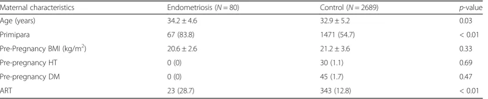
Table 1 shows the maternal characteristics in the metriosis and control groups. The patients in the endo-metriosis group were significantly older (34.2 ± 4.6 vs. 32.9 ± 5.2 years old,p= 0.03) and more likely to be prim-ipara (83.8% vs. 54.7%, p< 0.01) and conceive using ART (28.7% vs. 12.8%,p< 0.01) than the control group. No significant differences were observed in pre-pregnancy BMI, HT, and DM before pre-pregnancy between the two groups (Table1).
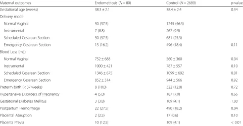
Maternal outcomes are shown in Table 2. There were significant differences in blood loss between the endo-metriosis and control groups (752 ± 688 mL vs. 560 ± 360 mL at normal vaginal delivery, p= 0.04, and 1346 ± 675 mL vs. 1099 ± 692 mL at scheduled cesarean section, p= 0.01). There were no significant differences in gesta-tional age, delivery mode, blood loss on instrumental de-livery, and emergency cesarean section between the two groups. Regarding pregnancy complications, the risk of placenta previa and PPH in the endometriosis group was significantly higher compared to the control group (12.5% vs. 4.1%, p< 0.01 and 27.5% vs. 18.2%, p= 0.04, respectively). A significant difference was not observed in the occurrences of preterm birth (< 37 weeks), HDP, GDM, and placental abruption between the two groups.
Neonatal outcomes are shown in Table3. All neonatal outcomes such as birth weight, height, SGA, Apgar score < 7 at 1 and 5 min, umbilical artery pH < 7.1, and
NICU admission rates were similar between the two groups.
Univariate and multivariate analyses were performed to identify the independent risk factors for placenta pre-via. The multivariate analysis was adjusted for endomet-riosis, pre-pregnancy BMI≥25 kg/m2, maternal age≥35 years old, ART, and multipara (Table 4). Endometriosis was identified as an independent risk factor for placenta previa (aOR, 3.19; 95% CI, 1.56–6.50,p< 0.01). ART was also considered to be an independent risk factor for pla-centa previa (aOR, 2.71; 95% CI, 1.70–4.31,p< 0.01).
To confirm whether endometriosis was an independ-ent risk factor for PPH, an additional multivariate ana-lysis was performed (Additional file 1: Table S1). We found that endometriosis was not an independent risk factor for PPH after adjusting for confounding factors including pre-pregnancy BMI≥25 kg/m2, maternal age≥ 35 years old, ART, primipara, placenta previa, and macrosomia (> 4000 g) (aOR, 1.14; 95% CI 0.66–1.98, p= 0.64).
An additional analysis was performed to identify pa-tients with a high risk for placenta previa among the endometriosis group (Table5). We obtained detailed in-formation on pre-pregnancy treatments such as surger-ies associated with endometriosis (cystectomy for ovarian endometriosis, ablation or excision of endomet-rial implants, salpingo-oophorectomy, adhesiolysis, and intestinal resection) and hormone therapies (oral contra-ceptives, progestogens, and GnRHa). The patients in the surgical treatment group showed a higher risk for pla-centa previa (crude odds ratio [OR], 4.62; 95% CI, 2.11– 10.10, p< 0.01) than the non-surgical treatment group. Particularly, patients with a gap of greater than 5 years between pregnancy and surgical treatment showed higher OR for placenta previa (crude OR, 5.92; 95% CI, 1.65–21.30, p< 0.01). On the contrary, patients in the non-surgical treatment group were not associated with an increased risk for placenta previa (crude OR, 1.63; 95% CI, 0.19–6.59,p= 0.36) (Table5).
[image:3.595.57.544.625.724.2]In patients with endometriosis, there was no significant difference in maternal and neonatal outcomes between the surgical treatment group and the non-surgical treat-ment group (Additional file1: Tables S2 and S3).
Table 1Maternal Characteristics in the Endometriosis and Control groups
Maternal characteristics Endometriosis (N= 80) Control (N= 2689) p-value
Age (years) 34.2 ± 4.6 32.9 ± 5.2 0.03
Primipara 67 (83.8) 1471 (54.7) < 0.01
Pre-Pregnancy BMI (kg/m2) 20.6 ± 2.6 21.2 ± 3.6 0.33
Pre-pregnancy HT 0 (0) 30 (1.1) 0.69
Pre-pregnancy DM 0 (0) 45 (1.7) 0.47
ART 23 (28.7) 343 (12.8) < 0.01
Discussion
This study aimed to investigate the effects of endometri-osis on maternal and neonatal outcomes and to demon-strate whether pre-pregnancy treatments, including surgical treatment and hormone therapies for infertility, ovarian endometriosis, and chronic pain due to endo-metriosis, affect perinatal outcomes. The main finding of our study was that endometriosis was an independent risk factor for placenta previa after the adjustment of several confounding factors such as fertility treatment, pre-pregnancy BMI, maternal age, and parity. In patients with endometriosis, we found that patients with a history of surgical treatment before pregnancy were associated
with an increased risk of placenta previa; on the con-trary, patients without pre-pregnancy surgical treatment such as patients who only received hormone therapies or patients coincidentally diagnosed with ovarian endo-metriosis in the first trimester of pregnancy were not as-sociated with an increased risk.
[image:4.595.57.539.100.350.2]Our findings are consistent with that of the previous studies that found an association between endometriosis and placenta previa [22–24]. Compared to the recent systematic reviews [17,25,26], endometriosis did not in-crease the risks of preterm birth, HDP, PPH, placental abruption, and SGA in this study. This discrepancy might have been caused by a difference in sample size, Table 2Maternal Outcomes in the Endometriosis and Control groups
Maternal outcomes Endometriosis (N= 80) Control (N= 2689) p-value
Gestational age (weeks) 38.3 ± 2.1 38.4 ± 2.4 0.34
Delivery mode
Normal Vaginal 30 (37.5) 1245 (46.3)
Instrumental 7 (8.8) 267 (9.9)
Scheduled Cesarean Section 30 (37.5) 681 (25.3)
Emergency Cesarean Section 13 (16.2) 496 (18.4) 0.11
Blood Loss (mL)
Normal Vaginal 752 ± 688 560 ± 360 0.04
Instrumental 1000 ± 421 787 ± 557 0.10
Scheduled Cesarean Section 1346 ± 675 1099 ± 692 0.01
Emergency Cesarean Section 852 ± 314 944 ± 566 0.92
Preterm birth (< 37 weeks) 8 (10.0) 322 (12.0) 0.72
Hypertensive Disorders of Pregnancy 4 (5.0) 187 (7.0) 0.66
Gestational Diabetes Mellitus 3 (3.8) 109 (4.1) 1.00
Postpartum Hemorrhage 22 (27.5) 490 (18.2) 0.04
Placental Abruption 2 (2.5) 17 (0.6) 0.10
Placenta Previa 10 (12.5) 109 (4.1) < 0.01
Data are presented as mean ± standard deviation orn(%). Postpartum Hemorrhage is defined as greater than 800 mL of estimated blood loss including amniotic fluid in a vaginal delivery or greater than 1500 mL of estimated blood loss including amniotic fluid in a cesarean section
Table 3Neonatal Outcomes in the Endometriosis and Control groups
Neonatal outcomes Endometriosis (N= 80) Control (N= 2689) p-value
Birth weight (g) 2851 ± 515 2899 ± 560 0.33
Low birth weight (< 2500 g) 13 (16.2) 352 (13.1) 0.51
Height (cm) 49.2 ± 3.3 49.1 ± 3.5 0.72
SGA 2 (2.5) 98 (3.6) 1.00
Male 45 (56.2) 1400 (52.1) 0.53
Apgar score at 1 min < 7 3 (3.8) 210 (7.8) 0.28
Apgar score at 5 min < 7 1 (1.2) 84 (3.1) 0.52
Umbilical artery pH < 7.1 2 (2.5) 26 (1.0) 0.19
NICU admission 17 (21.2) 521 (19.4) 0.78
[image:4.595.58.542.583.724.2]patients’ backgrounds, and study designs. Because our hospital specializes in high-risk pregnancies, in the present study, women in the control group may have higher risks for these complications compared to the control groups of the previous studies [12, 13,15], indicating that it might have been underpowered to show significant difference in maternal and neonatal outcomes.
The underlying mechanisms associating endometriosis to pregnancy complications remain largely unclear. However, several published papers have suggested that chronic inflam-mation (e.g., cyclooxygenase-2, interleukin-8, prostaglandin E2), adhesions, progesterone-resistant endometrium, and vascularized environment due to endometriosis could lead to various complications during pregnancy [16, 27, 28]. With regard to placenta previa, it has been hypothesized that hyperperistalsis of the uterus may be implicated in abnormal blastocyst implantation [29,30], and dense pelvic adhesions may inhibit the migration of the placenta away from the in-ternal ostium of uterus. Vercellini et al. reported that a higher incidence of placenta previa was observed in patients with rectovaginal lesions than in patients with peritoneal or ovar-ian lesions [30]. Additionally, severe cases such as deep infil-trating endometriosis (DIE) and revised American Society for Reproductive Medicine (rASRM) stage IV were signifi-cantly associated with a higher risk of placenta previa in sub-sequent pregnancies [31,32].
Presently, data on the efficacy of endometriosis treatments such as surgical treatment and hormone therapies on peri-natal outcomes in subsequent pregnancies are limited and unclear. To the best of our knowledge, only a few studies have demonstrated an association between pre-pregnancy surgery for endometriosis and the risk of placenta previa
[12, 31]. Berlac et al. showed that gynecological surgery for endometriosis before pregnancy had an increased risk for placenta previa in a singleton pregnancy using a national co-hort in Denmark (endometriosis, 2.2%; endometriosis with surgery, 3.4%; no endometriosis, 0.4%) [12]. Nirgianakis et al. demonstrated that patients with a history of surgical treatment for DIE presented a higher risk for placenta previa in subsequent pregnancies (endometriosis with surgery, 6.5%; no endometriosis, 0%) [31]. Although the exact reason why the risk for placenta previa increased despite a previous surgery for endometriosis remains unclear, it is possible that patients previously operated on before pregnancy might have exhibited severe endometriosis such as DIE, rASRM stage IV, and uncontrolled pain due to adhesion or ruptured ovarian endometriosis. Consistent with these previous re-ports [12,31], patients with a previous surgery in this study were likely to have higher score of rASRM based on limited patients’ data (data not shown). Previous report demon-strated that the recurrence rate of endometriosis after sur-gery is relatively high, estimated to be 21.5% at 2 years and 40–50% at 5 years [11], and was associated with the duration of follow-up after surgery and the rASRM stage of endomet-riosis at surgery [33]. In the present study, patients who had a gap of more than 5 years between an surgical treatment and pregnancy had a higher risk of placenta previa. There-fore, increased risk of placenta previa in patients with endo-metriosis with pre-pregnancy surgical treatment might be due to the higher rate of severe stage of endometriosis or a recurrence of endometriosis. Another possible explanation is that additional adhesions by operative manipulation might have some pathological role in a placenta previa. Addition-ally, the majority of patients with endometriosis in the non-Table 4Univariate and Multivariate Analysis of Risk Factors associated with Placenta Previa
Factors Crude OR (95% CI) Multi-adjusted OR (95% CI)
Endometriosis 3.38 (1.70–6.74) 3.19 (1.56–6.50)
Pre-pregnancy BMI≥25 (kg/m2) 0.62 (0.31–1.23) 0.65 (0.32–1.30)
Maternal age at delivery≥35 (years) 1.18 (0.82–1.71) 0.92 (0.62–1.36)
ART 2.55 (1.67–3.89) 2.71 (1.70–4.31)
Multipara 1.16 (0.80–1.67) 1.46 (0.99–2.15)
[image:5.595.55.538.100.182.2]The multivariate analysis was adjusted for each confounding factor including endometriosis, pre-pregnancy BMI≥25, maternal age at delivery≥35, ART and multipara by itself.BMIBody Mass Index,ARTAssisted Reproductive Technology,OROdds Ratio,CIConfidence Interval
Table 5Univariate Analysis of Pre-pregnancy Treatments associated with Placenta Previa in the Endometriosis group
Pre-pregnancy treatments With placenta previa (N= 10) Without placenta previa (N= 70) Crude OR (95% CI)
Surgical treatment group 8 41 4.62 (2.11–10.10)
Passed over 5 years at Pregnancy 3 12 5.92 (1.65–21.30)
Non-surgical treatment group 2 29 1.63 (0.19–6.59)
Total 10 70 3.38 (1.70–6.74)
[image:5.595.52.543.637.706.2]surgical treatment group were treated conservatively; there-fore, they may have had a milder variety of endometriosis compared to patients in the surgical treatment group.
Several limitations of this study should be acknowl-edged. First, the diagnosis of endometriosis in the non-surgical treatment group was based on ultrasound, MRI, and presence of symptoms, which are less reliable methods than laparoscopy (gold standard). Specifically, symptom-based diagnosis is associated with high risk of misclassification bias and can introduce bias in results es-pecially with a small sample size of 80 patients. Second, the sample size of this single-center retrospective study was smaller than the previous studies; therefore, further studies are required to test our findings in different popu-lations (e.g., races, and regions) [12,15]. Third, we could not collect sufficient data on the rASRM stage or the pres-ence of DIE for patients with a history of surgical treat-ment because some patients were operated on at another hospital. Thus, we could not identify the association be-tween placenta previa, pre-pregnancy treatments, and rASRM stage of the patients. Finally, the increased risk of placenta previa in the surgical treatment group may be partly due to the severity of endometriosis rather than sur-gical treatment itself. However, to determine the effect of surgery itself on pregnancy complications associated with endometriosis, a further study comparing another control group of women who have not been diagnosed with endo-metriosis but have had an abdominal surgery (e.g., appendectomy) is required.
Conclusions
Endometriosis is an independent risk factor for developing placenta previa. Additionally, patients with a history of surgical treatment for endometriosis before pregnancy, es-pecially patients with a gap of greater than 5 years between pregnancy and a previous surgery, were associated with increased risk of placenta previa. This may be due to the more severe stage of endometriosis found in these pa-tients. On the contrary, patients who had received only hormone therapies or who were coincidentally diagnosed with ovarian endometriosis in the first trimester of preg-nancy were not found to be at greater risk. Thus, patients with a history of surgery require particular attention for placenta previa during pregnancy. Hence, further studies are required to investigate the association between the site and stage of endometriosis or the nature of surgical treat-ment and placenta previa in a subsequent pregnancy.
Supplementary information
Supplementary informationaccompanies this paper athttps://doi.org/10. 1186/s12884-019-2514-1.
Additional file 1: Table S1.Univariate and Multivariate Analysis of Risk Factors associated with Postpartum Hemorrhage.Table S2.Maternal
Outcomes in the Surgical treatment and Non-surgical treatment groups. Table S3.Neonatal Outcomes in the Surgical treatment and Non-surgical treatment groups.
Abbreviations
ART:Assisted reproductive technology; BMI: Body mass index; CI: Confidence interval; DIE: Deep infiltrating endometriosis; DM: Diabetes mellitus; GDM: Gestational diabetes mellitus; GnRHa: Gonadotropin-releasing hormone agonist; HDP: Hypertensive disorders of pregnancy; HT: Hypertension; NICU: Neonatal intensive care unit; OR: Odds ratio; PPH: Postpartum hemorrhage; rASRM: Revised American Society for Reproductive Medicine; SGA: Small for gestational age
Acknowledgments
We thank all the participating patients for their generous contributions to this study. We would like to thank Editage (www.editage.jp) for the English language editing.
Authors’contributions
MM, TU, and TKot contributed to the concept and design of the study. MM and JW performed the statistical analyses. MM and TU drafted the first version of the manuscript. MM, TU, and TKot were involved in analyzing and interpreting the data. KI, YM, TKob, SO and FK contributed to interpreting the data, and gave critical feedback throughout the preparation of manuscript. TU and TKot critically reviewed the manuscript, and all authors gave approval for the final version of the manuscript.
Funding
The collection, analysis, interpretation of data and writing the manuscript were funded by a grant from Japan Society for the Promotion of Science (JSPS KAKENHI 19 K18637 and 15H02660) awarded to TU and FK, respectively.
Availability of data and materials
The datasets used and/or analyzed during the current study are available from the corresponding author on reasonable request.
Ethics approval and consent to participate
Ethics approval was obtained from the Institutional Review Board of Nagoya University (approval number: 2015–0415). Participants did not provide written informed consent for inclusion in the study since all data were anonymized and this was a retrospective study, in accordance to the ethical guidelines by the Japanese Ministry of Health, Labor and Welfare. The patients didn’t consent to analysis of their medical records and any further permission from the hospital was not required, however we informed patients about the study on the website of the hospital and there was an option for patients to opt out from their medical records being used in research.
Consent for publication Not applicable.
Competing interests
The authors declare that they have no competing interests.
Author details
1Department of Obstetrics and Gynecology, Nagoya University Graduate
School of Medicine, 65 Tsurumai-cho, Showa-ku, Nagoya 466-8550, Japan.
2Laboratory of Bell Research Center-Department of Obstetrics and
Gynecology collaborative research, Nagoya University Graduate School of Medicine, 65 Tsurumai-cho, Showa-ku, Nagoya 466-8550, Japan.3Center for
Maternal-Neonatal Care, Nagoya University Hospital, 65 Tsurumai-cho, Showa-ku, Nagoya 466-8560, Japan.4Department of Maternal and Perinatal
Received: 21 June 2019 Accepted: 16 September 2019
References
1. Vercellini P, Viganò P, Somigliana E, Fedele L. Endometriosis: pathogenesis and treatment. Nat Rev Endocrinol. 2014;10(5):261–75.
2. Dunselman GAJ, Vermeulen N, Becker C, Calhaz-Jorge C, D'Hooghe T, De Bie B, et al. ESHRE guideline: management of women with endometriosis. Hum Reprod. 2014;29(3):400–12.
3. Eisenberg VH, Weil C, Chodick G, Shalev V. Epidemiology of endometriosis: a large population-based database study from a healthcare provider with 2 million members. Bjog. 2018;125(1):55–62.
4. von Theobald P, Cottenet J, Iacobelli S, Quantin C. Epidemiology of endometriosis in France: a large, nation-wide study based on hospital discharge data. Biomed Res Int. 2016;2016:3260952.
5. Bhattacharya S, Porter M, Amalraj E, Templeton A, Hamilton M, Lee AJ, et al. The epidemiology of infertility in the north east of Scotland. Hum Reprod. 2009;24(12):3096–107.
6. Senapati S, Sammel MD, Morse C, Barnhart KT. Impact of endometriosis on in vitro fertilization outcomes: an evaluation of the Society for Assisted Reproductive Technologies Database. Fertil Steril. 2016;106(1):164–71.e1. 7. Zheng D, Zhou Z, Li R, Wu H, Xu S, Kang Y, et al. Consultation and
treatment behaviour of infertile couples in China: a population-based study. Reprod BioMed Online. 2019;38(6):917–25.
8. Kennedy S, Bergqvist A, Chapron C, D'Hooghe T, Dunselman G, Greb R, et al. ESHRE guideline for the diagnosis and treatment of endometriosis. Hum Reprod. 2005;20(10):2698–704.
9. Parasar P, Ozcan P, Terry KL. Endometriosis: epidemiology, diagnosis and clinical management. Curr Obstet Gynecol Rep. 2017;6(1):34–41.
10. Zanelotti A, Decherney AH. Surgery and endometriosis. Clin Obstet Gynecol. 2017;60(3):477–84.
11. Guo SW. Recurrence of endometriosis and its control. Hum Reprod Update. 2009;15(4):441–61.
12. Berlac JF, Hartwell D, Skovlund CW, Langhoff-Roos J, Lidegaard Ø. Endometriosis increases the risk of obstetrical and neonatal complications. Acta Obstet Gynecol Scand. 2017;96(6):751–60.
13. Shmueli A, Salman L, Hiersch L, Ashwal E, Hadar E, Wiznitzer A, et al. Obstetrical and neonatal outcomes of pregnancies complicated by endometriosis. J Matern Fetal Neonatal Med. 2019;32(5):845–50. 14. Bruun MR, Arendt LH, Forman A, Ramlau-Hansen CH. Endometriosis and
adenomyosis are associated with increased risk of preterm delivery and a small-for-gestational-age child: a systematic review and meta-analysis. Acta Obstet Gynecol Scand. 2018;97(9):1073–90.
15. Harada T, Taniguchi F, Onishi K, Kurozawa Y, Hayashi K, Group JECsS. Obstetrical Complications in Women with Endometriosis: A Cohort Study in Japan. PLoS One. 2016;11(12):e0168476.
16. Leone Roberti Maggiore U, Ferrero S, Mangili G, Bergamini A, Inversetti A, Giorgione V, et al. A systematic review on endometriosis during pregnancy: diagnosis, misdiagnosis, complications and outcomes. Hum Reprod Update. 2016;22(1):70–103.
17. Zullo F, Spagnolo E, Saccone G, Acunzo M, Xodo S, Ceccaroni M, et al. Endometriosis and obstetrics complications: a systematic review and meta-analysis. Fertil Steril. 2017;108(4):667–72.e5.
18. Itabashi K, Miura F, Uehara R, Nakamura Y. New Japanese neonatal anthropometric charts for gestational age at birth. Pediatr Int. 2014;56(5): 702–8.
19. Ishikawa G. Countermeasure for obstetric hemorrhage. Masui. 2010;59(3): 347–56.
20. Takeda S, Makino S, Takeda J, Kanayama N, Kubo T, Nakai A, et al. Japanese clinical practice guide for critical obstetrical hemorrhage (2017 revision). J Obstet Gynaecol Res. 2017;43(10):1517–21.
21. Kanda Y. Investigation of the freely available easy-to-use software 'EZR' for medical statistics. Bone Marrow Transplant. 2013;48(3):452–8.
22. Maggiore ULR, Inversetti A, Schimberni M, Vigano P, Giorgione V, Candiani M. Obstetrical complications of endometriosis, particularly deep endometriosis. Fertil Steril. 2017;108(6):895–912.
23. Lin H, Leng JH, Liu JT, Lang JH. Obstetric outcomes in Chinese women with endometriosis: a retrospective cohort study. Chin Med J. 2015;128(4):455–8. 24. Mannini L, Sorbi F, Noci I, Ghizzoni V, Perelli F, Di Tommaso M, et al. New
adverse obstetrics outcomes associated with endometriosis: a retrospective cohort study. Arch Gynecol Obstet. 2017;295(1):141–51.
25. Conti N, Cevenini G, Vannuccini S, Orlandini C, Valensise H, Gervasi MT, et al. Women with endometriosis at first pregnancy have an increased risk of adverse obstetric outcome. J Matern Fetal Neonatal Med. 2015;28(15):1795–8. 26. Lalani S, Choudhry AJ, Firth B, Bacal V, Walker M, Wen SW, et al.
Endometriosis and adverse maternal, fetal and neonatal outcomes, a systematic review and meta-analysis. Hum Reprod. 2018;33(10):1854–65. 27. Petraglia F, Arcuri F, de Ziegler D, Chapron C. Inflammation: a link between
endometriosis and preterm birth. Fertil Steril. 2012;98(1):36–40. 28. Patel BG, Rudnicki M, Yu J, Shu Y, Taylor RN. Progesterone resistance in
endometriosis: origins, consequences and interventions. Acta Obstet Gynecol Scand. 2017;96(6):623–32.
29. Kido A, Togashi K, Nishino M, Miyake K, Koyama T, Fujimoto R, et al. Cine MR imaging of uterine peristalsis in patients with endometriosis. Eur Radiol. 2007;17(7):1813–9.
30. Vercellini P, Parazzini F, Pietropaolo G, Cipriani S, Frattaruolo MP, Fedele L. Pregnancy outcome in women with peritoneal, ovarian and rectovaginal endometriosis: a retrospective cohort study. BJOG. 2012;119(12):1538–43. 31. Nirgianakis K, Gasparri ML, Radan AP, Villiger A, McKinnon B, Mosimann B, et al.
Obstetric complications after laparoscopic excision of posterior deep infiltrating endometriosis: a case-control study. Fertil Steril. 2018;110(3):459–66. 32. Fujii T, Wada-Hiraike O, Nagamatsu T, Harada M, Hirata T, Koga K, et al.
Assisted reproductive technology pregnancy complications are significantly associated with endometriosis severity before conception: a retrospective cohort study. Reprod Biol Endocrinol. 2016;14(1):73.
33. Busacca M, Marana R, Caruana P, Candiani M, Muzii L, Calia C, et al. Recurrence of ovarian endometrioma after laparoscopic excision. Am J Obstet Gynecol. 1999;180(3 Pt 1):519–23.
Publisher’s Note