Recent developments in choledochoscopy:
technical and clinical advances
Ming-Ming Xu Michel Kahaleh
Division of Gastroenterology and Hepatology, Weill Cornell Medical College, Cornell University, New York, NY, USA
Correspondence: Michel Kahaleh Division of Gastroenterology and Hepatology, Weill Cornell Medical College, Cornell University, 1300 York Avenue, New York, NY 10021, USA Tel +1 646 962 4000
Fax +1 646 962 0110 Email [email protected]
Abstract: Peroral cholangioscopy has become an important tool in the diagnosis and treatment of a variety of biliary diseases, ranging from indeterminate biliary strictures to bile duct stones. Although the first cholangioscopy was performed in the 1970s, recent technological advances have provided us with cholangioscopes that yield high-resolution images, possess single-operator capability, and have ultrathin design to allow easier maneuverability and detailed imaging of the biliary tract. We review here the currently available devices for peroral cholangioscopy, their clinical applications, limitations, and complications.
Keywords: peroral cholangioscopy, videocholangioscopy, single-operator cholangioscopy, SOC, electrohydraulic lithotripsy
Introduction
The mainstay method for evaluation of biliary pathology remains endoscopic retrograde cholangiopancreatography (ERCP), which relies on use of fluoroscopy to image the biliary tree. Peroral cholangioscopy (POC) or choledochoscopy allows direct visualiza-tion of the pancreatobiliary system for both diagnostic and therapeutic purposes. It has been well studied in the evaluation of indeterminate biliary strictures to improve the
diagnostic yield for biliary malignancy.1–6 Therapeutically, cholangioscopy has been
used for lithotripsy and extraction of large biliary stones. POC was first described in the 1970s, but the first-generation “mother–baby” cholangioscope was significantly limited by its cumbersome design with a need for two operators, scope fragility, limited
tip maneuverability, and prolonged procedure time.1,7 Recent technological advances
have provided us with cholangioscopes yielding high-resolution images and having ultrathin design that allows easier maneuverability and detailed imaging of the biliary tract. We review the currently available devices for POC, their clinical applications, limitations, and complications.
Technical advances
Videocholangioscope
Peroral videocholangioscopy, otherwise known as the “mother–baby” cholangioscope, uses a very slim video cholangioendoscope (CHF-B260; Olympus Medical System, Tokyo, Japan), with an outer diameter of 3.4 mm and an accessory channel diameter of 1.2 mm, which is introduced through the 4.2 mm working channel of a therapeutic duodenoscope (TJF 200 series; Olympus Medical System), and the entire system is
advanced into the bile duct.8 The bile duct is irrigated with saline continuously through
Clinical and Experimental Gastroenterology
Dove
press
R E V i E W open access to scientific and medical research
Open Access Full Text Article
Clinical and Experimental Gastroenterology downloaded from https://www.dovepress.com/ by 118.70.13.36 on 20-Aug-2020
For personal use only.
Number of times this article has been viewed
This article was published in the following Dove Press journal: Clinical and Experimental Gastroenterology
an accessory channel to maintain good visualization during the examination. Although this system provides good-quality images, with depth of field of 3–20 mm and narrow-band imaging (NBI) capability, its primary limitations prohibit-ing its widespread use were the need for two endoscopists
to perform every examination, its cost, and scope fragility.1,2
In one study,2 the cholangioscope broke down during its use
after 21 cases. Some of these limitations were addressed by the development of the next generation of cholangioscopes, the single-operator cholangioscope.
Single-operator cholangioscopy
The first single-operator cholangioscope, known as the Spyglass Direct Visualization System (Boston Scientific, Natick, MA, USA), allowed both direct visualization of the biliary tract and directed tissue sampling with a mini forceps (SpyBite). The single-operator cholangioscopy (SOC) system consists of a 10 Fr, 230 cm long access-and-delivery catheter (SpyScope), which can be inserted through the standard 4.2 mm working channel of a therapeutic duodenoscope. The system is introduced into the biliary tree with guide wire assistance after traditional ERCP-based biliary access. A reusable fiber optic probe fits through a 0.9 mm diameter channel within the SpyScope catheter. A 0.6 mm irrigation channel in the SpyScope catheter allows continuous flushing of the scope during the procedure to help maintain image clarity. Lastly, a disposable 3 Fr biliary biopsy forceps can be inserted into the 1.2 mm SpyScope working channel for visu-ally directed biopsies (SpyBite). The optical catheter provides 6,000 pixel images and has four-way tip maneuverability with a 30-degree view in each direction. The light source, video monitor, and irrigation pump are separate components that come with the first-generation system. The newest model is a digital system (Spyglass DS, Boston Scientific) that provides even higher-resolution images and slightly wider field of view with an integrated light source and camera unit.
Direct POC
An alternative method of POC is the use of an ultraslim endoscope (GIF-XP260NS; Olympus Co, Tokyo, Japan), initially designed for transnasal endoscopy and pediatric use, to directly enter the biliary system. However, a prior biliary sphincterotomy or sphincteroplasty is typically required for direct POC as the outer diameter of these ultraslim endo-scopes is 5–6 mm. Biliary cannulation can be technically challenging due to the ultrathin nature of the endoscope, frequent loop formation in the gastric fundus or the duo-denum during insertion, and difficulty in anchoring the scope
once it is in position. A few techniques have been reported to improve biliary access, including use of a stiff guidewire, intraductal balloon catheter, and overtube balloon-assisted cholangioscopy.
Larghi and Waxman9 first reported the use of a
0.035-inch-diameter super-stiff Jagwire (placed into the common bile duct during ERCP) to maintain biliary access after removal of the duodenoscope; the wire was then backloaded onto an ultraslim endoscope, which was advanced into the biliary tree. Wire dislodgement with loss of scope position is a com-mon drawback of this approach with variable reported rates of
success, some with ,50% success.10 Alternatively, a 5 Fr
bal-loon catheter can be inserted into the intrahepatic ducts (after guidewire biliary cannulation during ERCP) and inflated to anchor it within the duct and the ultraslim endoscope advanced over the balloon catheter. A small retrospective
cohort study10 comparing the guidewire versus intraductal
balloon approach reported a success rate of 45.5% versus 95.2%. Although other series have also confirmed a relatively
high success rate for the intraductal balloon approach,11,12 it
is important to note that an anchoring balloon made by Cook Medical (Winston-Salem, NC, USA) was taken off the market
after a case of fatal air embolism during cholangioscopy.11–13
Lastly, the use of an overtube, similar to those used for single- or double-balloon enteroscopy, to help prevent loop formation during scope placement has been reported, with
success rates of 83%.14–16 The overtube approach, however,
can be cumbersome as the inner dia meter of the overtubes used for enteroscopy is too large for an ultraslim endoscope. A modification with a smaller-diameter overtube for the ultraslim endoscope is still needed.
Clinical applications
Although ERCP has increasingly moved away from being a purely diagnostic tool toward becoming almost exclusively a therapeutic modality to treat biliary disease, the diagnosis of malignant biliary strictures remains a clinical challenge as our current standard tools, namely, ERCP with brushing and/or biopsy, have poor sensitivity in terms of detection of
malignancy.17 The development of direct cholangioscopy with
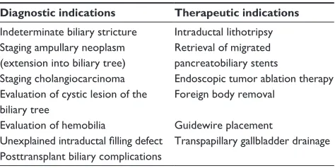
the ability to visualize the area of abnormality and conduct direct biopsies of abnormal regions is an advantage of this modality over standard techniques. The other less-common indications for cholangioscopy are listed in Table 1.
Biliary strictures
An indeterminate stricture is defined by the absence of a visible mass on imaging and/or nondiagnostic results from
Dovepress
Xu and Kahaleh
Clinical and Experimental Gastroenterology downloaded from https://www.dovepress.com/ by 118.70.13.36 on 20-Aug-2020
initial ERCP with brushing and/or biopsy. ERCP with brushing has a reported sensitivity of detecting malignancy between 23% and 56% despite the high specificity of
.95%.18–21 One of the possible causes of the poor diagnostic
yield of fluoroscopically guided brushing and biopsy is the imprecise targeting of the stricture because of the location of the stricture and/or difficulty with sampling in a tight area,
especially around the hilum.22,23 Cholangioscopy offers the
advantage of direct visual inspection of the suspected stric-ture to assess for visual characteristics of malignancy and target biopsies in those areas of interest. Various imaging characteristics have been proposed for malignant-appearing strictures on cholangioscopy, including dilated and tortuous “tumor vessels”, intraductal nodules or papillary projec-tions, ulcerated or infiltrative stricture, and irregular vascular
patterns with irregular mucosal surface.23 The strongest
feature suggestive of malignancy is the presence of dilated and tortuous vessels with a reported specificity and positive
predictive value of 100%.3,5,24 In Kim et al’s5 prospective
study of 63 patients with indeterminate strictures undergo-ing Spyglass cholangioscopy, usundergo-ing the tumor vessel sign as a predictor of malignancy had a sensitivity of 61% and specificity of 100% for malignancy. However, there have been some concerns raised about the interobserver agree-ment and reliability in assessing for malignancy based on the visual impression component alone. In a retrospective
study25 involving multiple, blinded, expert endoscopists
evaluating video clips of 38 cholangioscopies conducted for biliary strictures, the interobserver agreement on malignant versus benign strictures was only fair, although at the time of the study, no standard visual impression criteria had been established and the features used were slightly different than the more recently suggested criteria. The largest prospective, multicenter, observational study of the operating
characteris-tics of the SOC system by Chen et al6 included 226 patients
with biliary strictures (not all were cytology negative) and it reported sensitivity, specificity, positive predictive value,
and negative predictive value for malignancy of 78%, 82%, 80%, and 80%, respectively, based on the visual impression of stricture etiology. Visual impression had higher sensitivity compared to visually targeted biopsy, which was only 47%, although biopsy specificity was much higher at 98% with a positive predictive value of 100%. Other smaller prospective series of patients with indeterminate strictures have shown similar results, with sensitivity reported to be as high as 95% and specificity 79% for SOC visual impression and SpyBite
biopsies exhibiting 82% sensitivity and 82% specificity.26
The other novel component of the SOC system is the ability to perform visually directed biopsies. Only one
pub-lished study27 to date has directly compared the diagnostic
yield of SpyBite biopsy to brush cytology and fluoroscopy-based endobiliary biopsy. Although the study sample was small (26 patients), it clearly demonstrated the superior performance of cholangioscopy-guided biopsy to standard tissue sampling technique, with sensitivity for malignancy of 76.5% versus 29.4% for forceps biopsy, as well as diagnostic
accuracy of 84.6% versus 53.8% for forceps biopsy.27 These
results reflect the ability of cholangioscopy to better target the abnormal areas for biopsy relative to fluoroscopy-based sampling. Direct POC using ultraslim endoscope may have an advantage in providing larger biopsy samples than the miniforceps used in SOC, but there are currently no head-to-head data comparing the diagnostic yield of these two types of cholangioscopy.
One special population of patients in whom the evalua-tion of biliary strictures is particularly difficult is the group with primary sclerosing cholangitis (PSC). Cholangioscopy has been studied in a relatively small number of patients with PSC and further investigation of its role in this population is still needed. Although SOC can be helpful in identifying coincidental choledocholithiasis that presents as a dominant stricture, in many cases, the chronic inflammatory nature of PSC makes the visual distinction between cholangio carcinoma
(CCA) and a benign inflammatory stricture difficult.28 One
prospective series29 that included 53 patients with PSC with
dominant strictures evaluated by both standard ERCP and cholangioscopy found the visual impression of cholangio-scopy to have higher sensitivity (92% versus 66%), specificity (93% versus 51%), and accuracy (93% versus 55%) for CCA compared to ERCP cholangiogram impression. Large, multi-center studies using uniform visual impression criteria are still needed to confirm these published results.
Several adjunctive imaging techniques have also been investigated for concurrent use with SOC, including NBI and chromocholangioscopy, borrowing from the application of
Table 1 Clinical applications of cholangioscopy
Diagnostic indications Therapeutic indications
indeterminate biliary stricture intraductal lithotripsy Staging ampullary neoplasm
(extension into biliary tree)
Retrieval of migrated pancreatobiliary stents
Staging cholangiocarcinoma Endoscopic tumor ablation therapy Evaluation of cystic lesion of the
biliary tree
Foreign body removal
Evaluation of hemobilia Guidewire placement
Unexplained intraductal filling defect Transpapillary gallbladder drainage Posttransplant biliary complications
Dovepress Technical and clinical developments in choledochoscopy
Clinical and Experimental Gastroenterology downloaded from https://www.dovepress.com/ by 118.70.13.36 on 20-Aug-2020
these imaging techniques to detect dysplasia in other regions of the gastrointestinal tract. NBI uses the shorter bandwidth of spectral transmittance for optical imaging, resulting in the preferential enhancement of the pit and capillary
patterns of the superficial gastrointestinal mucosa.30 In the
biliary tract, NBI-enhanced cholangioscopy has been used to better delineate and detect neovascularization associated
with CCA, compared to white light cholangioscopy.31 The
largest prospective, multicenter case series32 of patients
who underwent POC with NBI for indeterminate strictures included 38 patients and found the diagnostic sensitivity of NBI-enhanced cholangioscopy for CCA to be 96%, specific-ity to be 80%, and accuracy to be 92%. However, a significant limitation of NBI cholangioscopy is that both bile and blood will appear dark or dark red on NBI, making their differentia-tion difficult and necessitating time-consuming lavage of the
biliary tree to achieve high-quality images.31 Additionally,
although NBI may improve the visualization of tumor vascu-larization, NBI-directed biopsies did not actually increase the diagnosis of malignancy or dysplasia compared to standard
POC in one prospective study33 of patients with PSC, raising
questions about the applicability of this technique.
Chromocholangioscopy is done with the injection of methylene blue (0.15%–0.1%) via an accessory channel into the biliary tract, followed by continuous lavage of the biliary epithelium to observe differences in surface staining between neoplastic and nonneoplastic tissues. The small number of case series describing this technique shows that normal biliary epithelium usually stains homogeneously, whereas neoplastic mucosa has heterogeneous uptake of the dye, resulting in patchy staining, and acutely inflamed tissue
stains intensely dark blue.34,35 Both of these image-enhanced
cholangioscopy techniques hold promising possibilities for better ways to visualize the contrast between normal and cancerous tissues in the biliary tract but are both still in the investigational stages of use, are not widely available, and require expertise in their image interpretation.
Bile duct stones
Although conventional ERCP with mechanical lithotripsy and stone retrieval remains the first-line therapy for bile duct stones, cholangioscopy-guided electrohydraulic lithotripsy (EHL) or laser lithotripsy has become a well-established technique for large stones and those that fail ERCP-based removal. Cholangioscopy can also detect bile duct stones that
were missed on cholangiogram during ERCP.26,28
Cholangio-scopy-guided lithotripsy has several distinct advantages: it can be performed during conventional ERCP as an adjunctive
therapy, the thinner probes can be maneuvered to reach intra-hepatic stones in otherwise-difficult locations, and it allows direct visualization of ductal clearance and examination for residual stones after stone extraction. The success and safety of cholangioscopy-guided EHL have been well established
in both retrospective and prospective studies,6,36–38 with stone
clearance rates between 90% and 100%.
Cholangioscopy-guided laser has also been described; in one series39 of patients
failing previous ERCP stone removal, complete stone clear-ance was achieved in 97% of patients with laser lithotripsy.
Other applications of choledochoscopy
Because choledochoscopy provides unprecedented visual and direct access to the biliary system, it has allowed endoscopists to expand its use to some unique clinical problems, including
evaluation of cystic lesions of the bile duct,40 tumor staging
in ampullary cancers,41 unexplained hemobilia,42 and
evalu-ation of post-liver-transplant strictures.43 Therapeutically,
cholangioscopy has been used in transpapillary gallbladder
drainage,44 retrieval of migrated biliary stents,45 foreign
body removal from the biliary tree,46 and tumor ablation
therapies using argon plasma coagulation and intraductal
radiotherapy,47 as well as photodynamic therapy.48
Complications and limitations
The major limitations to consider when using cholangio-scopy are the need for biliary sphincterotomy to advance the system into the biliary tree and the possible increase in rate
of complications. The largest prospective, multicenter study6
of diagnostic and therapeutic cholangioscopy reported seri-ous procedural complications in 7.5% and 6.1% of patients undergoing diagnostic cholangioscopy and cholangioscopy-guided EHL, respectively, with the most frequent adverse event being cholangitis, which occurred in 3.1% of patients.
A later study by Sethi et al49 confirmed a complication rate of
7.0% when cholangiopancreatoscopy was performed versus the conventional ERCP complication rate of 2.9%, with the difference attributed to a higher incidence of cholangitis; the rates of pancreatitis and perforation were not significantly
different between the two groups.49 Use of prophylactic
antibiotics is therefore recommended when cholangioscopy is performed. Finally, direct cholangioscopy is currently only used in select centers because of the high cost of the equip-ment and expertise required for its use.
Conclusion
The advent of direct cholangioscopy marks a significant step forward in the diagnosis and therapy of biliary diseases.
Dovepress
Xu and Kahaleh
Clinical and Experimental Gastroenterology downloaded from https://www.dovepress.com/ by 118.70.13.36 on 20-Aug-2020
It has allowed endoscopists to visualize the biliary system in unprecedented detail and deliver targeted therapy. POC is a promising adjunctive tool in the evaluation of indeterminate biliary strictures, which remains one of the most difficult clinical conundrums in biliary endoscopy. One of the main areas for future research is development of refined visual criteria for malignancy in SOC. It also remains to be seen whether cholangioscopy can gain widespread adoption given its current high cost, the level of expertise required for use, and other advanced imaging tools that are also in use or are being developed.
Disclosure
Dr Kahaleh has received consulting fee from Boston Scientific and research funding from Boston Scientific, Gore, Pinnacle, Cook, Maunakea and Xlumina. Dr Xu reports no conflicts of interest in this work.
References
1. Igarashi Y, Okano N, Ito K, Suzuki T, Mimura T. Effectiveness of peroral cholangioscopy and narrow band imaging for endoscopically diagnosing the bile duct cancer. Dig Endosc. 2009;21:S101–S102.
2. Parsi MA, Stevens T, Collins J, Vargo JJ. Utility of a prototype peroral video cholangioscopy system with narrow-band imaging for evaluation of biliary disorders (with videos). Gastrointest Endosc. 2011;74:1148–1151.
3. Nimura Y, Kamiya J. Cholangioscopy. Endoscopy. 1998;30:182–188. 4. Osanai M, Itoi T, Igarashi Y, et al. Peroral video cholangioscopy to
evaluate indeterminate bile duct lesions and preoperative mucosal cancerous extension: a prospective multicenter study. Endoscopy. 2013;45:635–642.
5. Kim HJ, Kim MH, Lee SK, Yoo KS, Seo DW, Min YI. Tumor vessel: a valuable cholangioscopic clue of malignant biliary stricture. Gastrointest Endosc. 2000;52:635–638.
6. Chen YK, Parsi MA, Binmoeller KF, et al. Single-operator cholangio-scopy in patients requiring evaluation of bile duct disease or therapy of biliary stones (with videos). Gastrointest Endosc. 2011;74:805–814. 7. Takekoshi T, Takagi K. Retrograde pancreatocholangioscopy [in
Japanese with English abstract]. Gastroenterol Endosc. 1975;17: 678–683.
8. Igarashi Y, Okano N, Sato D, et al. Peroral cholangioscopy using a new thinner videoscope (CHF-B260). Dig Endosc. 2005;17:S57–S59. 9. Larghi A, Waxman I. Endoscopic direct cholangioscopy by using an
ultra-slim upper endoscope: a feasibility study. Gastrointest Endosc. 2006;63:853–857.
10. Moon JH, Ko BM, Choi HJ, et al. Intraductal balloon-guided direct peroral cholangioscopy with an ultraslim upper endoscope (with videos). Gastrointest Endosc. 2009;70:297–302.
11. Meves V, Ell C, Pohl J. Efficacy and safety of direct transnasal cholangio-scopy with standard ultraslim endoscopes: results of a large cohort study. Gastrointest Endosc. 2014;79:88–94.
12. Pohl J, Meves VC, Mayer G, Behrens A, Frimberger E, Ell C. Prospective randomized comparison of short-access mother-baby cholangioscopy versus direct cholangioscopy with ultraslim gastroscopes. Gastrointest Endosc. 2013;78:609–616.
13. Efthymiou M, Raftopoulos S, Antonio Chirinos J, May GR. Air embolism complicated by left hemiparesis after direct cholangioscopy with an intraductal balloon anchoring system. Gastrointest Endosc. 2012;75:221–223.
14. Choi HJ, Moon JH, Ko BM, et al. Overtube-balloon-assisted direct peroral cholangioscopy by using an ultra-slim upper endoscope (with videos). Gastrointest Endosc. 2009;69:935–940.
15. Takaoka M, Shimatani M, Ikeura T, et al. Diagnostic and therapeutic procedure with a short double-balloon enteroscope and cholangioscopy in a patient with acute cholangitis due to hepatolithiasis. Gastrointest Endosc. 2009;70:1277–1279.
16. Matsushita M, Shimatani M, Takaoka M, Okazaki K. Effective peroral direct cholangioscopy with an ultraslim gastroscope in combination with a short double-balloon enteroscope for altered GI anatomy. Gastrointest Endosc. 2012;76:1075.
17. Navaneethan U, Njei B, Lourdusamy V, Konjeti R, Vargo JJ, Parsi MA. Comparative effectiveness of biliary brush cytology and intraductal biopsy for detection of malignant biliary strictures: a systematic review and meta-analysis. Gastrointest Endosc. 2015;81:168–176.
18. Burnett AS, Calvert TJ, Chokshi RJ. Sensitivity of endoscopic retro-grade cholangiopancreatography standard cytology: 10-y review of the literature. J Surg Res. 2013;184:304–311.
19. Ponchon T, Gagnon P, Berger F, et al. Value of endobiliary brush cytology and biopsies for the diagnosis of malignant bile duct stenosis: results of a prospective study. Gastrointest Endosc. 1995;42:565–572. 20. Glasbrenner B, Ardan M, Boeck W, Preclik G, Möller P, Adler G.
Prospec-tive evaluation of brush cytology of biliary strictures during endoscopic retrograde cholangiopancreatography. Endoscopy. 1999;31:712–717. 21. Jailwala J, Fogel EL, Sherman S, et al. Triple-tissue sampling at ERCP in
malignant biliary obstruction. Gastrointest Endosc. 2000;51:383–390. 22. De Bellis M, Sherman S, Fogel E, et al. Tissue sampling at ERCP
in suspected malignant biliary stricture. Gastrointes Endosc. 2002; 56:552–561.
23. Seo DW, Lee SK, Yoo KS, et al. Cholangioscopic findings in bile duct tumors. Gastrointest Endosc. 2000;52:630–634.
24. Itoi T, Neuhaus H, Chen YK. Diagnostic value of image-enhanced video cholangiopancreatoscopy. Gastrointest Endosc Clin N Am. 2009;19:557–566.
25. Sethi A, Widmer J, Shah NL, et al. Interobserver agreement for evalu-ation of imaging with single operator choledochoscopy: what are we looking at? Dig Liver Dis. 2014;46:518–522.
26. Ramchandani M, Reddy DN, Gupta R, et al. Role of single-operator peroral cholangioscopy in the diagnosis of indeterminate biliary lesions: a single-center, prospective study. Gastrointest Endosc. 2011;74:511–519.
27. Draganov PV, Chauhan S, Wagh MS, et al. Diagnostic accuracy of conventional and cholangioscopy-guided sampling of indeterminate biliary lesions at the time of ERCP: a prospective, long-term follow-up study. Gastrointest Endosc. 2012;75:347–353.
28. Awadallah NS, Chen YK, Piraka C, Antillon MR, Shah RJ. Is there a role for cholangioscopy in patients with primary sclerosing cholangitis? Am J Gastroenterol. 2006;101:284–291.
29. Tischendorf JJ, Krüger M, Trautwein C, et al. Cholangioscopic char-acterization of dominant bile duct stenoses in patients with primary sclerosing cholangitis. Endoscopy. 2006;38:665–669.
30. Gono K, Obi T, Yamaguchi M, et al. Appearance of enhanced tissue features in narrow-band endoscopic imaging. J Biomed Opt. 2004;9:568–577.
31. Itoi T, Sofuni A, Itokawa F, et al. Peroral cholangioscopic diagnosis of biliary tract diseases by using narrow-band imaging (with videos). Gastrointest Endosc. 2007;66:730–736.
32. Osanai M, Itoi T, Igarashi Y, et al. Peroral video cholangioscopy to evaluate indeterminate bile duct lesions and preoperative mucosal cancerous extension: a prospective multicenter study. Endoscopy. 2013;45:635–642.
33. Azeem N, Gostout CJ, Knipschield M, Baron TH. Cholangioscopy with narrow-band imaging in patients with primary sclerosing cholangitis undergoing ERCP. Gastrointest Endosc. 2014;79:773–779.
34. Hoffman A, Kiesslich R, Bittinger F, Galle PR, Neurath MF. Methylene blue-aided cholangioscopy in patients with biliary strictures: feasibility and outcome analysis. Endoscopy. 2008;40:563–571.
Dovepress Technical and clinical developments in choledochoscopy
Clinical and Experimental Gastroenterology downloaded from https://www.dovepress.com/ by 118.70.13.36 on 20-Aug-2020
35. Maetani I, Ogawa S, Sato M, Igarashi Y, Sakai Y, Shibuya K. Lack of methylene blue staining in superficial epithelia as a possible marker for superficial lateral spread of bile duct cancer. Diagn Ther Endosc. 1996;3:29–34.
36. Chen YK, Pleskow DK. SpyGlass single-operator peroral cholangio-pancreatoscopy system for the diagnosis and therapy of bile-duct disorders: a clinical feasibility study (with video). Gastrointest Endosc. 2007;65:832–841.
37. Farrell JJ, Bounds BC, Al-Shalabi S, et al. Single-operator duo-denoscope-assisted cholangioscopy is an effective alternative in the management of choledocholithiasis not removed by conventional methods, including mechanical lithotripsy. Endoscopy. 2005;37: 542–547.
38. Piraka C, Shah RJ, Awadallah NS, Langer DA, Chen YK. Transpapillary cholangioscopy-directed lithotripsy in patients with difficult bile duct stones. Clin Gastroenterol Hepatol. 2007;5:1333–1338.
39. Patel SN, Rosenkranz L, Hooks B, et al. Holmium-yttrium alumi-num garnet laser lithotripsy in the treatment of biliary calculi using single-operator cholangioscopy: a multicenter experience (with video). Gastrointest Endosc. 2014;79:344–348.
40. Warmann S, Meier PN, Kardorff R, Fuchs J. Cystic echinococcosis with perforation into the biliary tract in an eight-year-old girl. Eur J Pediatr Surg. 2002;12:134–137.
41. Cennamo V, Luigiano C, Fabbri C, et al. Cholangioscopy using a new type of cholangioscope for the diagnosis of biliary tract disease: a case series. Endoscopy. 2012;44:878–881.
42. Prasad GA, Abraham SC, Baron TH, Topazian MD. Hemobilia caused by cytomegalovirus cholangiopathy. Am J Gastroenterol. 2005;100:2592–2595.
43. Siddique I, Galati J, Ankoma-Sey V, et al. The role of choledochoscopy in the diagnosis and management of biliary tract diseases. Gastrointest Endosc. 1999;50:67–73.
44. Barkay O, Bucksot L, Sherman S. Endoscopic transpapillary gall-bladder drainage with the SpyGlass cholangiopancreatoscopy system. Gastrointest Endosc. 2009;70:1039–1040.
45. Kantsevoy SV, Frolova EA, Thuluvath PJ. Successful removal of the proximally migrated pancreatic winged stent by using the SpyGlass visualization system. Gastrointest Endosc. 2010;72:454–455. 46. Ransibrahmanakul K, Hasyagar C, Prindiville T. Removal of bile duct
foreign body by using spyglass and spybite. Clin Gastroenterol Hepatol. 2010;8(1):e9.
47. Lu XL, Itoi T, Kubota K. Cholangioscopy by using narrow-band imaging and transpapillary radiotherapy for mucin-producing bile duct tumor. Clin Gastroenterol Hepatol. 2009;7:e34–e35.
48. Talreja JP, DeGaetani M, Sauer BG, Kahaleh M. Photodynamic therapy for unresectable cholangiocarcinoma: contribution of single-operator cholangioscopy for targeted treatment. Photochem Photobiol Sci. 2011;10:1233–1238.
49. Sethi A, Chen YK, Austin GL, et al. ERCP with cholangio-pancreatoscopy may be associated with higher rates of complications than ERCP alone: a single-center experience. Gastrointest Endosc. 2011;73:251–256.
Clinical and Experimental Gastroenterology
Publish your work in this journal
Submit your manuscript here: http://www.dovepress.com/clinical-and-experimental-gastroenterology-journal Clinical and Experimental Gastroenterology is an international,
peer-reviewed, open access journal, publishing all aspects of gastroenterology in the clinic and laboratory, including: Pathology, pathophysiology of gastrointestinal disease; Investigation and treatment of gastointes-tinal disease; Pharmacology of drugs used in the alimentary tract;
Immunology/genetics/genomics related to gastrointestinal disease. This journal is indexed on CAS. The manuscript management system is completely online and includes a very quick and fair peer-review system. Visit http://www.dovepress.com/testimonials.php to read real quotes from published authors.
Dovepress
Dove
press
Xu and Kahaleh
Clinical and Experimental Gastroenterology downloaded from https://www.dovepress.com/ by 118.70.13.36 on 20-Aug-2020