1 Type of the Paper (Article)
1
Establishment of micro droplet digital polymerase
2chain reaction and real-time fluorescence
3quantitative polymerase chain reaction
4technologies for detecting Zika virus
5Yuan Hui1+, Zhiming Wu1+, Zhiran Qin1+, Li Zhu1+, Junhe Liang 2, Xujuan Li1+, Hanmin Fu1+, Shiyu 6
Feng1+, Weiwei Xiao1+, Qinghua Wu1+, Bao Zhang1+,*, Wei Zhao1,3* 7
1 Guangdong Provincial Key Laboratory of Tropical Disease Research, School of Public 8
Health, Southern Medical University, No. 1023, South Shatai Road, Guangzhou, 9
Guangdong Province 510515, 10 China;[email protected](Y.H.);[email protected](Z.M.W.);baobaofei666666@outlook 11 .com(Z.R.Q.);[email protected](L.Z.);[email protected](X.J.L.);[email protected](M.H.F.) 12 ;[email protected](S.Y.F.);[email protected](W.W.X.);[email protected](Q.H.W.) 13
2 Jiangmen Center for Disease Control and Prevention, No.10, Xinghe Roas, Jiangmen, 14
Guangdong Province 529085, China;[email protected](J.H.L) 15
3 Guangzhou Key Laboratory of Drug Research for Emerging Virus Prevention and 16
Treatment, School of Pharmacy, Southern Medical University, No. 1023, South Shatai Road, 17
Guangzhou, Guangdong Province 510515, China. 18
* Correspondence: [email protected](W.Z.) and [email protected](B.Z.); Tel.: 19
+86-020-6164-8567 20
+ These authors contributed equally to this work. 21
Abstract: Establishment of diagnostic methods with low detection limits plays a critical role 22
in the maintenance of early diagnosis, prevention of serious neurological complications, and 23
control of the spread of ZIKA. In this study, we established the micro-droplet digital 24
polymerase chain reaction (ddPCR) and real-time fluorescent quantification PCR (qPCR) 25
plasmid, the standard curve of R2 was 0.999, and the amplification efficiency was 92.203%, as 27
determined by qPCR. Both ddPCR and qPCR were positive for cell culture of Zika nucleic 28
acid.The minimum detection limit of ddPCR is 1–2 times lower than qPCR. Moreover, all 29
tests of Dengue virus (1–4 serotypes) were negative in cell culture. Overall, these results 30
suggested than ddPCR may have a lower limit of detection than qPCR. 31
Keywords: Zika virus; nucleic acid detection; micro-droplet digital polymerase chain reaction; 32
real-time fluorescence quantitative polymerase chain reaction 33
___________________________________________________________________________________ 34
1. Introduction 35
Zika virus (ZIKA) is a single-stranded RNA virus belonging to the family Flaviviride genus 36
Flavivirus. It was first isolated from a forest in Uganda in 1947 [1] and encodes three structural 37
proteins (capsid protein C, precursor protein pr M, and envelope protein E) and seven 38
nonstructural proteins (NS1, NS2A, NS2B, NS3, NS4A, NS4B, and NS5) [2]. 39
The NS5 gene is the largest gene in the ZIKA genome and encodes NS5 protein, which 40
has the highest relative molecular weight of all proteins encoded by ZIKA. NS5 contains an 41
RNA-dependent RNA polymerase active site at the N terminal and harbors a methylation site, 42
which is associated with the viral cap reaction of the viral RNA. The ZIKA genome has only 43
one open reading frame (ORF). NS5 is located at the 3′ end of the ORF and is a nucleic acid 44
detection target commonly used by the flavivirus [3]. Currently, several laboratories have 45
established nucleic acid detection technologies for ZIKA; the main targets of detection are the 46
E and NS5 genes [4]. The E gene has undergone several adaptive changes, whereas the NS5 47
gene is relativelystable. Therefore, the conserved sequence of NS5 gene may be more reliable 48
as a detection target. 49
albopictus[5], and can also spread between independent mosquitoes [6]. More importantly, the 51
virus can be transmitted from mother to fetus via amniotic fluid and fetal tissues [7] and can 52
be transmitted through sexual activity [8]. In humans, ZIKA infection is characterized by mild 53
fever (37.8–38.5 ), ℃ 3oint pain (particularly pain in the hands and feet), myalgia, headache, 54
orbital pain, con3unctivitis, and skin rash and can also cause microcephaly and Guillain-Barré 55
syndrome [9]. However, only 20% of infected individuals exhibit symptoms [10],and ZIKA 56
infection can be a benign, self-limiting disease [11]. As a result, the incidence of ZIKA 57
infection may be underestimated in epidemics or returning travelers. For the detection of 58
ZIKA, Giada Rossini etal [12] showed that the blood test period is longer after onset of the 59
disease than urine or whole blood tests. ZIKA can be detected in whole blood in about 3–26 60
days and in the plasma in 3–10 days [13]. 61
Accordingly, in this study, we aimed to establish micro-droplet digital polymerase chain 62
reaction (ddPCR) and real-time fluorescent quantification PCR (qPCR) protocols for the 63
detection of Zika virus based on the NS5 gene. 64
2.Materials and methods 65
2.1 Sample processing 66
Asian Zika virus Z16006 strain was provided by the Institute of Microbiology in the Center 67
for Disease Control and Prevention of Guangdong Province, China (GenBank no. 68
KU955589.1). Four serotypes of Dengue virus (DV1 Hawaii strain, DV2 NGC strain, DV3 H87 69
strain, and DV4 H241 strain) were preserved in our laboratory. The above viruses were all 70
repeatedly frozen and thawed three times, centrifuged and frozen at -80 . Clinically positive ℃ 72
ZIKA blood samples were obtained from the Centerfor Disease Control and Prevention of 73
Jiangmen,Guangdong Province, China. 74
2.2 Instruments and reagents 75
The micro-ddPCR instrument, amplification premixed reagent, Droplet PCR Supermix, and 76
micro-drop reagent were all from Bio-Rad (Hercules, CA, USA). Reverse transcription was 77
performed using a TAKARA Prime Script RT reagent Kit (Perfect Real Time kit; TaKaRa, 78
Shiga, Japan), Bestar qPCR Master Mix (TaqMan Probe) (DBI Bioscience, Germany), and a 79
QuantStudio 6 Flex Real-Time PCR System(ThermoFIsher, USA ). 80
2.3 Design of primers and probes 81
The sequences of 30 strains of ZIKA were downloaded from GenBank and compared. After 82
identifying conserved sequences, primers and probes were designed using Primer 5.0with the 83
Z16006 strain as the reference strain. The primers and probes were designed using Oligo 7. 84
2.4 RNA extraction and reverse transcription 85
Samples were brought to room temperature before use. Viral RNA was extracted using the 86
QIAamp® (Qiagen, Hilden, Germany), with a final RNA volume of 50 μL. The RNA was 87
separated into aliquots and preserved at -80 .℃ 88
For reverse transcription, the components were prepared on ice, as follows: 2 μL 89
PrimeScript RT Enzyme Mix I, 0.5 μL Oligo dT Primer, 2 μL Random 6-mers, 4 μL total RNA, 90
37 for 15 min, reverse transcriptase inactivation at 85 for 5 s, and a final hold at 4 .℃ ℃ ℃ 92
2.5 ddPCR 93
For ddPCR, the 20μL reaction system contained 10 μL ddPCR Supermix (no dUTP), 6 μL 94
primer probe premix (initial concentration of 10 μM upstream primer and 0.4 μL downstream 95
primer, probe 0.2 μL, deionized water 5 μL), and 4 μL nucleic acid extract. After mixing, 20 96
μL of the sample reaction system was added to the middle of a DGB cartridge. Next, 70 μL oil 97
was added to the bottom row of each lane to avoid the formation of bubbles, and the wells 98
were covered. The reaction system and droplet-forming cartridge were placed in a droplet 99
generator and sub3ected to micro-droplet treatment. 100
Droplets were generated in the top row of the wells, and the suction volume was ad3usted 101
to 40 μL. Samples were then slowly transferred to 96-well plates, and a preheated PX1 heat 102
sealing device was used as a sealing film (with the red line up) at 180°C for 5 s. The PCR 103
conditions were as follows: predenaturation at 95°C for 10 min, temperature change rate of 104
2°C/s; and 40 cycles of 94°C for 30 s and 55°C for 1 min, at 2°C/1°C/s. The 96-well plate 105
containing the PCR-amplified products was then placed on a QX200 micro-drop reader and 106
analyzed using Quanta Soft software. 107
2.6 Real-time fluorescent qPCR 108
For qPCR, reactions contained Bestar qPCR Master Mix (10 μL), upstream and downstream 109
primers (0.4 μL each), probe (0.2 μL), template DNA (4 μL), and ddH2O (5 μL). The 110
amplification conditions for qPCR were as follows: 95 denaturation for 2 min, follows by 40 ℃ 111
3.Results 113
3.1 Design and evaluation of primers and probes 114
We downloaded 30 popular ZIKA strains, compared them, and found conserved sequences. 115
Primers were designed according to the NS5 gene, keeping the GC content and Tm values as 116
similar as possible. The length of the target fragment was within 100 bp without hairpin 117
structures to avoid the formation of stable dimers and mismatches at the C-terminus. The 118
designed primers are shown in Table 1. 119
Table 1. Primers and probe for ZIKA detection 120
Primer/probe Sequence
ZK-F 5′-GGCRTTRGCCATCAGTCG-3′
ZK-R 5′-ATGGAGCATCCGKGAGACT-3′
Probe 5′-FAM-TGGCAGCTYCTTTATTTCCACARAAG-BHQ1-3′
121
3.2 Evaluation of the sensitivity of detection 122
Ten-fold dilutions of ZIKA plasmid standards (3.2 × 10¹⁰ copies/μL) were performed. 123
Detection by qPCR showed that the standard curve R2 value was 0.999, and the amplification 124
efficiency was 92.203%. The relationship between the Ct value and the copy number was as 125
127
Figure 1. The standard curve for fluorescent quantitative PCR. The concentration of each 128
point on the standard curve increased 10-fold from left to right. 129
The initial concentration of 7.8 × 10⁶ copies/μL was gradient diluted 5-fold from the cell 130
culture ZIKA solution, and qPCR was used to test each sample in order to evaluate the 131
detection method. The results are shown in Figure 2; a detection limit of about 100 copies/μL 132
was obtained. 133
134
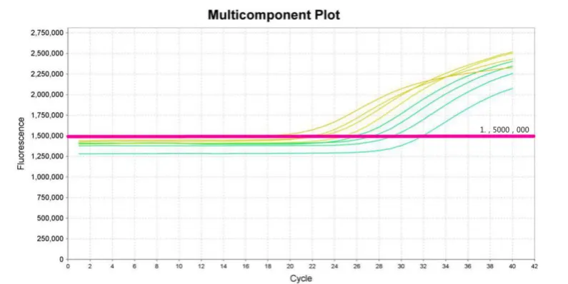
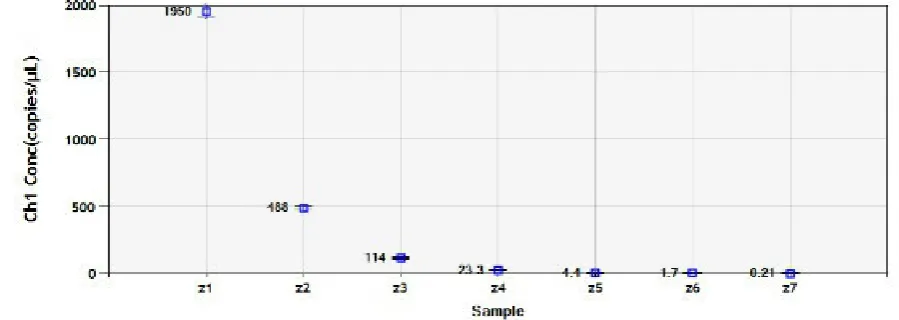
Next, we used ddPCR to detect ZIKA from cell cultures, as described above for qPCR. As 136
shown in Figure 3A, qPCR detected ZIKA with clear differences among sample 137
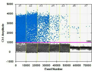
concentrations. In Figure 3B, the threshold was set to 1000, with positive nucleic acid droplets 138
showing values above 1000 and negative nucleic acid droplets showing values below 1000. As 139
indicated in Figure 3C, almost all samples generated more than 10,000 droplets, suggesting 140
that ddPCR may be more sensitive, with a detection limit of 1 copy/μL. 141
142
144
Figure 3B. Scatter plot showing event numbers. 145
146
Figure 3C. Histogram showing event numbers. 147
Figure 3. Results of ddPCR for 5-fold dilutions of cell culture. 148
3.3 Evaluation of the accuracy of qPCR and ddPCR 149
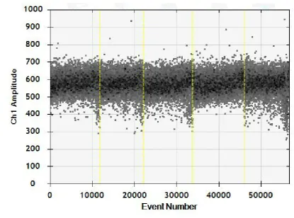
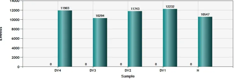
Next, we evaluated the accuracy of the assays using four Dengue virus serotypes. All of 150
nucleic acid concentrations detected by the absolute quantification method were all zero. 152
Moreover, only negative micro-droplets were observed (Figure 4B), and histogram analysis 153
showed that the total number of droplets was more than 10,000, of which none were positive 154
(Figure 4C).
155
156
Figure 4A. Absolute quantitative detection. 157
158
DV1, N; N is the negative control). 160
161
Figure 4C. Total number of micro-droplets, as shown in a histogram (positive on left and total 162
on right). 163
3.4 qPCR detection of Dengue virus samples in clinically positive blood samples 164
The lowest Ct value of clinically positive blood samples in qPCR analysis was 38.868 after 165
5-fold dilutions; this exceeded the detection range of qPCR (Ct values of 15–35; Figure 5). 166
167
Figure 5. Standard curve of qPCR. R2 = 0.998, amplification efficiency = 91.226%, Y = -3.552X + 168
39.06. The red dots are plasmid standards, and the blue dots are positive blood samples. 169
DdPCR analysis showed that the sample concentration was 14.2 copies/μL (Figure 6). 170
Figure 6A shows the scatter plot of the events, and Figure 6B shows the concentrations based 171
event number was 10923, with 131 positive events (Figure 6C). 173
174
Figure 6A. Scatter plot showing the event numbers. 175
176
Figure 6B. Concentrations determined by absolute quantification. 177
178
Figure 6. Limit of detection of ZIKA in blood samples by ddPCR. 180
4.Discussion 181
Approximately 20% of individuals infected with ZIKA may develop symptoms similar to 182
other flaviviruses [11], presenting a great challenge to clinical diagnosis. There are two types 183
of clinical diagnostic tests for ZIKA, serological and molecular detection. IgM specific 184
neutralizing antibodies and antibody capture enzyme-linked immunosorbent assays can be 185
used for qualitative detection of ZIKA IgM antibodies, but may crossreact with other 186
flaviviruses, making the results difficult to explain. Therefore, molecular biology diagnosis 187
has become a commonly used means of laboratory testing. 188
In molecular biology, traditional PCR can only roughly detect the amount of 189
amplification after the reaction has ended and cannot be used to quantitatively detect nucleic 190
acids in the sample. Real-time fluorescence qPCR technology utilizes changes in the 191
fluorescence intensity of chemical substances in the reaction system to realize quantitative 192
detection of nucleic acids. Xu et [1]al [14] used one-step SYBR Green real-time PCR for the 193
detection of ZIKA with a detection limit of at least 1.0 PFU/mL (1 PFU is approximately equal 194
to 2 × 10⁵ RNA genome copies). However, this method allows the simultaneous detection of 195
both specific and nonspecific PCR products and therefore produces false positives. Calvert et 196
al. [15] used reverse transcriptase loop-mediated isothermal amplification to detect RNA from 197
ZIKA as low as 1.2 copies/μL; however, they observed a very high false-negative rate. Our 198
laboratory uses a probe-based real-time PCR method with a detection limit of approximately 199
Micro-ddPCR detects nucleic acid molecules without relying on external standard curves 201
based on the Poisson distribution principle. With the advantage of absolute quantitative, this 202
method permits better accuracy at low concentrations without the need for preparing 203
standard curves [16]. 204
In this study, we designed specific primers and probes based on the NS5 gene of ZIKA and 205
established a method for ZIKA nucleic acid detection using ddPCR and qPCR. Our results 206
showed that good specificity could be obtained by designing primers and probes based on 207
the NS5 gene and that these primers/probes could be used for detection the differential ZIKA 208
and four serotypes of Dengue virus. Moreover, our findings demonstrated that ddPCR had 209
good sensitivity but was inaccurate for samples with high concentrations. Thus, ddPCR may 210
be more suitable for low viral loads. Thus, ZIKA, as well as four serotypes of Dengue virus, 211
can be detected by analyzing the NS5 gene at concentrations of about 1–105 copies/μL. 212
In summary, in clinical samples with low concentrations of ZIKA, such as those at 1–3 days 213
after infection, micro-droplet digital ddPCR may show the best diagnostic accuracy and 214
sensitivity, whereas in routine analyses laboratory of viral nucleic acid detection, including 215
analysis of clinical samples at more than 3 days after infection, fluorescence qPCR can be 216
used. 217
Acknowledgements : This study was supported by the National Natural Science Foundation of China
218
(No. 31470271, 81730110) and Guangzhou Science and Technology Program key pro3ects (No.
219
201508020263).We thank Changwen Ke, De Wu and Jiufeng Sun from Guang Dong Center for Disease
220
Control and Prevention for providing with Asian Zika virus Z16006 strain.
Author Contributions : Yuan Hui, Bao Zhang, Weiwei Xiao and Qinghua Wu conceived and designed
222
the experiments, Wei Zhao and Junhe Liang contributed reagents/materials/analysis tools; Zhiming Wu,
223
Zhiran Qin and Li Zhu performed the experiments, designed the experiments; Xu3uan Li, Hanmin Han
224
and Shiyu Feng performed part of the experiments, analyzed the data and wrote the paper.
225
Competing interests : The authors declare no conflicts of interest. 226
References 227
[1]Dick, G. W. A. (1952). Zika Virus (I). Isolations and serological specificity. Transactions of the Royal
228
Society of Tropical Medicine and Hygiene, 46(5), 509–520. https://doi.org/10.1016/0035-9203(52)90042-4
229
[2] Fontes-Garfias, C. R., Shan, C., Luo, H., Muruato, A. E., Medeiros, D. B. A., Mays, E., … Shi, P. Y.
230
(2017). Functional Analysis of Glycosylation of Zika Virus Envelope Protein. Cell Reports, 21(5),
231
1180–1190. https://doi.org/10.1016/3.celrep.2017.10.016 232
[3] Faye, O., Freire, C. C. M., Iamarino, A., Faye, O., de Oliveira, J. V. C., Diallo, M., … Sall, A. A. (2014).
233
Molecular evolution of Zika virus during its emergence in the 20(th) century. PLoS Neglected Tropical
234
Diseases, 8(1). https://doi.org/10.1371/3ournal.pntd.0002636 235
[4] Gourinat, A. C., O’Connor, O., Calvez, E., Goarant, C., & Dupont-Rouzeyrol, M. (2015). Detection of
236
zika virus in urine. Emerging Infectious Diseases, 21(1), 84–86. https://doi.org/10.3201/eid2101.140894 237
[5] Weaver, S. C., Costa, F., Garcia-Blanco, M. A., Ko, A. I., Ribeiro, G. S., Saade, G., … Vasilakis, N.
238
(2016). Zika virus: History, emergence, biology, and prospects for control. Antiviral Research.
239
https://doi.org/10.1016/3.antiviral.2016.03.010
240
[6] Chouin-Carneiro, T., Vega-Rua, A., Vazeille, M., Yebakima, A., Girod, R., Goindin, D., … Failloux, A.
241
B. (2016). Differential Susceptibilities of Aedes aegypti and Aedes albopictus from the Americas to Zika
242
Virus. PLoS Neglected Tropical Diseases, 10(3). https://doi.org/10.1371/3ournal.pntd.0004543 243
[7] Dupont-Rouzeyrol, M., Biron, A., OConnor, O., Huguon, E., & Descloux, E. (2016). Erratum:
244
Infectious Zika viral particles in breastmilk (The Lancet (2016) 387 (1051)). The Lancet.
245
https://doi.org/10.1016/S0140-6736(16)00663-2
246
[8] Calvet, G., Aguiar, R. S., Melo, A. S. O., Sampaio, S. A., de Filippis, I., Fabri, A., … de
247
Filippis, A. M. B. (2016). Detection and sequencing of Zika virus from amniotic fluid of fetuses with
248
microcephaly in Brazil: a case study. The Lancet Infectious Diseases, 16(6), 653–660.
249
https://doi.org/10.1016/S1473-3099(16)00095-5
250
[9] Chouin-Carneiro, T., Vega-Rua, A., Vazeille, M., Yebakima, A., Girod, R., Goindin, D., … Failloux, A.
251
B. (2016). Differential Susceptibilities of Aedes aegypti and Aedes albopictus from the Americas to Zika
252
Virus. PLoS Neglected Tropical Diseases, 10(3). https://doi.org/10.1371/3ournal.pntd.0004543
[10] Fellner, C. (2016). Zika Virus: Anatomy of a Global Health Crisis. P & T : A Peer-Reviewed Journal
254
for Formulary Management, 41(4), 242–53. Retrieved from
255
http://www.pubmedcentral.nih.gov/articlerender.fcgi?artid=4811255&tool=pmcentrez&rendertype=abst
256
ract
257
[11] Duffy, M. R., Chen, T.-H., Hancock, W. T., Powers, A. M., Kool, J. L., Lanciotti, R. S., … Hayes, E. B.
258
(2009). Zika virus outbreak on Yap Island, Federated States of Micronesia. The New England Journal of 259
Medicine, 360(24), 2536–2543. https://doi.org/10.1056/NEJMoa0805715 260
[12] Rossini, G., Gaibani, P., Vocale, C., Cagarelli, R., & Landini, M. P. (2017). Comparison of zika virus
261
(zikv) rna detection in plasma, whole blood and urine - case series of travel-associated zikv infection
262
imported to italy, 2016. Journal of Infect, 75(3), 242-245.https://doi.org/10.1016/3.3inf.2017.05.021
263
[13] Hayes, E. B. (2009). Zika virus outside Africa. Emerging Infectious Diseases.
264
https://doi.org/10.3201/eid1509.090442
265
[14] Xu, M. Y., Liu, S. Q., Deng, C. L., Zhang, Q. Y., & Zhang, B. (2016). Detection of Zika virus by SYBR
266
green one-step real-time RT-PCR. Journal of Virological Methods, 236, 93–97.
267
https://doi.org/10.1016/3.3viromet.2016.07.014
268
[15] Calvert, A. E., Biggerstaff, B. J., Tanner, N. A., Lauterbach, M., & Lanciotti, R. S. (2017). Rapid
269
colorimetric detection of Zika virus from serum and urine specimens by reverse transcription
270
loop-mediated isothermal amplification (RT-LAMP). PLoS ONE, 12(9).
271
https://doi.org/10.1371/3ournal.pone.0185340
272
[16] Strain, M. C., Lada, S. M., Luong, T., Rought, S. E., Gianella, S., Terry, V. H., … Richman, D. D. (2013).
273
Highly Precise Measurement of HIV DNA by Droplet Digital PCR. PLoS ONE, 8(4).
274
https://doi.org/10.1371/3ournal.pone.0055943