© 2019, IJCSMC All Rights Reserved 120
International Journal of Computer Science and Mobile Computing
A Monthly Journal of Computer Science and Information Technology
ISSN 2320–088X
IMPACT FACTOR: 6.199
IJCSMC, Vol. 8, Issue. 7, July 2019, pg.120 – 128
Classification Approach for
Brain Tumor Detection
Mansha
1M.Tech Scholar
USET.RBU Mohali
[email protected]
Kiranpreet Kaur
2Assistant Professor
USET.RBU Mohali
[email protected]
Abstract:- In the previous approach, weight based algorithm is used to classify the normal and cancer cells and it is been analyzed that weight based algorithm taken long time to classify the data. To classify the data in minimum amount of time HMM classifier is used for classification. The second issue with weight based algorithm is of accuracy. As due to weight calculation accuracy of classification is less which can be improved with the use of Bayesian classifier In the feature selection part on three features are used which are mass, density and margin . In the improvement more features like tissue color will be added which improve detection rate The simulation is performed in MATLAB and it is been analyzed that proposed technique performs well in terms of certain parameters.
Keywords: HMM, Brain Tumor Detection, MRI, Classification.
I. INTRODUCTION
© 2019, IJCSMC All Rights Reserved 121
the tumor generating cells move to other parts of the body is known as metastasis. From the regular tissue reinstate, the tumor initiates. The different types of brain tumors are meningioma and glioma. If the brain tumor is recognized at very early stage, it can be cured and treated. The intracranial pressure can be maximized due to which the brain can be destroyed permanently [5]. On the basis of size of tumor, location and its type, the symptoms of brain tumor reply. With the help of MRI and CT scan, the tumor can be detected. Within the brain, the brain angiogram procedure is applied within the blood vessels. The tumor part is then fed with blood. There are tissues or sample of cells included from the brain as per the procedure of biopsy. This is utilized at the time of surgical treatment such that the benign of cancerous brain tumor can be predicted. Due to few symptoms it is possible sometimes to delay or misuse the cancer diagnosis. Certain techniques are utilized for the detection of brain tumor at early age or in the later stage [6]. There are different factors that influence the reason for it furthermore there are various techniques proposed for its detection and removal. Here, there are certain image processing strategies that have been utilized as a part of distinguishing the tumor. The principle set-up systems for brain tumor control depend on prime counteractive action alongside early detection. There are two methods for brain tumor spreads in our body [7]. Tumor shapes in the tissues of brain. Early detection and appropriate medicinal registration are mandatory however in the meantime legitimate diet and sustenance additionally battles with malignant tumor cells. In human body, new cells are framed and old cells are vanishing every day. Sometimes new cells are created, become wildly and structure abnormal cell structure called tumor cells. Brain tumor diagnosis has turned into the need of great importance and examination works around there is highly testing [8]. These days there are many propelled procedures incorporated into mammography. Image enhancement is the fundamental step in image processing. Principle motivation behind image enhancement is to deliver better and quality image. Smoothing is utilized to diminish the noise, less blurring, produce a less pixelated image cleaning for the same size with no image size modification in the data. Filter is utilized to evacuate some unwanted signal or component in the image. Filters expel certain frequencies to smother meddling signals and lessen background noise. Noise is unwanted signals in the image [9]. Noise is not generally arbitrary and irregularity is a simulated term. Two plagues image acquisition are (1) Noise. (2) Blur (Out of center, Movements, Bad weather). Noise is dependably not terrible. To conquer these issues we utilize denoise. Denoise is to rectify the issue of outwardly obnoxious, awful compression and terrible examination. An image histogram offers graphical representation of tonal dispersion of gray values in digital picture. Enhancing the image quality is by utilizing the histogram equalization. Histogram equalization demonstrates the intensity values along aggregate range of value should be possible in images and background and foreground are splendid at a time.
II. LITERATURE REVIEW
Devendra Somwanshi, et.al (2016) presented brain tumor is a fatal disease whose detection and diagnosis at the early stage is required due to which a computer based image processing technique is utilized in this paper [10]. This computer based technique is MRI in which segmentation of images is done using which tumor size, location and shape can be detected easily. In the image processing, segmentation can be done using various techniques. There is an entropy based algorithmic techniques present in the threshold technique using which brain tumor can be detected at the early stage. They found threshold selection of images based on entropy methods approach very useful and effective in the diagnosis of brain tumor. On the basis of simulation results, they analyzed and compared techniques and concluded that havrda-charvat entropy performs better than any other entropy algorithms.
© 2019, IJCSMC All Rights Reserved 122
Luxit Kapoor, et.al (2017) presented with the advent in the technology, there is growth and demand of biomedical image processing field. The extraction of meaningful information and accurate information from these images with the least error possible is the main objective of medical imaging. MRI is considered as the most reliable and safe technique among various types of medical imaging processes available currently [12]. Therefore, the tumor can be segmented easily after processing MRI in it. There are several different techniques present in the tumor segmentation. In the four categories the whole process is categorized in order to detect brain tumor from an MRI such as Pre- Processing, Segmentation, Optimization and Feature Extraction. They surveyed in detailed and analyzed all the process in this paper.
Sanjivani Salwe, et.al (2016) proposed a new approach in order to detect affected mass in magnetic resonance images (MRI) [13]. Therefore, using this adaptive threshold component is further threshold. On the basis of obtained components, the original image is again reconstructed using proposed segmentation. At level one, the approximation component is unaltered. There is false detection of brain tumor due to presence of light in-homogeneity and due to hard tissues based on window based threshold for which windowing technique is used by which it is eliminated. On the basis of performed experiments, it is concluded images showed complete black region in the final segmented image showed by the normal patient while the region of interest after segmentation showed in malignant images after segmentation process.
Smita.A.Nagtode, et.al (2016) presented in this paper the utilization of discrete wavelet transform and probabilistic neural network by which brain tumor can be detected and classified. The appropriate results are provided by the variance of illumination and probabilistic neural network for classification due to the robustness of GW features against local distortions [14]. The proposed Gabor Wavelet approach is used to study all other images such as practical images in which advantage of wavelet transform used for face recognition that give exact outcomes. They proposed a two dimensional Gabor wavelet analysis application for brain images in this paper. In order to identify tumor at early stage, they classified a method for it in which images are classified into non-cancerous (benign) brain tumor and cancerous (malignant) brain tumor.
Rahul Singh, et.al (2016) presented the extraction of brain tumor in magnetic resonance (MR), for which an innovative and robust image segmentation approach was proposed in this paper. In order to classify a given MR brain image as benign or malignant, a novel technique is proposed by them. They implemented the wavelet transform in the initial stage followed by Laplacian Eigen maps (LE) in order to extract the features from given MR brain tumor image. This is done to restrict the dimensions of extracted features [15]. The Leave-one-out crosses validation (LOOCVCV) strategy also utilized by them in order to enhance generalization of K-SVM. On the basis of performed experiments, it is concluded that proposed method has better performance as compared to previously utilized methods in terms of computational and qualitative aspect. Therefore, with the help of this proposed method doctors can easily examine whether the tumors is benign or malignant.
III. RESEARCH METHODOLOGY
© 2019, IJCSMC All Rights Reserved 123
sequence. In the various areas application of HMMs can be found in signal processing such as in speech processing. In the low level NLP tasks this process can be utilized which provide successful results such as part-of-speech tagging, phrase chunking, and extracting target information from documents. In the weighted automaton, a Markov chain is a special case according to which weights are probabilities. The state of the automaton it passing through is uniquely determined by the input sequence. This Markov chain is only useful for assigning probabilities to explicit sequences as inherently ambiguous issues are not represented by it. For both observed events and hidden events, one can provide its view that what one can think of causal factors in the probabilistic model as it is allowed by this model. It is the method which provides the probability of transitioning between any two states due to which it is called as fully connected or ergodic HMM. There are some cases in HMM in which zero probability exists in various transitions beBakis network tween states. For instance, from left to right the transition state proceed in the left-to-right HMMs also called as Bakis. There are no transitions happen from a higher-numbered state to a lower-numbered state in case of above mentioned method. There is zero probability if transitions occur from a higher-numbered state to a lower numbered state. With the help of this temporal processes like speech can be modeled by it.
No
Yes
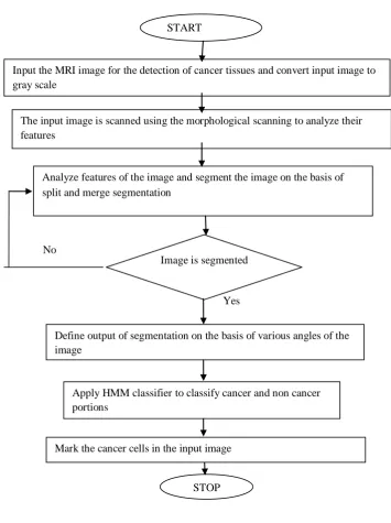
Figure 1: Proposed Flowchart
START
Input the MRI image for the detection of cancer tissues and convert input image to gray scale
The input image is scanned using the morphological scanning to analyze their features
Analyze features of the image and segment the image on the basis of split and merge segmentation
Image is segmented
Define output of segmentation on the basis of various angles of the image
Apply HMM classifier to classify cancer and non cancer portions
Mark the cancer cells in the input image
© 2019, IJCSMC All Rights Reserved 124 IV. EXPERIMENTAL RESULTS
The proposed research is implemented in MATLAB and the proposed and existing approaches are compared with each other to perform a simulation analysis.
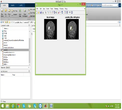
Figure 2: Tumor detection from the MR image
As shown in the figure 2, the image which is given as input is segmented to detect boundary of the image. The segmented image is given as input to remove noise from the image. The input image is analyzed horizontally and vertically to analyze image features. In the last step technique of neural networks is applied to detect tumor portion from the image.
Figure 3: HMM Classifier
© 2019, IJCSMC All Rights Reserved 125
Figure 4: Segmentation of Tumor
As shown in figure 4, the HMM classifier is applied in this work which can scan whole image diagonally and detect segment portion. The segmented portion is marked with the yellow color.
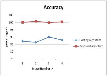
Figure 5: Accuracy analysis
As shown in figure 10, the accuracy of the proposed and existing algorithm is compared for the performance analysis.
© 2019, IJCSMC All Rights Reserved 126
Figure 6: Specificity analysis
As shown in figure 6, the specificity of the proposed and existing algorithm is compared for the performance analysis.
The specificity of proposed algorithm is due to use of HMM classifier as compared to existing algorithm.
© 2019, IJCSMC All Rights Reserved 127
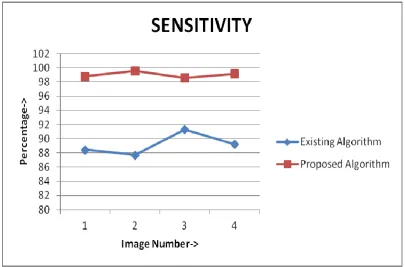
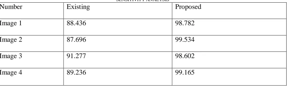
As shown in figure 7, the sensitivity of the proposed and existing algorithm is compared for the performance analysis.
The sensitivity of proposed algorithm is due to use of HMM classifier as compared to existing algorithm.
TABLE I ACCURACY ANALYSIS
TABLE II SPECIFICITY ANALYSIS
Number
Existing
Proposed
Image 1
88.526
98.342
Image 2
87.786
99.094
Image 3
91.367
98.162
Image 4
89.326
98.725
TABLE III SENSITIVITY ANALYSIS
Number
Existing
Proposed
Image 1
88.436
98.782
Image 2
87.696
99.534
Image 3
91.277
98.602
Image 4
89.236
99.165
Number
Existing
Proposed
Image 1
87.206
100.
Image 2
86.466
100.
Image 3
90.047
99.
© 2019, IJCSMC All Rights Reserved 128 V. CONCLUSION
In this work, it is been concluded that to detect breast cancer various techniques has been proposed in the previous times. The most efficient technique of brain cancer detection is based on morphological scanning, split and merge segmentation and on nearest neighbor classifier. In this work, to improve efficiency of the breast cancer detection SVM classifier is replaced with HMM classifier. The split and merge segmentation will split the input image on the basis of their properties. The output of split and merge segmentation is given as input to HMM classifier which will classify the features of the basis of their properties. The cancer and non cancer cells are marked with different colors. The simulation is performed in MATLAB and it is been analyzed that proposed technique performs well in terms of fault detection rate and accuracy.
REFERENCES
[1]. Rangaraj M. Rangayyan, Liang Shen, Yiping Shen, J. E. Leo Desautels, Heather Bryant, Timothy J. Terry, Natalka Horeczko, and M. Sarah Rose,” Improvement of Sensitivity of Breast Cancer Diagnosis with Adaptive Neighborhood Contrast Enhancement of Mammograms’, 1997, IEEE TRANSACTIONS ON INFORMATION TECHNOLOGY IN BIOMEDICINE, VOL. 1, NO. 3.
[2]. AO yan-li, Bayin Guoleng, Xinjiang,” Introduction to digital image pre-processing and segmentation”, 2015 IEEE. [3]. R. Guzmán-Cabrera, J. R. Guzmán-Sepúlveda, M. Torres-Cisneros, D. A. May-Arrioja, J. Ruiz-Pinales, O. G. Ibarra-Manzano, G. Aviña-Cervantes, A. González Parada,” 2013, Int J Thermophys 34:1519–1531.
[4]. Craig K. Abbey, Roger J. Zemp, Jie Liu, Karen K. Lindfors, and Michael F. Insana,” Observer Efficiency in Discrimination Tasks Simulating Malignant and Benign Breast Lesions Imaged With Ultrasound”, 2006, IEEE TRANSACTIONS ON MEDICAL IMAGING, VOL. 25, NO. 2.
[5]. Hang Song, Hayata Knon, Yuji Seo, Afreen Azhari, Junichi Somei, Eiji, Suematsu, Yuichi Watarai, Toshihiko Ota, HIromasa Watanabe, Yoshinori HIramatsu, Akihiro Toya, Xia Xiao and Takamaro Kikkawa,” A Radar-Based Breast Cancer Detection System Using CMOS Integrated Circuits”, 2015, IEEE, volume 3.
[6]. Ranjeet Singh Tomar, Tripty Singh, Dr Sulochana Wadhwani and Dr. Sarita Singh Bhadoria,” Analysis of Breast Cancer Using Image Processing Techniques”, 2009, IEEE, 978-0-7695-3886-0/09.
[7]. R. Ramani, Dr. S. Suthanthiravanitha, S. Valarmathy,” A Survey of Current Image Segmentation Techniques for Detection of Breast Cancer”, 2012, Vol. 2, Issue 5, pp.1124-1129.
[8]. Anuj Kumar Singh and Bhupendra Gupta,” A Novel Approach for Breast Cancer Detection and Segmentation in a Mammogram”, 2015, Elsvier, IMCIP.
[9]. Inam ul Islam Wani, M. C Hanumantharaju and M. T Gopalakrishna,” Review of Mammogram Enhancement Techniques for Detecting Breast Cancer”, 2014, International Journal of Computer Applications (0975 – 8887).
[10] Devendra Somwanshi, Ashutosh Kumar, Pratima Sharma, Deepika Joshi, “An efficient Brain Tumor Detection from MRI Images using Entropy Measures”, IEEE International Conference on Recent Advances and Innovations in Engineering (ICRAIE-2016), December 23-25, 2016.
[11] Manu Gupta, Prof. B.V.V.S.N.Prabhakar Rao, Dr.Venkateswaran Rajagopalan, “Brain Tumor Detection in conventional MR Images based on Statistical Texture and Morphological Features”, IEEE, 2016.
[12] Luxit Kapoor, Sanjeev Thakur, “A Survey on Brain Tumor Detection Using Image Processing Techniques”, IEEE, 2017.
[13] Sanjivani Salwe, Ranjana Raut, “Brain Tumor Pixels Detection using Adaptive Wavelet Based Histogram Thresholding and Fine Windowing”, IEEE, 2016.
[14] Smita.A.Nagtode, Bhakti B. Potdukhe, Pradnya Morey, “Two dimensional Discrete Wavelet Transform and Probabilistic Neural Network used for Brain Tumor Detection and Classification”, IEEE, 2016.