ABSTRACT
TANG, WENXING. New Solid-State NMR Methodologies for Structure Determination of Membrane Proteins: Sensitivity Enhancement, Spectroscopic Assignment, and Macroscopic Alignment. (Under the direction of Professor Alexander A. Nevzorov.)
Oriented-sample NMR (OS NMR) has emerged as a powerful tool for structure
determination of membrane proteins in their native lipid environment. In the present work,
we address three issues of critical importance for OS NMR of membrane proteins: sensitivity
enhancement, spectroscopic assignment, and macroscopic alignment. (i). A repetitive-contact
cross-polarization (REP-CP) scheme was developed that allows one to fully transfer the
thermodynamic limit of polarization from the abundant protons to low-gamma nuclei. As a
result, a factor of two (2) enhancement in sensitivity was gained over the single-contact
traditional CP, whereas 45% average intensity gain is achieved as compared to CP-MOIST,
currently the most widely used CP scheme in OS NMR. (ii). We have also developed a
strategy for assigning solid-state NMR spectra of Pf1 coat protein reconstituted in
magnetically aligned bicelles, and obtained a two-dimensional spin-exchanged version of the
SAMPI4 spectrum correlating 15N chemical shift and 15N-1H dipolar couplings. Combining the spin-exchanged version with the original SAMPI4 experiment makes it possible to
establish sequence-specific assignments, and this technique is generally applicable to other
membrane proteins. Notably, only a single uniformly labeled protein sample is required as
opposed to multiple selectively labeled samples currently employed for spectroscopic
assignment. Using sensitivity enhancement techniques such as REP-CP assists in further
elucidating cross peaks and allows the establishment of correlations between the adjacent
residue along the backbone. Simulation accurately predicts the optimal MMHH condition as
method to align membrane proteins into the lipid bilayers and obtained high-resolution OS
NMR spectra of the uniformly 15N labeled transmembrane domain of Pf1 coat protein. Nanoporous anodic aluminum oxide (AAO) sheets have been proven to be an alternative
alignment system for the study of membrane proteins in their native environments by means
of solid-state NMR. AAO-supported bilayers have the potential of providing an alternative
membrane mimic of highly flexible composition for the structure-function studies of
© Copyright 2013 Your Name
New Solid-State NMR Methodologies for Structure Determination of Membrane Proteins: Sensitivity Enhancement, Spectroscopic Assignment, and Macroscopic Alignment
by Wenxing Tang
A dissertation submitted to the Graduate Faculty of North Carolina State University
in partial fulfillment of the requirements for the degree of
Doctor of Philosophy
Chemistry
Raleigh, North Carolina
2013
APPROVED BY:
_______________________________ ______________________________ Professor Alexander A. Nevzorov Professor Edmond F. Bowden
Committee Chair
ii DEDICATION
BIOGRAPHY
Early in one morning in February, 1982, a new born child took his first look at this
world in a city called Tianjin in northern China. His overjoyed parents were both majored in
Literature and hence named him Wenxing, which direct translates as “a rising star in
literature”. None of them know at that point that, some day he will go to the United States.
Neither did they expect that, he will major in Chemistry and focus on solid-state NMR.
Time flies. At the age of 18, Wenxing was accepted by one of the oldest university in
China, Tianjin University. He applied to be majored in Architecture or Civil Engineering, but
was, somehow, enrolled by the Department of Chemistry.
Wenxing was really depressed, and his way of fighting back was to skip all the
classes. Till the 4th year. One of the mandatory classes, Biochemistry, was offered by Prof.
Wang, who received his Ph.D at Columbia University and was the vice dean of Department
of Pharmaceutical Science. Wenxing became so fascinated by his class that, he, for the first
time, found bio-related subject interesting. It was also at that time that, he made up his mind
to go to the United States.
The summer of 2008 will always be remembered, because it was when Wenxing’s
dream finally came true: he was enrolled by the Department of Chemistry, again. Only this
time it was by his own will. He chose to join Prof. Nevzorov’s group to study membrane
proteins with solid-state NMR, which he had absolutely no idea about.
During the years, he made a lot of friends, had a lot of fun. He met with a beautiful
girl on his first visit to DMV and married her.
iv And now, after 4 years of study, he thinks he could finally say he knows a little bit
about what he’s been doing, even though he’s still trying to pick up quantum mechanics.
ACKNOWLEDGMENTS
This work would not have been possible without the support of many people. First
and foremost, I would like to express my deep and sincere gratitude to my adviser, Professor
Alexander A. Nevzorov. He patiently trained me when I first joined the group, and gradually
guided me into the field of solid-state NMR. Throughout the years, his enlightening
discussion over different topics has truly broadened my knowledge and outlook. His unique
way of thinking not only helped me in solving research related problems, but also
appreciating more on what’s normally viewed as basic. It was his encouragement, motivation
and support that made my Ph.D. experience productive and joyful. His clear vision both in
research and life inspires me. I could not have asked for a better mentor and friend.
Many people on the faculty and staff of the Department of Chemistry assisted and
encouraged me in various ways during my course of studies. I am especially grateful to Prof.
Tatyana I. Smirnova, Prof. Reza A. Ghiladi, Prof. Edmond F. Bowden and Prof. Hanna
Gracz for being my committee members.
My deepest gratitude is also due to other group members for their constant support
and laughs. They provided a great atmosphere for research and study.
Last but not least, I would like to thank my family and friends for their constant love
and support. They have always been there at every turn of my life. And I must thank my wife
vi TABLE OF CONTENTS
LIST OF TABLES ...x
LIST OF FIGURES ... xi
CHAPTER 1 INTRODUCTION ...1
1.1 Membrane proteins and the challenges in their structure determination ...1
1.2 Most widely used pulse sequences by Oriented-Sample NMR ...4
1.2.1 Polarization Inversion Spin Exchange at the Magic Angle (PISEMA) ...4
1.2.2 SAMMY pulse sequence with π/4 pulse correction(SAMPI4)...5
1.3 Polarity Index Slant Angle (PISA) wheels ...7
1.4 Sample alignment in the study of membrane proteins by OSNMR...9
1.4.1 The use of glass plates ...9
1.4.2 The use of bicelles ...11
1.5 Filamentous bacteriophage and Pf1 coat protein ...14
CHAPTER 2 REPETITIVE CROSS-POLARIZATION CONTACTS VIA EQUILIBRATION-RE-EQUILIBRATION OF THE PRTON BATH: SENSITIVITY ENHANCEMENT FOR NMR OF MEMBRANE PROTEINS RECONSTITUTED IN MAGNETICALLY ALIGNED BICELLES ...19
2.1 Abstract ...19
2.2 Introduction ...20
2.3 Materials and methods ...22
2.4 Results and discussion ...26
CHAPTER 3 A SPECTROSCOPIC ASSIGNMENT TECHNIQUE FOR MEMBRANE
PROTEINS RECONSTITUTED IN MAGNETICALLY ALIGNED BICELLES ...34
3.1 Abstract ...34
3.2 Introduction ...35
3.3 Materials and methods ...39
3.4 Results and discussion ...42
3.4.1 Simulations of spin dynamics for the perpendicular bicelles ...42
3.4.2 Experimental results...47
3.5 Conclusions ...54
CHAPTER 4 ANODIZED ALUMINUM OXIDE NANOPORES AS AN ALTERNATIVE ALIGNMENT MEDIA FOR THE STUDY OF MEMBRANE PROTEINS USING ORIENTED-SAMPLE NMR ...56
4.1 Abstract ...56
4.2 Introduction ...57
4.3 Materials and methods ...61
4.3.1 Preparation of AAO stripes ...61
4.3.2 Preparation of Pf1 coat protein and nanopore-supported lipid bilayers ...62
4.3.3 NMR spectroscopy...63
4.4 Results and discussion ...63
4.5 Conclusions ...70
CHAPTER 5 SUMMARY AND FUTURE WORK ...72
viii 5.2 Future work ...74
REFERENCES ...76 APPENDICES ...88 Appendix A Pulse sequences used in the study of REP-CP. A. Single CP contact; B. CP-MOIST; C. Multiple Contacts; D. REP-CP. ...89
Appendix B Pulse program of one-dimensional 1H decoupled 31P experiment. ...90 Appendix C Pulse program of one-dimensional 1H decoupled 15N experiment. ...91 Appendix D Pulse program of SAMPI4_multicont (for the SLF spectrum with only main peaks, REP-CP included). ...93
Appendix E Pulse program of NN_TSAR2d_sammy_multicont (for the spin exchange version of SAMPI4, to get both main- and cross-peaks). ...96
Appendix F Pulse program of NN_TSAR2d_multicont (for the N-N correlation spectrum with both main- and cross-peaks). ...99
Appendix G Pulse program of NN_PDSD_multicont (for the N-N correlation spectrum with PDSD instead of MMHH). ...102
Appendix H Crosspeaks from the N-N homonuclear exchange experiment for Figure 3.3. ... ...105
Appendix I Main peaks from the SAMPI4 experiment (Figs. 3.3) and their assignment. ..106 Appendix J Cross-peaks from the spin-exchanged SAMPI4 experiment (Figs. 3.3). ...107 Appendix K Matlab (Mathworks, Inc.) script to simulate spectra from assigned PISEMA data. ...108
Appendix M Protocol for reconstituting Pf1 bacteriophage coat protein into the
DMPC/DHPC (q=3.2) bicelles. ...116
x LIST OF TABLES
APPENDICES
Table S1 Crosspeaks from the N-N homonuclear exchange experiment for Figure 3.3A, C ...
...105
Table S2 Main peaks from the SAMPI4 experiment (Figs. 3.3B, D) and their assignment. ...
...106
LIST OF FIGURES CHAPTER 1 INTRODUCTION
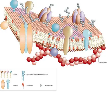
1.1 A cartoon representation of the cell membrane showing its essential components. (Image taken from http://www.nature.com/scitable/topicpage/protein-function-14123348) ...2
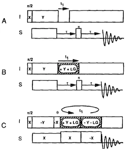
1.2 Pulse sequences for two-dimensional heteronuclear dipolar/chemical-shift spectroscopy. (A) Simplest version of the SLF experiment. (B) SLF experiment with Lee-Goldburg
homonuclear decoupling applied during the t1 period. (C) PISEMA experiment. ...5
1.3 The pulse sequence of SAMMY. The heteronuclear interaction evolves during section 1 and 3. During section 2, the S spins are decoupled from the abundant I spins. To achieve optimal decoupling, the duration of section 1, 2 and 3 requires experimental adjustments. ....6
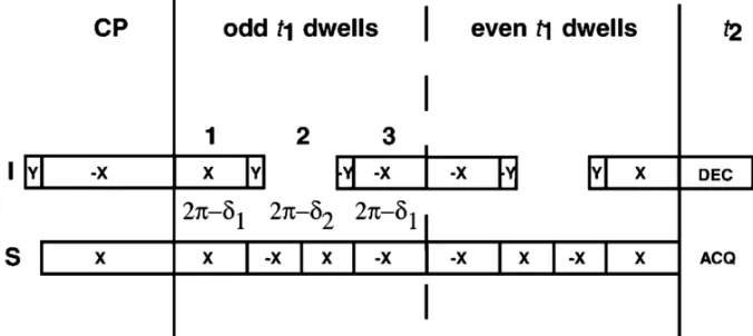
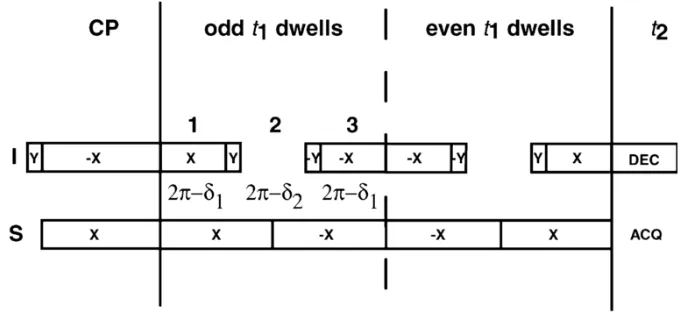
1.4 The SAMPI4 pulse sequence. Compared with SAMMY in Figure 1.3, only one phase alteration on the S spin channel is used. A π/4 pulse correction is applied to δ1 and δ2. ...7
1.5 PISEMA spectra calculated for a 19-residue α-helix with 3.6 residues per turn and uniform dihedral angles (φ= -65, ψ= -40) at various helix tilt angles relative to the bilayer normal...9
1.6 A demonstration of reconstituting membrane proteins into lipid bilayers supported by glass plates ...10
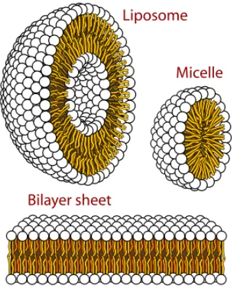
1.7 Structures that can be formed by phospholipids in aqueous solutions. (Image taken from http://en.wikipedia.org/wiki/Micelle) ...12
1.8 Schematic representations of magnetically aligned bicelles. (a) Perpendicular or
xii 1.9 An illustrative demonstration of a phage assembly. The thin eclipse in red represents the circular viral DNA, whereas the small eclipses in dark gray represent the main coat protein subunits. The rectangles on the bottom are other proteins related to the phage life cycles. ...15
1.10 A demonstration of the structure of the coat protein in Pf1 bacteriophage in its different forms. (a) and (d) on the top shows the phase difference mapped on the helical wheel
diagrams of the C-terminal region of the protein in bicelles (a) and bacteriophage particles (d). (b) demonstrates the Pf1 coat protein in a membrane environment. (c) 159° rotation of panel (b) to the vertical axis. (e) is the monomer structure of Pf1 coat protein in intact bacteriophage particles. (f) and (g) are the top and side views of a model structure of Pf1 bacteriophage particles. (Figure taken from Opella, et al, 2008) ...17
CHAPTER 2 REPETITIVE CROSS-POLARIZATION CONTACTS VIA EQUILIBRATION-RE-EQUILIBRATION OF THE PROTON BATH: SENSITIVITY ENHANCEMENT FOR NMR OF MEMBRANE PROTEINS RECONSTITUTED IN MAGNETICALLY ALIGNED BICELLES
2.1 The REP-CP pulse sequence employing repetitive CP contacts during the preparation period and experimentally observed enhancement for NMR spectra for Pf1 coat protein reconstituted in magnetically aligned bicelles. Single-contact CP (dotted line); CP-MOIST (dashed line); REP-CP (solid line). A two-fold gain in the signal-to-noise ratio is obtained for the case of REP-CP as compared to the conventional CP, and a 45% gain over CP-MOIST. All experiments have equal total experimental times; 50 Hz exponential linebroadening has been applied. ...23
2.2 A. 15N Spectra of Pf1 phage: CP-MOIST (solid line) and multiple contacts (dashed line). All phage spectra were measured at -4 oC, 1 ms single contact time was used and three (3) CP contacts were employed for the multiple contact experiment; 64 transients with a 6 sec recycle delay were acquired for each spectrum, and the acquisition time was 5 ms. The signal-to-noise ratio for the CP-MOIST experiment is 48:1; whereas for the multiple contacts it is 38:1. B. 15N Spectra of Pf1 phage: single-contact CP (dotted line, 256 scans, 1 ms contact, 10 ms acquisition time); CP-MOIST (dashed line, all parameters are the same as for the single-contact CP); REP-CP (solid line, 6 contacts, 200 µs each, 1 sec z-filter delay, other parameters are the same). About 50% intensity gain is obtained for the case of REP-CP as compared to the conventional CP, and 25% relative to CP MOIST. ...27
spins were cross-polarized and detected. The T1ρ relaxation time was determined by fitting the integrals of the spectra corresponding to the transmembrane region of Pf1 coat protein (from 55 to 110 ppm) yielding T1ρ =4.3 msec. B. 15N-detected proton T1Z experiments for Pf1 coat protein reconstituted in magnetically aligned bicelles using the inversion recovery pulse sequence as shown. A 1.3 sec spin-lattice relaxation time for the protons was determined from the crossover...28
2.4 Comparison of REP-CP spectrum (black) vs. direct-excitation echo-detected 15N spectrum (red). 990 scans were used for the REP-CP experiment with 6 contacts each having 200 µs contact time, and the z-filter was set to 0.04 s. For the direct-excitation echo-detected 15
N spectrum, the number of scans was 1024, and a 300 s relaxation delay was utilized. The peaks at 75 and 85 ppm are due to the mobile Arg side-chains which exhibit small dipolar couplings in the 2D spectrum (cf. Fig. 2.5), and can interfere with the assessment of the enhancement factor for this spectral range. The enhancement effect for the transmembrane helical region (105 to 65 ppm) varies from 9 to 14 depending on the position of the peak in the spectrum due to a very low signal-to-noise ratio. Note that the peaks between 110 and 130 ppm, which correspond to the more dynamic N-terminal residues (1-18), are efficiently excited by a single pulse, but are absent from the CP spectrum where only residues 19-43 are observed. ...30
2.5 SAMPI4 experiments for Pf1 coat protein reconstituted in magnetically aligned bicelles acquired at 500 MHz proton frequency. (A) REP-CP enhanced SAMPI4 (14 scans for each t1 increment); (B) CP-MOIST enhanced SAMPI4 (16 scans). Representative slice along 3 kHz in the dipolar dimension shows that that superior sensitivity is obtained in part A (with 6 REP-CP contacts, 300 μs each, 0.15 sec z-filter time) as compared to CP-MOIST with a single 1 ms contact (part B). The experimental time was 1 hour and 42 minutes for each experiment...31
2.6 Comparison of the signal-to-noise ratios for 21 resolved residues in the α-helical transmembrane region of Pf1 coat protein. Solid line: REP-CP enhanced SAMPI4 (128 scans, other parameters are as in Fig. 2.2); dashed line: CP-MOIST enhanced SAMPI4 (128 scans). For select peaks the gain is up to 60% with an average gain of 35%. ...32
CHAPTER 3 A SPECTROSCOPIC ASSIGNMENT TECHNIQUE FOR MEMBRANE PROTEINS RECONSTITUTED IN MAGNETICALLY ALIGNED BICELLS
xiv sequence utilizing MMHH. (B) Spin-exchanged SAMPI4 pulse sequence (C) 15N-15N correlation pulse sequence utilizing PDSD. Either conventional cross-polarization, CP, or REP-CP can be used to enhance the initial 15N magnetization. A 5 ms contact time was chosen for all the MMHH-based experiments, whereas a mixing time of 3 seconds was used for the PDSD experiment. For further details of the pulse sequences cf. the text.. ...40
3.2 Simulations of the efficiency of magnetization transfer between the rare 15N spins under the MMHH conditions. (A) N = 12 spin simulations including single pairs of amide 15N spins for Pf1 reconstituted in bicelles (with the coordinates for residues I22-M42 taken from PDB ID 1ZN5, cf. the text) comparing the (i, i+1) and (i, i+2) magnetization exchange pathways at 20, 40, and 60 kHz 15N spin-lock B1 fields (marked by vertical dotted lines). Values were averaged across the transmembrane region (residues 22-42). At 20 kHz 15N spin lock B1 field, the optimal transfer is enhanced by ca. 5.5% and 7.5% over the 40 kHz and 60 kHz 15N spin lock rf fields respectively (cf. Traaseth et al, 2010). (B) Three-15N spin (N = 13) simulations examining the effects of spin competition on the long-range (i, i+2) magnetization transfer in the presence of the more preferential (i, i+1) pathway. The (i, i+2) magnetization transfer between two representative residues A36 and L38 (in the presence of a competing nitrogen spin at G37; gray line) is compared to the A36-G37 (i, i+1) transfer (black line). The efficiency of the (i, i+2) transfer in the presence of the competing (i, i+1) pathway is even lower than in part (A). (C) Three-15N spin (N = 13 total number of spins) simulations comparing the magnetization build up and demonstrating the effect of competing spins on the (i, i+1) and (i+1, i) cross-peak intensities as a function of the rf amplitude mismatch. The transfer from the nitrogen spin at G37 to that of L38 (in the presence of A36; black line) and the transfer from L38 to G37 (in the presence of I39; gray line) have been considered. The A36-G37 transfer from part B is also shown here (black dashed line) to compare it to the L38-G37 transfer. ...45
3.3 Spectra of Pf1 coat protein reconstituted in magnetically aligned DMPC/DHPC bicelles at T = 38°C. (A) Experimental 15N‐15N spin correlation spectrum: 512 scans; MMHH at 45.5 kHz 15N B1 rf field and 51.5 kHz 1H B1 field, 0.5 s Z-filter and 5 ms mixing time; 64 complex
shown by the red boxes. For clarity, a representative assignment pathway for residues V35-M42 is shown in the digital versions of the spectra (parts C and D). ...49
3.4 A comparison between (A) 15N-15N MMHH spin-exchange spectrum at 512 scans, 20 kHz 15N B1 rf field and 25 kHz 1H B1 field, and 0.5 s Z-filter; and (B) 15N-15N PDSD spectrum: 256 scans and 3 s mixing time. The REP-CP pulse sequence with 5 contacts of 300
µs length each was used to enhance magnetization on the 15N spins (Tang and Nevzorov, 2011), 80 complex t1 points, 20 ms total acquisition time in the direct dimension, and a 6 s recycle delay were used in both spectra. In part B, the peaks are slightly broader, and some of them (both the main and cross peaks) are missing in the spectrum. ...51
3.5 A zoomed-in view of the symmetrized 15N-15N MMHH exchange spectrum from Figure 3.4A overlaid with the simulated crosspeaks (red crosses) calculated using the chemical shifts from the main peaks of the SAMPI4 spectrum (from Fig. 3D) and the previous assignment (Opella et al, 2008; Park, 2010). All simulated cross peaks have underlying intensity, which proves the correctness of the previous assignment. Moreover, the cross peaks generally follow the (i, i+1) connectivity pattern. ...53
CHAPTER 4 ANODIZED ALUMINUM OXIDE NANOPORES AS AN ALTERNATIVE ALIGNMENT MEDIA FOR THE STUDY OF MEMBRANE PROTEINS USING ORIENTED-SAMPLE NMR
4.1 A schematic demonstration of membrane proteins reconstituted in lipid bilayers with the support of nanoporous AAO slides. ...60
4.2 SEM images of the produced AAO sheets following the procedure as described in the text. A) The top view. B) The side view. (Photo courtesy of Dr. Antonin Marek) ...64
4.3 Distribution of the pore size of various homemade AAO samples as compared to the commercially available WhatmanTM AAOs at 200 nm diameter. ...65
4.4 CD spectrum of the coat protein of Pf1 bacteriophage reconstituted in the lipid bilayers before the deposition into the AAO nanopores. ...66
xvi scans was used for the 31P experiment, whereas 8192 scans with a 90° pulse duration of 4.9 µs was used for the 15N experiment. More details are covered in the text. ...67
CHAPTER 1 INTRODUCTION
1.1Membrane proteins and the challenges in their structure determination
The proteins that span the cell membrane perform an array of functions that are vital
to our lives. These functions include, but are not limited to: ion transport, signal transduction,
energy regulation, pathway activation, and molecular recognition. It has been proven that, the
misfolding of the secondary structure generally leads to the malfunction of the membrane
protein, some of which are directly related to serious diseases such as cancer. In addition, the
vital role of membrane proteins could also be reflected by the fact that they are the targets of
more than 50% of the modern drugs (1). However, despite their critical importance and the
fact that ~30% of all proteins in eukaryotic cells are membrane proteins (2), only 371 unique
membrane protein structures have been solved to date
(http://blanco.biomol.uci.edu/mpstruc/query), which accounts for less than 1% of the total
protein structures deposited in the Protein Data Bank (PDB). This is mainly due to the fact
that high-resolution structures of integral membrane proteins in phospholipid bilayers have
been notoriously difficult to obtain by either X-ray diffraction or solution NMR methods.
Despite its success in solving the structures of soluble proteins, X-ray crystallography
(XRC) of membrane proteins, which is generally achieved with the aid of detergents, might
still be considered a high art. And the reason is that, the method itself requires the sample to
be trapped in a crystal conformation. However, this requirement introduces a few
2 would either get trapped in some energetically favorable intermediate conformations, or
would result in a blurred region representing the average of all possible conformations.
Second, potential loss of structural lipids, due to the existence of detergents, may lead to
protein misfolding or denaturation (3). Finally, many membrane proteins are extremely
difficult or even impossible to be crystallized due to their amphiphilic nature of having both
the hydrophobic and hydrophilic regions (4).
Solution-state NMR is another highly utilized experimental method; however, it
requires the proteins to rapidly reorient in solution. Large vesicular assemblies such as
liposomes have proven to be much too large for their reorientation time to be fast enough for
the solution-state NMR time scale (5). Nevertheless, some progress has been made in the
structural studies of membrane proteins by utilizing phospholipid micelles, which rapidly
reorient in aqueous solutions (6). However, micelles represent less than an ideal structure
since their highly curved geometry significantly differs from that of the biological
membranes. Furthermore, many membrane proteins require a full bilayer in order to be
reconstituted in a native-like conformation (7,8) and their structure in micelles can be
considerably different from that in bicelles which mimic more closely the native lipid
environment (9,10).
In contrast, solid-state NMR (SSNMR) spectroscopy has become an effective
alternative approach for studying membrane proteins in their native-like functional
environment. There are two different approaches in the realm of SSNMR: magic-angle
spinning (MAS) and oriented-sample (OS) NMR. The first involves spinning the sample at a
specific angle, which is 54.7°, with regard to the external magnetic field B0, to average out
the anisotropic terms such as dipolar couplings, and obtain spectra at high resolution.
Whereas the latter approach involves macroscopic sample alignment, either mechanically or
magnetically, the OS NMR spectrum directly contains structural information of the target
protein. Proteins whose structures have been successfully investigated by SSNMR include,
4 M2 domain (14), M2 domain of the influenza A virus (15), phospholamban (16), Vpu from
HIV-1(17), and MerF (18).
1.2Most widely used pulse sequences by oriented-sample NMR
1.2.1 Polarization Inversion Spin Exchange at the Magic Angle (PISEMA)
Ever since its introduction in 1994 by C. H. Wu, A. Ramamoorthy and S. J. Opella
(19), PISEMA has become one of the most powerful tools for high-resolution SSNMR of
membrane proteins. The main idea is to combine polarization inversion of the S spin (e.g.
nitrogens) with the flip-flop Lee-Goldburg homonuclear decoupling on the abundant I spins
(e.g. protons) following spin-lock cross polarization to the dilute S spins during the t1 evolution. In this way, the experiment yields a two-dimensional correlation of the chemical
shift of the sparse spin S with the heteronuclear IS dipolar coupling. As a result, linewidths
are remarkably reduced in the dipolar dimension compared to that of the conventional
separated-local-field (SLF) spectrum. Pulse sequences for the conventional SLF experiment
as well as the PISEMA experiments are depicted in Figure 1.2. Furthermore, dipolar splitting
and chemical-shift frequencies could also be used in combination in three-dimensional
Figure 1.2 Pulse sequences for two-dimensional heteronuclear dipolar/chemical-shift spectroscopy. (A) Simplest version of the SLF experiment. (B) SLF experiment with Lee-Goldburg homonuclear decoupling applied during the t1 period. (C) PISEMA experiment. Figure taken from Ref. (19).
1.2.2 SAMMY pulse sequence with π/4 pulse correction (SAMPI4)
Though providing heteronuclear dipolar coupling spectra with higher resolution
compared to conventional separated local-field experiments, PISEMA, being sensitive to the
choice of 1H carrier frequency, suffers from unstable performance when the range of 1H frequencies is broad. This is intrinsic to PISEMA because it is based on the off-resonance
6 environments, since such a complex system would give rise to a wide range of 1H resonance frequencies.
To overcome this problem, a pulse sequence based on the Average Hamiltonian
Theory (AHT) was implemented by Nevzorov and Opella in 2003 (21). In order to remove
the homonuclear interactions between various 1H spins, a “magic sandwich” pulse scheme was incorporated (hence the name “SAMMY”), instead of the frequency-switched
Lee-Goldburg irradiation. The pulse program is diagrammed in Figure 1.3.
Figure 1.3 The pulse sequence of SAMMY. The heteronuclear interaction evolves during section 1 and 3. During section 2, the S spins are decoupled from the abundant I spins. To achieve optimal decoupling, the duration of section 1, 2 and 3 requires experimental adjustments. Figure taken from Ref. (21)
Unfortunately, spectroscopic studies have shown that the linewidths for most
resonances in the heteronuclear dipolar coupling dimension of two-dimensional SAMMY
Combining the broadband capability of SAMMY with the line-narrowing ability of PISEMA
in a single experiment has then become the next goal.
In 2007, an improved version of SAMMY, which is termed as SAMPI4, pulse
scheme was proposed (22). And the pulse diagram is depicted in Figure 1.4. Generally, it
yields sub-200 Hz linewidths in the heteronuclear dipolar coupling dimension while covering
the broad ranges of frequencies given rise from the aligned samples of membrane proteins
reconstituted in lipid bilayers under high magnetic fields.
Figure 1.4 The SAMPI4 pulse sequence. Compared with SAMMY in Figure 1.3, only one phase alteration on the S spin channel is used. Aπ/4 pulse correction is applied to δ1 and δ2. Figure taken from Ref.(22).
1.3Polarity Index Slant Angle (PISA) Wheels
Resonance patterns observed in two-dimensional Oriented-Sample NMR spectra
8 the PISEMA sequence gives high-resolution 1H-15N dipolar coupling/15N chemical shift separated-local-field (SLF) spectra where the individual resonances contain orientational
constraints for structure determination (19). It has been shown that the PISEMA spectra of
membrane proteins reconstituted in the magnetically aligned lipid bilayers are good
indicators of α-helical structure (23,24). For helical proteins, these spectra exhibit
characteristic wheel-like patterns of resonances, which are termed as “Polar Index Slant
Angle” (PISA) wheels, and reflect helical wheel projections of residues (25). These wheels
are useful both in assigning the resonances and in determining the general orientation of
α-helices, as well as β-sheets (26), with respect to the magnetic field.
Figure 1.5 is a demonstration of the calculated PISA wheels caused by the variation
of the helix tilt angle with respect to the bilayer normal. One could easily tell that the
resonance frequencies dramatically depend on the helical orientation.
Together with the high resolution of PISEMA or SAMPI4 spectra, these achievements make
Oriented-Sample NMR a powerful tool for obtaining structural information about membrane
Figure 1.5 PISEMA spectra calculated for a 19-residue α-helix with 3.6 residues per turn and uniform dihedral angles (φ= -65, ψ= -40) at various helix tilt angles relative to the bilayer normal.
1.4Sample alignment in the study of membrane proteins by OSNMR
1.4.1 The use of glass plates
Pioneered by Seelig and Gally (27), the use of glass plates as a media to mechanically
align NMR sample could be dated back to the 1970s, and is now widely used in the study of
membrane proteins in their native environments. The idea is to refold membrane proteins into
10 Figure 1.6 A demonstration of reconstituting membrane proteins into lipid bilayers supported by glass plates
molecules are oriented in the same way with regard to the external magnetic field. By doing
so, the uniformity of the alignment, instead of molecular motions, narrows down the
spectroscopic linewidths, and hence benefits the structural study of membrane proteins with
SSNMR. Figure 1.6 is a demonstration of such a scheme. One of the greatest advantages of
mechanically aligned samples on glass plates is that, NMR spectra can be acquired as a
function of sample orientation when necessary. However, the drawbacks of such a design are
the glass plates, which limits the maximum amount of lipid molecules as well as the target
proteins that could be deposited. On the other hand, it’s hard to maintain the surface of a
sample fully hydrated since it is directly exposed to the environment.
1.4.2 The use of bicelles
In the past decades, researchers have developed various models to investigate the
structure and dynamics of membrane proteins in their native-like environments such as
micelles (6), multilamellar vesicles (28), unilamellar vesicles (29) or mechanically oriented
bilayers between glass plates (30,31). Recently, an alternative system, which represents a
combination of micelles and vesicles, became widely used. The foundation for this system
was laid in the pioneering work of Roberts and co-workers on “bilayered micelles” in the
early 1980s (32). In 1992, Sanders and Schwonek have made an interesting discovery that the
mixture of DMPC and DHPC has the ability to spontaneously align in an external magnetic
field (33). This phenomenon makes the system extremely valuable for the NMR study of
membrane proteins. Further improvement of this model system has lead to a change in the
terminology to “bicelles” (7) in 1995.
Nowadays, bicelles are normally referred to as the aqueous suspension of the mixture
of aliphatic long chain lipids (between 12 and 16 carbons) and short chain lipids (6–8
carbons). The most recognized organization is a nanodisc with the long chain lipids forming
the disc plane and the short chain lipids mainly distributed in the torus of the disc.
The reason that bicelles align in the presence of the external magnetic field is mainly
12 between the parallel (χ∥) and the perpendicular (χ⊥) magnetic susceptibility to the long lipid
axis: Δχ = χ∥ - χ⊥). Hence, it is possible to “flip” the bicelles by adding a component with a
positive Δχ.
Figure 1.7. Structures that can be formed by phospholipids in aqueous solutions. (http://en.wikipedia.org/wiki/Micelle)
The most widely used method now is the addition of a small amount of lanthanide ions, Yb3+ for example. Spectra studies of various proteins reconstituted in magnetically aligned bicelles
in both orientations have been published (18,34). A comparison of the spectra obtained for
and, in some cases, can even provide a method for assigning solid-state NMR spectra of
membrane proteins (35).
The use of magnetically aligned bicelles in the structural studies of membrane
proteins provides numerous benefits, the most important one being relatively easy sample
preparation. In addition, the use of a sealed tube containing a liquid sample not only prolongs
the life-time of the sample but also enables its placement inside a solenoid coil, which allows
for optimal NMR probe performance. Furthermore, the availability of instant control over
different sample parameters, such as pH and solvent composition, makes it possible to
perform many standard procedures, such as hydrogen/deuterium exchange experiments
(17,36). However, bicelles are not perfect; they also have their disadvantages. First, the
choices of lipid combinations are limited, since not all lipids align in an external magnetic
field. Second, each lipid combination has its own restricted temperature range. However,
experimental solutions to these problems have been found by introducing mixed long-chain
14 Figure 1.8 Schematic representations of magnetically aligned bicelles. (a) Perpendicular or “unflipped” bicelles: the bilayer normal is perpendicular to the direction of the external static magnetic field. (b) Parallel or “flipped” bicelles: in the presence of YbCl3, the bilayer normal is parallel to the main magnetic field. Figure taken from Ref. (38).
1.5Filamentous bacteriophages and Pf1 coat protein
A bacteriophage (also called phage or bacterial virus) is a virus that infects and
multiplies within bacteria, and is mainly composed of coat proteins encapsulating a DNA or
RNA genome. Their discovery could be dated back to the beginning of the 20th century.
Bacteriophages are categorized based on their morphology and the nucleic acid within the
core by the International Committee on Taxonomy of Viruses (ICTV). Of all the known
bacteriophages, filamentous bacteriophages are of special interest due to their relatively
simple structures and broad applications (39,40).
Although their DNA genomes could be dramatically different from one to another,
cycle. Such a virion is a flexible protein rod that is normally 800-2000 nm in length with a
diameter of around 6 nm, comprised of thousands of identical α-helical major coat protein
subunits, with a circular single-stranded DNA (ssDNA) in the core. Each of these subunits is
about 7 nm long by 1 nm diameter, with their long axes oriented at a small angle to the virion
axis. Shown in Figure 1.9 is an illustrative demonstration of a filamentous bacteriophage
16 assembly. Upon infection, viral DNA enters the host cell cytoplasm with the help of the
major coat protein subunits after virion uncoating, and is converted to a double-stranded
replicative form by host enzymes. It is then replicated and assembled with a viral
replication-assembly protein. The replication-replication-assembly protein is then replaced by capsid proteins at the
cell membrane as the virion is extruded without killing the host.
The most widely used strains of filamentous bacteriophage in scientific research are f1, fd,
M13 in class I, which are almost identical filamentous phages that infect bacteria with F-pili,
due to their ease to cultivate and high reproductive rate. Pf1 in class II, on the other hand,
gives by far the highest resolution X-ray diffraction patterns known for filamentous
bacteriophage, and is very well studied not only by XRD, but also by solid-state NMR.
The structure of the main coat protein of Pf1 bacteriophage has been thoroughly
studied in not only intact virus particle but also membrane-bound form by different groups
(41-45). Figure 1.10 is a demonstration of the structure of the main coat protein of Pf1
bacteriophage in different environments. Hence, it is a perfect system to test newly
Figure 1.10 A demonstration of the structure of the coat protein in Pf1 bacteriophage in its different forms. (a) and (d) on the top shows the phase difference mapped on the helical wheel diagrams of the C-terminal region of the protein in bicelles (a) and bacteriophage particles (d). (b) demonstrates the Pf1 coat protein in a membrane environment. (c) 159° rotation of panel (b) to the vertical axis. (e) is the monomer structure of Pf1 coat protein in intact bacteriophage particles. (f) and (g) are the top and side views of a model structure of Pf1 bacteriophage particles. (Figure taken from Opella, et al, 2008)
In summary, in the present Dissertation, three (3) issues of critical importance of
OSNMR of membrane proteins will be addressed. The first one is the intrinsic low sensitivity
of SSNMR. To circumvent the problem, a pulse sequence termed as REP-CP is developed. A
18 sequence will be discussed in Chapter 2. The second issue is to develop a strategy to assign
the OSNMR spectra of membrane proteins. Unlike the conventional method which requires
multiple selectively labeled samples and assumes ideal secondary structure of the protein, a
novel spectroscopic assignment scheme is proposed, which requires only one single
uniformly labeled sample. Last but not least, an alternative approach for macroscopic sample
alignment is established. The first two-dimensional SSNMR study of a uniformly 15N labeled membrane protein reconstituted in lipid bilayers of various composition supported by AAO
nanopores is accomplished. More details are covered in Chapter 4. It will be demonstrated
that these achievement have considerably increased the utility of the OSNMR for the
CHAPTER 2
REPETITIVE CROSS-POLARIZATION CONTACTS VIA EQUILIBRATION-RE-EQUILIBRATION OF THE PROTON BATH: SENSITIVITY ENHANCEMENT FOR NMR OF MEMBRANE PROTEINS RECONSTITUTED IN MAGNETICALLY
ALIGNED BICELLES*
*This chapter is based on the publication entitled “Repetitive cross-polarization contacts via
equilibration-re-equilibration of the proton bath: Sensitivity enhancement for NMR of
membrane proteins reconstituted in magnetically aligned bicelles” by Wenxing Tang and
Alexander A. Nevzorov.
2.1 Abstract
Thermodynamic limit of magnetization corresponding to the intact proton bath often
cannot be transferred in a single cross-polarization contact. This is mainly due to the finite
ratio between the number densities of the high- and low-gamma nuclei, quantum-mechanical
bounds on spin dynamics, and Hartmann-Hahn mismatches due to rf field inhomogeneity.
Moreover, for fully hydrated membrane proteins refolded in magnetically oriented bicelles,
short spin-lock relaxation times (T1ρ) and rf heating can further decrease cross polarization efficiency. Here we show that multiple equilibrations-re-equilibrations of the high- and
low-spin reservoirs during the preparation period yield an over two-fold gain in the magnetization
20 as compared to the mismatch-optimized CP-MOIST scheme for bicelle-reconstituted
membrane proteins. This enhancement is achieved by employing the differences between the
spin-lattice relaxation times for the high- and low-gamma spins. The new technique is
applicable to systems with short T1ρ’s, and speeds up acquisition of the multidimensional solid-state NMR spectra of oriented membrane proteins for their subsequent structural and
dynamic studies.
2.2 Introduction
As for virtually every spectroscopic measurement, signal-to-noise ratio plays perhaps
the most critical role in the acquisition and interpretation of solid-state NMR spectra of
macroscopically aligned samples. This method has recently demonstrated the capability of
providing remarkable detail about the conformations of membrane proteins in their
native-like, fully hydrated lipid environment at nearly atomic resolution (46). Magnetically aligned
bicelles (47,48) have the potential of advancing this technique even further since they
provide superior spectral resolution as compared to glass plates (49-51). However, the
necessity to detect dilute spins in such strongly proton-coupled systems is inherently
connected with the problem of low sensitivity. As a result, the acquisition of
multidimensional solid-state NMR spectra can take several days. Usually, magnetization
enhancement for the dilute spins during the preparation period is achieved via the
cross-polarization (CP) method (52) under the Hartmann-Hahn matching conditions (53).
CP-MOIST (55,56), CP involving simultaneous phase inversion (57), variable-amplitude CP
(58), selective-excitation RELOAD-CP technique (59), frequency-modulated CP (60), and
CP-COMPOZER (61). The latter two techniques have shown their robustness with respect to
Hartmann-Hahn mismatches and the capability of improving the signal to noise ratio by up to
20-25% for aligned samples (60,62). However, only a part of the overall magnetization is
transferred from the abundant proton bath to the dilute spins in a single cross-polarization
contact (63). This can be due to the finite ratio of the total number of protons with respect to
that of the low-gamma spins, as well as relaxation effects. In addition, universal or
quantum-mechanical bounds on spins dynamics (64) may play a role, thus further limiting the amount
of the maximum transferred quasi-stationary magnetization (55,65). The classical
multiple-contact scheme (52) can be employed to further enhance the magnetization transfer from the
protons to the dilute spins in static and spinning solids. However, for membrane proteins
reconstituted in magnetically aligned bicelles (49), this scheme may not be appropriate due to
the relatively short T1ρ relaxation times (66) (typically up to several milliseconds) inherent to the liquid-like bilayers and uniaxially rotating membrane proteins (67,68). In such samples,
the proton spin-lock responsible for the successive enhancement of magnetization would be
lost during the 10 ms of the first acquisition period. Moreover, substantial heating of the
sample would take place if the protons are irradiated for 30 ms or longer. Therefore,
CP-MOIST and ramped CP currently remain the most widely used methods to enhance 15N magnetization in uniaxially aligned membrane protein systems. Here we employ an
alternative scheme (69) based on repetitive short CP contacts during the preparation period
22 short T1ρ’s. This pulse sequence (which we term here as REP-CP) yields more than a factor 2
enhancement of magnetization as compared to a single-contact CP for membrane proteins
refolded in bicelles, and up to 45% on average improvement as compared to CP-MOIST.
2.3 Materials and Methods
Uniformly 15N-labeled Pf1 phage sample was purchased from Hyglos GmbH (Regensburg, Germany). To isolate the protein, the sample was dissolved in 1 ml of TFE
(50%)/TFA (0.1%) in order to remove the DNA, and the soluble fraction was isolated and
lyophilized. About 6 mg of pure lyophilized protein was reconstituted in DMPC/DHPC (at
3:1 molar ratio) bicelles as previously described (38). All experiments have been performed
on a Bruker Avance II spectrometer operating at 500 MHz 1H frequency with Topspin 2.0 software. A commercial Bruker 5 mm round low-E coil probe was used. For the
bicelle-reconstituted protein, the sample temperature was maintained at 38oC, 6 sec recycle delay and 40.3 kHz B1 fields were used. The z-filter time was chosen as short as possible to shorten the overall length of the experiment, on the one hand, and to minimize the losses of
15
N magnetization due to proton-driven spin diffusion, on the other. At the same time,
however, the z-filter should be sufficiently long to let the proton bath equilibrate to the lattice
temperature after each flip-back pulse. Optimal z-filter times of less than 1 sec have been
found. Increasing the z-filter time to greater than 1 sec yielded considerable loss in intensity
The REP-CP sequence is based on multiple equilibration-re-equilibrations of the two
spin reservoirs, and is depicted in Fig. 2.1. Consider NI abundant and NS dilute spins (e.g. 15N
nitrogens) with the gyromagnetic ratios γI and γS, respectively. After each CP-contact
followed by two flip-back pulses and the z-filter, the protons are re-equilibrated to the lattice
temperature. After two simultaneous 90-degree pulses followed by the application of
radiofrequency irradiation with the amplitude B1I for the protons and B1S for the nitrogens,
24 the spin temperature of the latter equilibrates with that of the proton bath, initially at the
temperature T0 (in the tilted frame). Assuming that the two spin systems are at thermodynamic equilibrium at all times, we write for the conservation of energy in the
doubly tilted rotating frame (63) after each contact:
−β0CIB12I −βn−1CSB12S = −βn(CIB12I+CSB12S) (Eqn. 2.1)
Here the symbol βn denotes the inverse spin temperature in the tilted frame after the n-th
contact, β0 = /kBT0, and CI = 1/3γI2I(I+1)NI and CS = 1/3γS2S(S+1)NS are the Curie
constants for the I and S spins, respectively. In establishing the above relation, we have also
assumed that the spin-lattice relaxation time of the dilute spins is much longer than that of the
high-gamma abundant spins. Consequently, the inverse spin temperature of the low-gamma
spins, βn-1, remains constant after the previous contact if the z-filter is sufficiently short. At
the exact Hartmann-Hahn match for S = I = 1/2, γIB1I = γSB1S, the above equation can be
rewritten as:
βn = β0+εβn−1
1+ε (Eqn. 2.2)
where ε = NS / NI < 1. The final spin temperature of the I-S system after n contacts can be
obtained by summing up a geometric progression, which yields:
βn = β0
1+ε
ε 1+ε
k=0
n−1
∑
k =β0 1− ε 1+ε n
→β0,n→ ∞ (Eqn. 2.3)
In contrast to the original multiple-contact experiment (52), the convergence (albeit a very
fast one for small values of ε) is achieved to the inverse spin temperature β0 instead of being
This simplified thermodynamic treatment implies that even if the ratio between the number
of protons and that of the dilute spins is finite, one can nevertheless equilibrate them to the
same spin temperature corresponding to the intact proton bath. Having equal amounts of
magnetization on both nitrogen and protons is advantageous for multidimensional separated
local field (SLF) experiments in order to minimize the positive or negative zero-frequency
peaks such as those observed in PISEMA (19) or SAMPI4 (22). For instance, if ε = 0.13 (7-8
protons per nitrogen spin, the ratio typical for proteins), after one CP contact resulting in the
nitrogen spin-locking temperature β0(1+ε)-1 only 88% of the intact proton bath temperature
will be transferred. However, after just two re-equilibrations this number will increase to
99%. It should be noted that T1ρ relaxation and Hartmann-Hahn mismatches due to rf field
inhomogeneity have not been explicitly taken into account by Eqn. 2.1, which may yield
even lower magnetization transferred in each contact. Moreover, there is an additional factor
that may limit the final amount of magnetization. A recent many-body quantum-mechanical
treatment (69) has shown that a single nitrogen spin never achieves full thermodynamic
contact with the proton bath even under perfect Hartmann-Hahn matching conditions.
Briefly, for a system consisting of a single nitrogen spin and NI protons, the density matrix
ρ(t) obeys unitary evolution in the doubly tilted rotating frame:
ρ(t)=e−iHTtI ze
iHTt (Eqn. 2.4)
Here the truncated CP Hamiltonian for the IS system, HT, is given by:
HT =1
4 an(S+I−
(n)+ S−I+(n)) n=1
NI
∑
−12 bij 3 2Iz
(i)
Iz(j)−1
2I
(i)
I(j)
i<j NI
26 where the an are the coupling constants describing the dipolar interactions between the S spin
(nitrogen) and the I spins (protons), and bij are the coupling constants for the homonuclear
interactions. The quasistationary amount of normalized transferred magnetization is
calculated as (69):
MS(∞)=lim
t→∞
Tr(Sze
−iHTt
Ize iHTt
)
Tr(Sz2
) =1−2
−NI+1
k' Sz k 2
k
∑
<1 (Eqn. 2.6)where the summation is carried over the values of k together with the indices k’ that
correspond to the same degenerate eigenvalues of the Hamiltonian, HT. Many-spin
simulations have shown that the quasi-stationary limit of the transferred magnetization, Eq.
(6), converges to a value of around 0.84 (69). Therefore, the nitrogen spin temperature as
given by Eqn. 2.2 should be corrected by this factor at each transfer step. The combination of
the above quantum-mechanical (84%) and thermodynamic bounds (88%) would result in the
single-step CP enhancement of around 74% relative to the “ideal” (i.e. γH /γN) value.
Hartmann-Hahn mismatches and loss of spin-lock due to T1ρ relaxation could lower this
amount even further. Disregarding the additional losses, the theoretical gain factor for
REP-CP is thus estimated to be 1.35 relative to a single-contact REP-CP.
2.4 Results and Discussion
The method of multiple equilibrations of high- and low-gamma spins has been applied to
Pf1 coat protein both in the phage form and reconstituted in magnetically aligned bicelles
shorter than the acquisition period (typically about 10 ms), the original multiple-contact
scheme (52) is not very efficient. Experiments on 15N-labeled Pf1 phage, as is shown in Figure 2.2, have demonstrated that for the same number of transients, the signal-to-noise
ratio is even less than for the single-contact CP-MOIST experiment (55,56) since more noise
than signal is acquired during the subsequent contacts. For Pf1 coat protein reconstituted in
magnetically aligned bicelles, 15N-detected proton T1ρ experiment (see Figure 2.3) has
yielded the T1ρ relaxation time of 4.3 msec, which would make the multiple contacts also
prohibitive for such systems. The spin-lattice relaxation time for the protons has been
determined by an 15N-detected proton inversion recovery, which yielded T1Z = 1.3 sec. While the experimentally found optimal time of the z-filter for the bicelle-reconstituted protein
28 (0.15 sec) is much shorter than 1H T1Z, in combination with the flip-back pulses it appears to be sufficient to re-equilibrate the proton bath to the lattice temperature, and thus achieve the
successive 15N magnetization enhancement. This is due to the fact that the amide protons, which donate most of the magnetization to the nitrogen spins, are quickly re-equilibrated by
the rest of the proton bath by spin diffusion. A very similar mechanism of an accelerated T1 relaxation was observed for the carbonyl carbons in the RELOAD-CP experiment employing
selective excitation pulses (59). Figure 2.1 shows an overlay of the REP-CP experiment (5
CP-MOIST contacts each having 300 µs contact time, 0.15 sec z-filter time), CP-MOIST,
and CP (with a 300 µs contact time). (The parameters for REP-CP may need to be optimized
depending on the type of sample; fewer numbers of contacts can also be used since most of
the magnetization is transferred from the protons to the nitrogen spins after as little as 3-4
contacts.) One thousand twenty-four transients were acquired in the conventional CP and
CP-MOIST experiments; whereas 930 scans were acquired for the REP-CP experiment (resulting
in equal total times for each experiment of 1 hour and 42 minutes). It should be noted that the
single-contact CP-MOIST sequence (55,56) already yields an over 50% enhancement as
compared to the conventional CP (the integral ratio between the two spectra is 1.55). This
observation may indicate that in magnetically aligned bicelles it is difficult to satisfy the
exact Hartmann-Hahn match for every protein species present in the sample, possibly due to
rf field inhomogeneity and/or sample heating (62). Notably, more than a factor-two gain in
intensity is achieved by the REP-CP experiment as compared to the single-contact CP
experiment (the integral ratio between the two spectra is 2.2), and about a 45% gain in the
signal-to-noise ratio when compared to the CP-MOIST. A comparison of the REP-CP
spectrum with direct-excitation 15N spectrum for the transmembrane helix region, as is shown in Figure 2.4, has yielded from 9 to 13-fold intensity enhancement depending on the
spectral position, with the integral ratio taken over the helical regions equal to 9.8. Taking
into account the very low sensitivity obtainable by direct 15N excitation, these gain values on
average correspond to the ratio γH /γN. It should also be noted that for Pf1 phage the
enhancement factor for the REP-CP was only 1.51 as compared to a single-contact CP, and
1.25 as compared to CP-MOIST (cf. Fig 2.2). This would mean that, for the more rigid phage
samples, a single-contact CP is more efficient than for the more dynamic
30 Figure 2.4 Comparison of REP-CP spectrum (black) vs. direct-excitation echo-detected 15N spectrum (red). 990 scans were used for the REP-CP experiment with 6 contacts each having 200 µs contact time, and the z-filter was set to 0.04 s. For the direct-excitation echo-detected 15
N spectrum, the number of scans was 1024, and a 300 s relaxation delay was utilized. The peaks at 75 and 85 ppm are due to the mobile Arg side-chains which exhibit small dipolar couplings in the 2D spectrum (cf. Fig. 2.5), and can interfere with the assessment of the enhancement factor for this spectral range. The enhancement effect for the transmembrane helical region (105 to 65 ppm) varies from 9 to 14 depending on the position of the peak in the spectrum due to a very low signal-to-noise ratio. Note that the peaks between 110 and 130 ppm, which correspond to the more dynamic N-terminal residues (1-18), are efficiently excited by a single pulse, but are absent from the CP spectrum where only residues 19-43 are observed.
magnetic field. An even lower (<20%) enhancement was previously observed for a single
crystal of n-aceyl Leucine (69), which has a much higher proton-to-nitrogen ratio (15:1), on
the one hand, and virtually no dynamics on the other, thus yielding greater single-contact CP
efficiencies. This issue merits additional investigation as it may allow one to study
transferred cross-polarization as compared to the single-contact CP and, whenever feasible,
to direct excitation of the low-gamma nuclei.
Such an enhancement can appreciably speed up the acquisition of multidimensional
NMR data. Figure 2.5 shows two-dimensional SAMPI4 (22) spectra for the
bicelle-reconstituted Pf1 coat protein acquired with only 14 transients per each of the 64 t1 increments for the REP-CP and 16 scans for the CP-MOIST enhanced SAMPI4 (resulting in
less than 2 hour total time per experiment). It can be seen that the application of repetitive
CP contacts during the preparation period is sufficient to detect all peaks in the spectrum;
32 whereas if the initial enhancement is made via CP-MOIST more scans would be necessary. A
comparison of the signal-to-noise ratios for 21 fully resolved resonances in the
transmembrane alpha-helical region (50) of the Pf1 protein (performed at 128 scans) is
shown in Fig. 2.6. As can be seen, the use of the REP-CP scheme of Fig. 2.1 has yielded up
to 60% technique (70,71) even greater signal-to-noise enhancements can be expected in the
SLF experiments. In addition, the use of the above methods in combination with
paramagnetic T1 relaxation enhancers (72) may considerably reduce the z-filter lengths and the overall acquisition time for NMR of oriented membrane proteins.
2.5 Conclusion
In conclusion, the REP-CP cross-polarization scheme based on multiple
equilibration-re-equilibrations of the high- and low-gamma reservoirs has yielded a significant gain in the
signal-to-noise ratio as compared to conventional cross-polarization. Contrary to only a
marginal (12%) enhancement expected from the conservation of energy argument under the
assumption of thermodynamic equilibrium, and experimentally observed in single crystals
(69), a two-fold enhancement has been obtained for Pf1 coat protein reconstituted in
magnetically aligned bicelles. This is primarily due to a combination of thermodynamic
bounds (finite ratios of the numbers of high- and low-gamma spins in proteins) and quantum
mechanical bounds on spin dynamics resulting in reduced quasi-equilibrium magnetization
transferred to the low-gamma spins in a single CP contact. Furthermore, Hartmann-Hahn
mismatches due to rf field inhomogeneity, protein and bilayer dynamics, rf heating, and T1ρ
relaxation, can further considerably reduce the amount of the transferred magnetization for
fully hydrated biological samples. Regardless of the type of losses, however, repetitive CP
contacts during the preparation period would still bring the two spin reservoirs to the spin
temperature corresponding to the intact proton bath. The proposed REP-CP scheme can also
34 CHAPTER 3
A SPECTROSCOPIC ASSIGNMENT TECHNIQUE FOR MEMBRANE PROTEINS RECONSTITUTED IN MAGNETICALLY ALIGNED BICELLS*
*This chapter is based on the publication entitled “A spectroscopic assignment technique for
membrane proteins reconstituted in magnetically aligned bicelles” by Wenxing Tang, Robert
W. Knox and Alexander A. Nevzorov.
3.1 Abstract
Oriented-sample NMR (OS-NMR) has emerged as a powerful tool for the structure
determination of membrane proteins in their physiological environments. However, the
traditional spectroscopic assignment method in OS NMR that uses the “shotgun” approach,
though effective, is quite labor- and time- consuming as it is based on the preparation of
multiple selectively labeled samples. Here we demonstrate that, by using a combination of
the spin exchange under mismatched Hartmann-Hahn conditions and a recent
sensitivity-enhancement REP-CP sequence, spectroscopic assignment of solid-state NMR spectra of Pf1
coat protein reconstituted in magnetically aligned bicelles can be significantly improved.
This method yields a two-dimensional spin-exchanged version of the SAMPI4 spectrum
correlating the 15N chemical shift and 15N-1H dipolar couplings, as well as spin-correlations between the (i, i±1) amide sites. Combining the spin-exchanged SAMPI4 spectrum with the
technique is generally applicable to other uniaxially aligned membrane proteins. Inclusion of
a 15N-15N correlation spectrum into the assignment process helps establish correlations between the peaks in crowded or ambiguous spectral regions of the spin-exchanged SAMPI4
experiment. Notably, unlike the traditional method, only a uniformly labeled protein sample
is required for spectroscopic assignment with perhaps only a few selectively labeled “seed”
spectra. Simulations for the magnetization transfer between the dilute spins under
mismatched Hartmann Hahn conditions for various B1 fields have also been performed. The results adequately describe the optimal conditions for establishing the cross peaks, thus
eliminating the need for lengthy experimental optimizations.
3.2 Introduction
Magnetically oriented bicelles (38,47,48) provide a high degree of macroscopic
alignment thus yielding sharp resonance lines that can be used for structure determination of
bicelle-reconstituted membrane proteins. Moreover, bicelles provide a native-like
environment for the embedded proteins including complete hydration, high lipid-to-protein
ratios, and a near-physiological temperature range. In their natural aligned state, the
DMPC/DHPC bicelles orient so that their membrane normals are perpendicular to the
aligning magnetic field. Though it is possible to ‘flip’ the bicelles with the addition of
lanthanide ions (73), thus making the bicelle normals parallel to the external magnetic field,
unflipped bicelles yields sharper resonance lines. This is due to the fact that the range for the
36 the perpendicular orientation, thus making proton decoupling more efficient and the effect of
the mosaic spread less pronounced. Here the structural information is provided by the
uniaxially averaged chemical shift anisotropies and dipolar couplings due to the fast
rotational diffusion of membrane proteins about their alignment axis (the bilayer normal)
(68,74). Previous studies (68,75) have estimated the correlation time of the uniaxial diffusion
to be on the microsecond time scale.
Pf1 coat protein is composed of 46 amino acids (4.6 kDa), and is the major protein of
Pf1 bacteriophage. It has a relatively simple structure: two alpha helices connected by a loop
with the Q16–A46 region spanning the membrane (50). In its membrane-bound form, it
assists the virus exit from the infected bacterial cells and its assembly by coating the phage
virion (41). The structure of Pf1 coat protein reconstituted in magnetically aligned bicelles
has been recently reported (50). Two-dimensional solid-state 15N NMR spectra of Pf1 were found to contain resolved resonances for the residues Q16-A46 (50). Spectroscopic
assignment of the 15N NMR spectra has been accomplished by detecting the positions of the resonances for selectively labeled samples followed by the application of the “shotgun”
approach (50). The latter still remains the current principal assignment method in
oriented-sample NMR (OS NMR) as it provides nearly absolute assignment. However, it is generally
restricted to the main secondary-structure elements such as alpha-helix (23,24) and beta-sheet
(26). In addition, this method requires preparation of multiple selectively labeled samples,
making it time consuming and expensive. Hence, a purely spectroscopic method of
assignment utilizing uniformly labeled samples would greatly increase the value of OS NMR
cross-referencing of the anisotropic chemical shifts at the perpendicular and parallel sample
orientations has been proposed (34,76). However, this method requires the knowledge of the
corresponding isotropic chemical shifts for each residue, thus necessitating additional
solution NMR experiments. Alternatively, proton-driven spin diffusion (PDSD) (77) can be
used to establish cross-correlations between the neighboring spins, thus providing a
spectroscopic assignment method similar to those routinely used in solution and MAS NMR.
In recent years, various spectroscopic methods based on dilute spin exchange (78,79) have
been implemented to establish sequence-specific resonance assignments. Very recently, the
15
N OS NMR spectrum of sarcolipin has been assigned in “flipped” bicelles (80), where the
membrane normal is parallel to the external magnetic field. In order to achieve higher
spectral resolution, a three-dimensional pulse sequence employing PDSD was utilized.
However, the PDSD-based methods suffer from the requirement of long mixing intervals (up
to several seconds) in order to establish detectable spin-exchange signals among the weakly
coupled dilute 15N nuclei, thus potentially resulting in missing cross peaks. As an alternative to circumvent the above issue, an analog of the proton-assisted recoupling experiment (81)
has been developed for aligned samples (82) with the potential of providing a general
spectroscopic assignment method in OS NMR. This method is based on the transfer of the
magnetization between the rare spins (15N or 13C) under mismatched Hartmann-Hahn (MMHH) conditions. The magnetization transfer utilizing MMHH conditions is
accomplished with the assistance of the proton spin bath, and does not depend on the direct
coupling between the low spins (82). Under the MMHH conditions, the Zeeman order of
38 the Zeeman order of the neighboring 15N spins (83), thus establishing cross peaks in a multidimensional NMR spectrum. Based on the results obtained for an n-acetyl leucine
(NAL) single crystal, this scheme is capable of establishing correlations among the dilute
spins (15N) separated by as far as 6.7Å. Such an ability would be of great value for the spectroscopic assignment of solid-state NMR spectra of oriented membrane proteins.
Additionally, the contact time in the MMHH scheme is only several milliseconds, which
dramatically shortens the overall experiment time as compared to PDSD-based spin
exchange, which require the contact times of several seconds. Furthermore, the MMHH
method has been extended to the measurement of heteronuclear dipolar couplings by
inclusion of the SAMPI4 pulse sequence (22) in the indirect dimension, thus yielding a
spin-exchanged high-resolution separated local-field spectrum (84). In a previous study, the
MMHH method has demonstrated its applicability for the uniformly labeled Pf1 phage when
the axis of the sample alignment is parallel to the magnetic field (85). In principle, the
method is applicable to any macroscopically aligned dilute-spin system bridged by a strong
proton dipolar network, thus providing a general strategy for the sequential assignment of
resonances in NMR spectra of oriented membrane proteins.
In the present work, we apply the MMHH technique to establish spin-correlations for
membrane proteins at the perpendicular uniaxial alignment provided by magnetically
oriented bicelles. Three (3) two-dimensional experiments have been performed using the