ARTICLE OPEN ACCESS
Whole genome sequence analyses of brain
imaging measures in the Framingham Study
Chlo´e Sarnowski, PhD, Claudia L. Satizabal, PhD, Charles DeCarli, MD, Achilleas N. Pitsillides, PhD, L. Adrienne Cupples, PhD, Ramachandran S. Vasan, MD, James G. Wilson, MD, Joshua C. Bis, PhD, Myriam Fornage, PhD, Alexa S. Beiser, PhD, Anita L. DeStefano, PhD, Jos´ee Dupuis, PhD,
and Sudha Seshadri, MD, NHLBI Trans-Omics for Precision Medicine (TOPMed) Consortium, On behalf of the TOPMed Neurocognitive Working Group
Neurology
®
2018;90:e188-e196. doi:10.1212/WNL.0000000000004820Correspondence
Dr. Sarnowski [email protected]
Abstract
ObjectiveWe sought to identify rare variants influencing brain imaging phenotypes in the Framingham Heart Study by performing whole genome sequence association analyses within the Trans-Omics for Precision Medicine Program.
Methods
We performed association analyses of cerebral and hippocampal volumes and white matter hyperintensity (WMH) in up to 2,180 individuals by testing the association of rank-normalized residuals from mixed-effect linear regression models adjusted for sex, age, and total intracranial volume with individual variants while accounting for familial relatedness. We conducted gene-based tests for rare variants using (1) a sliding-window approach, (2) a selection of functional exonic variants, or (3) all variants.
Results
We detected new loci in 1p21 for cerebral volume (minor allele frequency [MAF] 0.005,p= 10−8) and in 16q23 for hippocampal volume (MAF 0.05,p= 2.7 × 10⁻⁸). Previously identified associations in 12q24 for hippocampal volume (rs7294919,p= 4.4 × 10⁻⁴) and in 17q25 for WMH (rs7214628,p= 2.0 × 10⁻³) were confirmed. Gene-based tests detected associations (p≤ 2.3 × 10⁻⁶) in new loci for cerebral (5q13, 8p12, 9q31, 13q12-q13, 15q24, 17q12, 19q13) and hippocampal volumes (2p12) and WMH (3q13, 4p15) including Alzheimer disease– (UNC5D) and Parkinson disease–associated genes (GBA). Pathway analyses evidenced en-richment of associated genes in immunity, inflammation, and Alzheimer disease and Parkinson disease pathways.
Conclusions
Whole genome sequence–wide search reveals intriguing new loci associated with brain measures. Replication of novel loci is needed to confirm thesefindings.
From the Department of Epidemiology (C.S., L.A.C., A.S.B., A.L.D., J.D.), Boston University School of Public Health; Boston University and the NHLBI’s Framingham Heart Study (C.L.S., A.N.P., L.A.C., R.S.V., A.S.B., A.L.D., J.D., S.S.); Departments of Neurology (C.L.S., A.S.B., A.L.D., S.S.) and Cardiology, Preventive Medicine & Epidemiology (R.S.V.), Boston University School of Medicine, Boston, MA; Department of Neurology and Center for Neuroscience (C.D.), University of California at Davis; Department of Physiology and Biophysics (J.G.W.), University of Mississippi Medical Center, Jackson; Cardiovascular Health Research Unit (J.C.B.), Department of Medicine, University of Washington, Seattle; and Institute of Molecular Medicine (M. F.), University of Texas Health Science Center, Houston.
Go to Neurology.org/N for full disclosures. Funding information and disclosures deemed relevant by the authors, if any, are provided at the end of the article. The Article Processing Charge was funded by NIH.
NHLBI Trans-Omics for Precision Medicine (TOPMed) Consortium coinvestigators are listed at links.lww.com/WNL/A120; TOPMed Neurocognitive Working Group coinvestigators are listed at links.lww.com/WNL/A121.
Glossary
AD= Alzheimer disease;CHARGE= Cohorts for Heart and Aging Research in Genomic Epidemiology;dbGaP= Database of Genotype and Phenotype; eQTL = expression quantitative trait locus; FHS= Framingham Heart Study; FLAIR= fl uid-attenuated inversion recovery; GWAS = genome-wide association studies; HPV = hippocampal volume; LD = linkage disequilibrium;MAF= minor allele frequency; MAGENTA= Meta-Analysis Gene-set Enrichment of Variant Associations; QC= quality control;SKAT= sequence kernel association test;SNV= single nucleotide variant;TCBV= total cerebral brain volume;TCV= total intracranial volume;TOPMed= Trans-Omics for Precision Medicine Program;WGS= whole genome sequence;WMH= white matter hyperintensity.
Brain imaging phenotypes (cerebral and hippocampal atrophy or white matter hyperintensity [WMH] burden) are accepted endophenotypes of Alzheimer disease (AD) and vascular brain injury.1 Identifying genetic loci that influence these measures could lead to the discovery of new biological mechanisms underlying these diseases. Recently, large genome-wide association studies (GWAS) have successfully identified and replicated asso-ciations between genetic variants and brain imaging phenotypes.2–7
Two regions were consistently reported by association studies (17q25, WMH and 12q24, hippocampal atrophy).2,4,6,7The 17q25 region was originally discovered by a GWAS of WMH in stroke-free European individuals from the Cohorts for Heart and Aging Research in Geno-mic Epidemiology (CHARGE) consortium (n = 9,361) with replication in 2 cohorts (n = 3,024).7 This locus encompasses 6 genes (WBP2,TRIM65,TRIM47,MRPL38, FBF1, and ACOX1).7
The 12q24 region was initially reported by a GWAS of dementia-free individuals from the CHARGE consortium (n = 9,232) with replication in 2 samples (n = 2,318) and external validation in the En-hancing Neuroimaging Genetics through Meta-Analysis (ENIGMA) consortium (n = 7,794). These associations implicate genes related to apoptosis (HRK) and ubiquiti-nation (FBXW8).2
These prior GWAS have identified common genetic var-iants (minor allele frequency [MAF]≥5%) with modest effect sizes that resided mostly in noncoding regions. Be-sides, the causal variant and the functional basis of asso-ciations are unclear at many loci. Thus, more detailed scans of low frequency and rare variants (MAF≤5%) available from whole genome sequence (WGS) are needed, partic-ularly in GWAS loci and coding regions.
GWAS are performed using single nucleotide variants (SNVs) that are available on commercial genotyping chips or in ref-erence panels. They focus on SNVs that are common in a population (MAF≥5%). WGS can capture other types of genetic variations such as repeats or indels (insertions/ deletions) and have afiner resolution compared to GWAS. Indeed, all genetic variations from an individual can be detected, including rare and individual-specific variations.
Thus, WGS association analyses can help to detect new sig-nals that could have been missed by GWAS.
In this study, we sought to identify low-frequency and rare variants associated with brain measures in the Framingham Heart Study by performing WGS association analyses within the Trans-Omics for Precision Medicine Program (TOPMed).
Methods
Description of the Framingham Heart Study We included participants from the 3 generations of the Fra-mingham Heart Study (FHS). Briefly, the FHS is a pro-spective, population-based study that began in 1948 to study the determinants of cardiovascular disease. The study has followed participants from the town of Framingham, Massa-chusetts, whose population was almost entirely white at the beginning of the study. Thefirst generation (original cohort/ Gen1) has been followed since 1948 and included 5,209 participants; survivors are still receiving examinations bian-nualy.8The second generation (offspring cohort/Gen2) has been followed since 1971 and comprises 5,124 offspring and spouses of the offspring, including 3,514 biological offspring; they have received examinations once every 4–8 years.9The third generation (Gen3) was enrolled in 2002 and included 4,095 children from the largest families of the offspring co-hort; they have received 2 examinations 4 years apart and a third examination is currently underway.10All cohorts are under active surveillance for cardiovascular events.
Attendees of the 26th Gen1 and 7th Gen2 examinations were invited to participate in brain MRI between March 1999 and June 2005; attendees of the 2nd Gen3 examination were similarly invited to undergo brain MRI from 2009. Among 4,772 indi-viduals with brain MRI data, 96 participants were excluded for stroke or TIA, 73 for dementia, and 102 because of other neu-rologic conditions such as multiple sclerosis, meningiomas, pri-mary or metastatic brain tumors, or significant head trauma. The remaining 4,501 participants constitute our sample for this study.
Standard protocol approvals, registrations, and patient consents
MRI protocol and phenotyping
Brain MRI acquisition measures and image processing methods have been described in detail elsewhere.11 Briefly, participants were imaged on a 1T (1999–2005) or 1.5T (after 2005) Magnetom scanner (Siemens Medical, Erlangen, Germany). We used 3D T1-weighted coronal spoiled gradient-recalled echo acquisition scans for all participants. In addition, we used T2 fluid-attenuated inversion recovery (FLAIR) sequences for scans acquired after 2005. All MRI were transferred to the University of California–Davis Med-ical Center for centralized reading. We used QUANTA 6.2 (Ultra 5 workstation, Sun Microsystems, Santa Clara, CA) for image analyses and interpretation was performed blinded to participants’demographic and clinical characteristics in ran-dom order.
The semiautomated segmentation protocols for quantifying total intracranial volume (TCV), total cerebral brain volume (TCBV), hippocampal volume (HPV), and WMH have been described elsewhere.12Briefly, TCV was determined by out-lining the intracranial vault lying above the tentorium.13To accurately distinguish CSF from brain matter, we used a semiautomated analysis of MRI pixel distributions for CSF, gray matter, and white matter.14 HPV was computed by a semiautomatic multiatlas hippocampal segmentation algo-rithm.15For analyses of WMH, we included the subsample of participants who were imaged on a 1.5T scanner and had FLAIR sequences, and used a semiautomated procedure13 with high inter-rater reliability16 for the segmentation and quantification of WMH on FLAIR. TCBV is expressed rela-tive to TCV to account for differences in head size. No sub-stantial bias was observed when compared participants with and without MRI.11
Whole genome sequencing
WGS was part of the National Heart, Lung and Blood Insti-tute’s TOPMed (nhlbiwgs.org) that serves as an initial step for a larger Precision Medicine Initiative. TOPMed phase I consisted of 11 different studies covering a range of heart, lung, blood, and sleep disorders phenotypes and a total of
;20,000 samples. Phase I began in October 2014 and com-pletion of sequencing production was done in February 2016. The FHS is part of this phase I with 4,148 sequenced indi-viduals (372 Gen1, 2,194 Gen2, and 1,582 Gen3). The samples were sequenced at >×30 depth of coverage at the Broad Institute of the Massachusetts Institute of Technology and Harvard. Individual genetic variations across the genome were identified in a joint calling of all samples performed by the TOPMed Informatics Resource Center (University of Michigan). Centralized read mapping and genotype calling, along with variant quality metrics andfiltering of variants and samples that failed to meet these quality metrics, was also completed by the TOPMed Informatics Research Center. Phenotype harmonization, data management, quality control (QC) to ensure correct sample identification, and general study coordination were provided by the TOPMed Data Coordinating Center. Methods for WGS data acquisition and
QC are described in a document that is publicly available on the Database of Genotype and Phenotype (dbGaP) website (ncbi.nlm.nih.gov/projects/gap/cgi-bin/GetPdf.cgi? id=phd006969.1). We excluded variants with a read depth less than 10.
Statistical analysis
We used mixed-effect linear regression models to examine associations between WGS SNVs and brain MRI measures. We adjusted models for age, sex, and TCV for WMH, age, sex, age2, and TCV for HPV and age, sex, and age2 for TCBV. Brain volumes and WMH are related to TCV, and vary with age and between men and women. We created the residuals of the original phenotypes adjusted for covariates and inverse normal transformed them. Then, we tested the association between rank-normalized residuals from these models and individual SNVs while taking into account fa-milial relatedness using an empirical kinship matrix based on analysis of actual genotype similarities between partic-ipants. Wefiltered the results including only variants with an allele count of 10 or greater and used a threshold ofp= 1.5 × 10⁻⁸to consider an SNV association as genome-wide significant.17
In further analyses, we performed conditional analyses on the previous reported associations in 2 GWAS loci: 12q24 for HPV and 17q25 for WMH.
Within each gene, we also conducted gene-based tests to test the association between rare variants (MAF≤1%) and MRI measures using 2 different multimarker methods (i.e., sequence kernel association test [SKAT] and burden test [T1]).18 These methods aggregate individual score test statistics of all rare genetic variations in a gene or a region. SKAT tests are robust to the presence of rare variants with either risk-increasing or risk-decreasing effects. In the T1 test, the summed aggregate effect is considered and only rare variants (MAF <1%) are included to assign a score to each gene or region, and hence this test is most powerful when all variants are either increasing or decreasing risk.
used a more stringent threshold ofp= 1.8 × 10⁻⁷to declare a gene association as genome-wide significant to account for overlapping windows (correction for the number of tests performed in each gene, 0.05/279,713). We performed functional annotations of the SNVs with the publicly avail-able variant function predicting software Annovar.20All as-sociation analyses were carried out using EPACTS 3.2.6 software (University of Michigan) with the EMMAX test. Finally, we used the Meta-Analysis Gene-set Enrichment of Variant Associations (MAGENTA) method to explore pathway-based associations using single-SNV association results. MAGENTA implements a gene set enrichment analysis-based approach, as previously described.21 In this method, a gene score is calculated for each gene based on the SNV with the lower associationpvalue in a 110 kb upstream, 40 kb downstream window around the gene. This score is corrected for confounding factors (gene size, SNV density, or linkage disequilibrium [LD] between SNVs in the gene) and each gene is ranked on its score. In each pathway, an empiricalpvalue is calculated corresponding to the observed number of genes with a rank above a given significance threshold (95th or 75th percentiles of all gene scores) compared to 106randomly permuted pathways of the same size.
In total, 10,992 pathways from the Gene Ontology, PAN-THER, KEGG, Ingenuity, Reactome, and Biocarta databases
were tested for enrichment of associations with each brain MRI phenotype.
Results
We included a total of 2180, 2,170, and 1,667 individuals from FHS in the WGS association analyses of TCBV, HPV, and WMH, respectively (table 1). Most participants were women (;55%) and the mean age (SD) of individuals was 61.8 years (13.6) for TCBV and HPV and 59.7 years (13.3) for WMH.
SNV associations
QQ plots and Manhattan plots for the WGS association analyses of TCBV, HPV, and WMH are presented infigures e-1 and e-2 (links.lww.com/WNL/A32). WGS association analyses revealed at the genome-wide level a new locus in 1p21 for TCBV located between the RWDD3 and PTBP2 genes (top SNV rs181221422, MAF = 0.005,p= 10−8, table 2). A second new locus in 16q23 for HPV within LOC102724084(top SNV rs9921114, MAF = 0.05,p= 2.7 × 10⁻⁸, table 2) was close to the genome-wide level. We
con-firmed previously identified associations in 12q24 for HPV (rs7294919, MAF = 0.11,p = 4.4 × 10⁻⁴) and in 17q25 for WMH (rs7214628, MAF = 0.19,p= 2.0 × 10⁻³) (table e-1, links.lww.com/WNL/A33). Our best 12q24 HPV association was rs7132910 (MAF = 0.15,p= 3.4 × 10⁻⁵) in modest LD
Table 1 Main characteristics of the individuals included in the whole genome sequence association analyses of total cerebral brain volume (TCBV), hippocampal volume (HPV), and white matter hyperintensity (WMH)
TCBV (n = 2,180) HPV (n = 2,170) WMH (n = 1,667)
Men, n (%) 985 (45.18) 980 (45.16) 765 (45.89)
Age, y, mean (SD) 61.80 (13.59) 61.79 (13.60) 59.74 (13.33)
Age, y, median (25%–75%) 62.2 (52.46–71.57) 62.17 (52.37–71.56) 60.69 (49.92–69.33)
TCV, mean (SD) 1,235.90 (126.22) 1,236.10 (126.18) 1,237.87 (125.41)
Phenotype, mean (SD) 81.78 (6.08) 6.64 (0.76) 3.25 (5.77)
Abbreviation: TCV = total cranial volume.
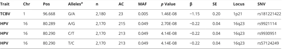
Table 2 Single nucleotide variants (SNVs) in main loci associated with total cerebral brain volume (TCBV) and hippocampal volume (HPV) in single-variant analyses atp≤5 × 10⁻⁸
Trait Chr Pos Allelesa n AC MAF pValue β SE Locus SNV
TCBV 1 96.668 G/A 2,180 23 0.005 1.46E-08 −1.15 0.20 1p21 rs181221422
HPV 16 80.289 A/G 2,170 215 0.049 2.70E-08 −0.22 0.04 16q23 rs9921114
HPV 16 80.290 C/T 2,170 213 0.049 4.14E-08 −0.22 0.04 16q23 rs9930951
HPV 16 80.290 T/C 2,170 213 0.049 4.14E-08 −0.22 0.04 16q23 rs57124249
with the previously reported association (r2
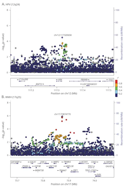
= 0.49, D’= 0.94 with rs7294919). Conditional analysis revealed that the as-sociation observed with rs7132910 was distinct from the one observed with rs7294919 (p = 1.2 × 10⁻⁵). The SNP rs7132910 lies at 1.4 kb from the 59region ofHRKgene and it is an expression quantitative trait locus (eQTL) forHRKand FBXW8genes in blood.22Strong promoter histone marks are described in brain tissues.22Our best 17q25 WMH association was rs9889965 (MAF = 0.15,p= 1.2 × 10⁻⁶) in modest LD with the previously reported association (r2
= 0.68, D’= 0.88 with rs7214628). Conditional analysis revealed that the as-sociation observed with rs9889965 was distinct from the one observed with rs7214628 (p = 3.0 × 10⁻⁵). The SNP rs9889965 lies at 113 bp from the 59region ofTRIM47gene and it is an eQTL forTRIM65gene in nerve tibial tissue and TRIM47 in brain, skin, and blood tissues.22 Strong active transcription starting site histone marks are described in brain tissues.22Single-SNV results in 12q24 and 17q25 regions are presented in table e-1 and regional plots are presented in
figure 1.
Gene-based tests
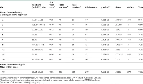
The main gene-based test results are presented in table 3 and regional plots for each gene are provided infigure e-3 (links. lww.com/WNL/A32). Using a sliding-window approach within genes, we detected genome-wide or suggestive asso-ciations in new loci for TCBV with the SKAT test (FCHO2-5q13,UNC5D-8p12, CCDC33-15q24) and with the burden test (C9orf84-9q31, UBL3-13q12-q13, SYT3-19q13), for HPV with the SKAT test (LRRTM4-2p12), and for WMH with the burden test (ALCAM-3q13,GBA3-4p15). Using all SNVs within genes, we detectedSOCS7(17q12) significantly associated with TCBV using the SKAT test. No significant results were found when selecting functional exonic SNVs only.
Pathway analyses
MAGENTA analyses revealed interesting pathways associ-ated with brain MRI phenotypes (table e-2, links.lww.com/ WNL/A33). Some of them are linked to immunity or
in-flammation (B lymphocyte pathway, interleuk4 and in-terleukin-6 signaling, antigen presentation pathway) or to AD pathology such as the presenilin pathway. These analyses also confirmed the importance of the ubiquitin proteasome pathway that was found associated with WMH (p= 4 × 10⁻⁴, false discovery rate = 0.04 for the 75th percentile enrichment cutoff).
Discussion
We investigated low-frequency or rare SNVs influencing brain MRI phenotypes by performing WGS association analyses in the FHS within the TOPMed.
Using single-variant analyses, we identified rare or low-frequency variants in 1p21 (TCBV) and 16q23 (HPV). In 1p21, SNVs were located at 159 kb from PTBP2
(polypyrimidine tract binding protein 2), a brain-specific homologue ofPTBP1. Both genes regulate differentiation of neural precursor cells and promote the proliferation and mi-gration of glioma cell lines.23In 16q23, the SNVs were located within an ncRNA geneLOC102724084.
Using gene-based tests, we identified 10 potential new genes associated with brain MRI phenotypes. Several of these genes are particularly relevant for brain-related diseases and some are also strongly expressed in the brain.
UNC5D(unc-5 netrin receptor D), related to TCBV, encodes a UNC-5 netrin receptor that plays a role in the regulation of axon guidance and it is strongly expressed in the developing sensory areas of the neocortex in mice24and in human brain.25 Interestingly, a gene of the same family,UNC5C, was reported associated with AD.26 Moreover, UNC5C genotypes have been found associated with the middle temporal volume and may alter the atrophy of AD regions such as hippocampus and precuneus.27The expression ofC9orf84(chromosome 9 open reading frame 84), related to TCBV, was found to be upre-gulated in the hippocampus of individuals with major de-pression.28UBL3(ubiquitin like 3) belongs to the ubiquitin pathway that is implicated in the pathogenesis of neurode-generative disorders.29,30 UBL3is strongly expressed in the brain.25SYT3(synaptotagmin 3) is highly expressed in cortex, frontal cortex, anterior cingulate cortex, hippocampus, cere-bellum, and the cerebral hemispheres. It belongs to a family of brain-specific proteins present on the membrane of synapse vesicles that play a role in the secretion of neurotransmitter.31 LRRTM4 (leucine-rich repeat transmembrane neuronal 4) belongs to the leucine-rich repeat proteins that are important regulators of synapse development and function.32LRRTM4 was found to regulate excitatory synapse formation in cul-tured hippocampal neurons.32 LRRTM4 is strongly expressed in the brain.25 ALCAM(activated leukocyte cell adhesion molecule or CD166) is a ligand for CD6 and regulates leukocyte extravasation in the inflamed CNS. ALCAMis expressed on human CNS microvascular endo-thelium, particularly during neuroinflammatory processes, and plays a key role in the recruitment and migration of leukocytes into the brain.33GBA3(glucosylceramidaseβ3) has been associated with Gaucher disease34and GBA genes have been associated with late-onset Parkinson and Lewy body dementia.35–37 Furthermore, genetic variants near GBA3have been found associated with white matter lesion progression.38 SOCS7(suppressor of cytokine signaling 7) belongs to a family of proteins that play a role in preventing inflammation in the brain. SOCS7 levels were found to be increased in AD human brains.39 SOCS7 is noticeably expressed in murine brain and mice with disruptedSOCS7 gene showed defects in CSF homeostasis.40
Figure 1Regional association plots
suggesting that this gene may have a role early in the patho-genesis but not in patients with dementia.
The strengths of this study are the population-based sample, the use of quantitative MRI techniques, as well as sequenced data and the fact that we focused on endophenotypes that have substantial variance in our population. This study also has several limitations. First we performed our analyses in a single study. However, the access to datasets with brain MRI phe-notypes and WGS data is limited and, to this date, FHS was the only TOPMed phase 1 study with sequenced data and brain MRI measures available. Byfine-mapping GWAS regions, we confirmed the association of SNVs that were found associated with the imaging endophenotypes in other studies. A replica-tion or validareplica-tion in an independent cohort is necessary to confirm the new associations (variants or genes) identified in this study. We were also limited by our sample sizes to discover rare variants (2,180, 2,170, and 1,667 individuals analyzed for TCBV, HPV, and WMH, respectively). Finally, the pre-dominantly European origin of our sample limits the general-ization of these results to other ethnic groups.
This FHS WGS-wide search for brain MRI measures reveals rare variants in intriguing new loci associated with brain vol-umes. Replication of these results in the phase 2 TOPMed studies, whose sequencing is ongoing, is planned to confirm these findings. Further investigation of these loci, such as
biological experiments, functional studies, or animal models, will have the potential to validate ourfindings.
Web resources
TOPMed websitesnhlbi.nih.gov/research/resources/nhlbi-precision-medicine-initiative/topmed
nhlbiwgs.org/
Methods for WGS data acquisition and QC in the FHS are described in a document that is publicly available on dbGaP
ncbi.nlm.nih.gov/projects/gap/cgi-bin/GetPdf.cgi?id=phd 006969.1
Author contributions
Chlo´e Sarnowski: acquisition and analysis of data, drafting a significant portion of the manuscript orfigures. Claudia L. Satizabal: acquisition and analysis of data, drafting a significant portion of the manuscript orfigures. Charles DeCarli: acquisi-tion and analysis of data. Achilleas N. Pitsillides: acquisiacquisi-tion and analysis of data. L. Adrienne Cupples: acquisition and analysis of data. Ramachandran S. Vasan: acquisition and analysis of data. James G. Wilson: acquisition and analysis of data. Joshua C. Bis: conception and design of the study, acquisition and analysis of
Table 3Top genes associated with total cerebral brain volume (TCBV), hippocampal volume (HPV), and white matter hyperintensity (WMH) in gene-based tests atp≤2.5 × 10⁻⁶
Chr
Positions (build 37, Mb)
Frac with rarea
Total markersb
Pass
markersc Allele count pValued Gene Method Trait
Genes detected using a sliding-window approach
2 77.67–77.68 0.05 73 50 116 1.66E-06 LRRTM4 SKAT HPV
3 105.14–105.15 0.10 74 44 164 1.58E-06 ALCAM T1 WMH
4 22.81–22.82 0.12 80 54 194 1.46E-06 GBA3 T1 WMH
5 71.25 0.03 45 29 63 5.01E-08 FCHO2 SKAT TCBV
8 35.59 0.06 58 41 131 2.30E-06 UNC5D SKAT TCBV
9 114.50–114.51 0.06 52 36 131 1.67E-06 C9orf84 T1 TCBV
13 30.41–30.42 0.07 60 39 144 6.85E-07 UBL3 T1 TCBV
15 74.57 0.06 81 46 131 2.15E-06 CCDC33 SKAT TCBV
19 51.12–51.13 0.08 68 50 168 8.79E-07 SYT3 T1 TCBV
Genes detected using all SNVs within gene
17 36.51–36.56 0.56 835 585 279 1.58E-06 SOCS7 SKAT TCBV
Abbreviations: Chr = chromosome; SKAT = sequence kernel association test; SNV = single nucleotide variant. aFraction of individuals carrying rare variants below the maximum minor allele frequency (MAF) threshold. bNumber of all variants defining the group.
data. Myriam Fornage: conception and design of the study, acquisition and analysis of data. Alexa S. Beiser: conception and design of the study, acquisition and analysis of data. Anita L. DeStefano: conception and design of the study, acquisition and analysis of data. Jos´ee Dupuis: conception and design of the study, drafting a significant portion of the manuscript orfigures. Sudha Seshadri: conception and design of the study, acquisition and analysis of data, drafting a significant portion of the man-uscript orfigures. Review of the manuscript: all authors.
Acknowledgment
The authors thank the Framingham Heart Study participants, as well as the study team (especially the investigators and staffof the neurology team), for their contributions; whole genome sequencing (WGS) for the Trans-Omics in Precision Medicine Program (TOPMed) was supported by the National Heart, Lung and Blood Institute (NHLBI); WGS for “NHLBI TOPMed: Whole Genome Sequencing and Related Pheno-types in the Framingham Heart Study”(phs000974.v1.p1) was performed at the Broad Institute of MIT and Harvard (HHSN268201500014C); centralized read mapping and genotype calling, along with variant quality metrics andfiltering, were provided by the TOPMed Informatics Research Center (3R01HL-117626-02S1); phenotype harmonization, data man-agement, sample-identity QC, and general study coordination were provided by the TOPMed Data Coordinating Center (3R01HL-120393-02S1); the authors thank the studies and participants who provided biological samples and data for TOPMed; Goncalo Abecasis for his work that made the TOPMed data available; Cashell Jaquish for her leadership that has catapulted TOPMed into the largest WGS project and for her support of Framingham within TOPMed; and the contributions of the investigators of the NHLBI TOPMed Consortium (nhlbiwgs.org/topmed-banner-authorship).
Study funding
Supported by the National Heart, Lung and Blood Institute’s Framingham Heart Study (contract N01-HC-25195 and HHSN268201500001I); and grants from the National In-stitute on Aging (R01s AG033193, AG008122, AG054076, AG049505, AG016495, AG049607, NS017950) and the Na-tional Heart, Lung and Blood Institute (UH2 NS100605, R01 HL093029, HL096917). Dr. DeCarli is supported by the UCD Alzheimer’s Disease Center (P30 AG 010129).
Disclosure
C. Sarnowski, C. Satizabal, C. DeCarli, A. Pitsillides, L. Cupples, R. Vasan, J. Wilson, J. Bis, M. Fornage, A. Beiser, and A. DeStefano report no disclosures relevant to the man-uscript. J. Dupuis reports grants from NIH during the conduct of the study. S. Seshadri reports no disclosures relevant to the manuscript. Go to Neurology.org/N for full disclosures.
Received March 14, 2017. Accepted infinal form September 22, 2017.
References
1. Braskie MN, Ringman JM, Thompson PM. Neuroimaging measures as endopheno-types in Alzheimer’s disease. Int J Alzheimers Dis 2011;2011:490140.
2. Bis JC, DeCarli C, Smith AV, et al. Common variants at 12q14 and 12q24 are associated with hippocampal volume. Nat Genet 2012;44:545–551.
3. Hibar DP, Stein JL, Renteria ME, et al. Common genetic variants influence human subcortical brain structures. Nature 2015;520:224–229.
4. Stein JL, Medland SE, Vasquez AA, et al. Identification of common variants associated with human hippocampal and intracranial volumes. Nat Genet 2012;44:552–561. 5. Traylor M, Zhang CR, Adib-Samii P, et al. Genome-wide meta-analysis of cerebral
white matter hyperintensities in patients with stroke. Neurology 2016;86:146–153. 6. Verhaaren BF, Debette S, Bis JC, et al. Multiethnic genome-wide association study of
cerebral white matter hyperintensities on MRI. Circ Cardiovasc Genet 2015;8: 398–409.
7. Fornage M, Debette S, Bis JC, et al. Genome-wide association studies of cerebral white matter lesion burden: the CHARGE consortium. Ann Neurol 2011;69: 928–939.
8. Dawber TR, Kannel WB. The Framingham Study: an epidemiological approach to coronary heart disease. Circulation 1966;34:553–555.
9. Feinleib M, Kannel WB, Garrison RJ, McNamara PM, Castelli WP. The Framingham Offspring Study: design and preliminary data. Prev Med 1975;4:518–525. 10. Splansky GL, Corey D, Yang Q, et al. The third generation cohort of the National
Heart, Lung, and Blood Institute’s Framingham Heart Study: design, recruitment, and initial examination. Am J Epidemiol 2007;165:1328–1335.
11. DeCarli C, Massaro J, Harvey D, et al. Measures of brain morphology and infarction in the Framingham Heart Study: establishing what is normal. Neurobiol Aging 2005;26: 491–510.
12. Kaur B, Himali JJ, Seshadri S, et al. Association between neuropathology and brain volume in the Framingham Heart Study. Alzheimer Dis Assoc Disord 2014;28: 219–225.
13. DeCarli C, Fletcher E, Ramey V, Harvey D, Jagust WJ. Anatomical mapping of white matter hyperintensities (WMH): exploring the relationships between periventricular WMH, deep WMH, and total WMH burden. Stroke 2005;36:50–55.
14. DeCarli C, Miller BL, Swan GE, et al. Predictors of brain morphology for the men of the NHLBI twin study. Stroke 1999;30:529–536.
15. Aljabar P, Heckemann RA, Hammers A, Hajnal JV, Rueckert D. Multi-atlas based segmentation of brain images: atlas selection and its effect on accuracy. Neuroimage 2009;46:726–738.
16. Carmichael O, Mungas D, Beckett L, et al. MRI predictors of cognitive change in a diverse and carefully characterized elderly population. Neurobiol Aging 2012;33: 83–95.
17. Xu C, Tachmazidou I, Walter K, Ciampi A, Zeggini E, Greenwood CM; UK10K Consortium. Estimating genome-wide significance for whole-genome sequencing studies. Genet Epidemiol 2014;38:281–290.
18. Lin WY, Lou XY, Gao G, Liu N. Rare variant association testing by adaptive com-bination of P-values. PLoS One 2014;9:e85728.
19. Tang R, Feng T, Sha Q, Zhang S. A variable-sized sliding-window approach for genetic association studies via principal component analysis. Ann Hum Genet 2009;73: 631–637.
20. Wang K, Li M, Hakonarson H. ANNOVAR: functional annotation of genetic variants from high-throughput sequencing data. Nucleic Acids Res 2010;38:e164. 21. Segre AV; DIAGRAM Consortium, MAGIC Investigators, Groop L, Mootha VK,
Daly MJ, Altshuler D. Common inherited variation in mitochondrial genes is not enriched for associations with type 2 diabetes or related glycemic traits. PLoS Genet 2010;6:e100105.
22. Ward LD, Kellis M. HaploReg v4: systematic mining of putative causal variants, cell types, regulators and target genes for human complex traits and disease. Nucleic Acids Res 2016;44:D877–D881.
23. Cheung HC, Hai T, Zhu W, et al. Splicing factors PTBP1 and PTBP2 promote proliferation and migration of glioma cell lines. Brain 2009;132:2277–2288. 24. Takemoto M, Hattori Y, Zhao H, et al. Laminar and areal expression of unc5d and its
role in cortical cell survival. Cereb Cortex 2011;21:1925–1934.
25. GTEx Consortium. Human genomics: the genotype-tissue expression (GTEx) pilot analysis: multitissue gene regulation in humans. Science 2015;348:648–660. 26. Wetzel-Smith MK, Hunkapiller J, Bhangale TR, et al. A rare mutation in UNC5C
predisposes to late-onset Alzheimer’s disease and increases neuronal cell death. Nat Med 2014;20:1452–1457.
27. Sun JH, Wang HF, Zhu XC, et al. The impact of UNC5C genetic variations on neuroimaging in Alzheimer’s disease. Mol Neurobiol 2016;53:6759–6767. 28. Kim S, Hwang Y, Webster MJ, Lee D. Differential activation of immune/inflammatory
response-related co-expression modules in the hippocampus across the major psy-chiatric disorders. Mol Psychiatry 2016;21:376–385.
29. Debette S, Ibrahim Verbaas CA, Bressler J, et al. Genome-wide studies of verbal declarative memory in nondemented older people: the cohorts for heart and aging research in genomic epidemiology consortium. Biol Psychiatry 2015;77:749–763. 30. Ying Z, Wang H, Wang G. The ubiquitin proteasome system as a potential target for
the treatment of neurodegenerative diseases. Curr Pharm Des 2013;19:3305–3314. 31. Li C, Ullrich B, Zhang JZ, Anderson RG, Brose N, Sudhof TC. Ca(2+)-dependent and -independent activities of neural and non-neural synaptotagmins. Nature 1995;375:594–599. 32. de Wit J, O’Sullivan ML, Savas JN, et al. Unbiased discovery of glypican as a receptor for
LRRTM4 in regulating excitatory synapse development. Neuron 2013;79:696–711. 33. Cayrol R, Wosik K, Berard JL, et al. Activated leukocyte cell adhesion molecule
promotes leukocyte trafficking into the central nervous system. Nat Immunol 2008;9: 137–145.
35. Gamez-Valero A, Prada-Dacasa P, Santos C, et al. GBA mutations are associated with earlier onset and male sex in dementia with Lewy bodies. Mov Disord 2016;31: 1066–1070.
36. Geiger JT, Ding J, Crain B, et al. Next-generation sequencing reveals substantial genetic contribution to dementia with Lewy bodies. Neurobiol Dis 2016;94:55–62. 37. Swan M, Doan N, Ortega RA, et al. Neuropsychiatric characteristics of
GBA-associated Parkinson disease. J Neurol Sci 2016;370:63–69.
38. Hofer E, Cavalieri M, Bis JC, et al. White matter lesion progression: genome-wide search for genetic influences. Stroke 2015;46:3048–3057.
39. Walker DG, Whetzel AM, Lue LF. Expression of suppressor of cytokine signaling genes in human elderly and Alzheimer’s disease brains and human microglia. Neu-roscience 2015;302:121–137.
SOURCE ARTICLE NPub.org/pqojyh
Whole genome sequence analyses of brain
imaging measures in the Framingham Study
Chlo´e Sarnowski, PhD, Claudia L. Satizabal, PhD, Charles DeCarli, MD, Achilleas N. Pitsillides, PhD, L. Adrienne Cupples, PhD, Ramachandran S. Vasan, MD, James G. Wilson, MD, Joshua C. Bis, PhD, Myriam Fornage, PhD, Alexa S. Beiser, PhD, Anita L. DeStefano, PhD, Jos´ee Dupuis, PhD,
and Sudha Seshadri, MD, NHLBI Trans-Omics for Precision Medicine (TOPMed) Consortium, On behalf of the TOPMed Neurocognitive Working Group
Cite as:Neurology
®
2018;90:e188–e196. doi:10.1212/WNL.0000000000004820Correspondence
Dr. Sarnowski [email protected]
Study question
Are there are any low-frequency and rare variants affecting brain imaging phenotypes in the Framingham Study?
Summary answer
Low-frequency or rare variants were identified in 1p21 and 16q23 for total cerebral volume (TCBV) and for hippocampal volume (HPV), respectively, and 10 novel genes were iden-tified to be associated with brain MRI phenotypes.
What is known and what this article adds Associations exist between genetic variants and brain imaging phenotypes, and common genetic variants in noncoding regions with modest effect sizes have been reported. This study identifies new rare and low-frequency variants and novel genes associated with TCBV, HPV, and white matter hyper-intensity (WMH), which may have clinical relevance.
Participants and setting
The study population was drawn from the 3 generations of the Framingham Heart Study (FHS), a prospective, population-based study of determinants of cardiovascular disease that has been following participants from Framing-ham, Massachusetts, since 1948.
Design, size, and duration
In total, 4,501 participants were recruited, after excluding cases of stroke or TIA, dementia, and other neurologic con-ditions associated with the relevant imaging abnormalities.
Whole genome sequencing (WGS) was conducted during FHS Phase I; TCBV, HPV, and WMH analyses were con-ducted for 2,180, 2,170, and 1,667 participants, respectively.
Main results and the role of chance
Rare or low-frequency variants were identified in 1p21 (159 kb fromPTBP2[polypyrimidine tract binding protein 2], a brain-specificPTBP1homologue) and 16q23 (within an ncRNA gene LOC102724084) for TCBV and HPV, respectively. Previously identified associations for HPV (12q24) and WMH (17q25) were also confirmed. Furthermore, 10 novel genes potentially associated with MRI phenotypes were identified on 5q13, 8p12, 9q31, 13q12-q13, 15q24, 17q12, and 19q13 for cerebral vol-umes; 2p12 for hippocampal volvol-umes; and 3q13 and 4p15 for WMH, including genes associated with Alzheimer disease and Parkinson disease.
Bias, confounding, and other reasons for caution
The analyses were performed for a single cohort, and the sample sizes were low for the discovery of rare variants.
Generalizability to other populations
The predominantly European origin of the study sample may limit generalizability to other ethnic groups.
Study funding/potential competing interests The study was funded by a group of university and foundation grants. Go to Neurology.org/N for full disclosures.
TableSingle nucleotide variants (SNVs) in loci associated with TCBV and HPV in single-variant analyses atp≤5 × 10⁻⁸
Trait Chr Pos Allelesa n AC MAF pValue β SE Locus SNV
TCBV 1 96.668 G/A 2,180 23 0.005 1.46E-08 −1.15 0.20 1p21 rs181221422
HPV 16 80.289 A/G 2,170 215 0.049 2.70E-08 −0.22 0.04 16q23 rs9921114
HPV 16 80.290 C/T 2,170 213 0.049 4.14E-08 −0.22 0.04 16q23 rs9930951
HPV 16 80.290 T/C 2,170 213 0.049 4.14E-08 −0.22 0.04 16q23 rs57124249
Abbreviations: AC = allele count; Chr = chromosome; MAF = minor allele frequency; Pos = position in Mb on Build 37. aCoded/noncoded.
A draft of the short-form article was written by E. Feric, a writer with Editage, a division of Cactus Communications. The authors of the full-length article and the journal editors edited and approved thefinal version.
108 Copyright © 2018 American Academy of Neurology
DOI 10.1212/WNL.0000000000004820
2018;90;e188-e196 Published Online before print December 27, 2017
Neurology
Chloé Sarnowski, Claudia L. Satizabal, Charles DeCarli, et al.
Whole genome sequence analyses of brain imaging measures in the Framingham Study
This information is current as of December 27, 2017
Services
Updated Information &
http://n.neurology.org/content/90/3/e188.full
including high resolution figures, can be found at:
References
http://n.neurology.org/content/90/3/e188.full#ref-list-1
This article cites 40 articles, 8 of which you can access for free at:
Citations
http://n.neurology.org/content/90/3/e188.full##otherarticles
This article has been cited by 2 HighWire-hosted articles:
Subspecialty Collections
http://n.neurology.org/cgi/collection/mri
MRI
http://n.neurology.org/cgi/collection/association_studies_in_genetics
Association studies in genetics
http://n.neurology.org/cgi/collection/alzheimers_disease
Alzheimer's disease following collection(s):
This article, along with others on similar topics, appears in the
Permissions & Licensing
http://www.neurology.org/about/about_the_journal#permissions
its entirety can be found online at:
Information about reproducing this article in parts (figures,tables) or in
Reprints
http://n.neurology.org/subscribers/advertise
Information about ordering reprints can be found online:
ISSN: 0028-3878. Online ISSN: 1526-632X.
Wolters Kluwer Health, Inc. on behalf of the American Academy of Neurology.. All rights reserved. Print 1951, it is now a weekly with 48 issues per year. Copyright Copyright © 2017 The Author(s). Published by
® is the official journal of the American Academy of Neurology. Published continuously since