1 Neurologic and inflammatory manifestations in Sjögren’s
syndrome: the role of tryptophan/kynurenine pathway
Fabíola Reis de Oliveira1, Marina Zilio Fantucci1, Leidiane Adriano1,
Valéria Valim2, Thiago Mattar Cunha1, Paulo Louzada Junior1,
Eduardo Melani Rocha1
1Ribeirao Preto Medical School, Ribeirao Preto, University of Sao
Paulo, Ribeirao Preto, SP, Brazil.
2Espírito Santo Federal University, Vitoria-ES, Brazil.
Financial Support: FAPESP, CNPQ, FAEPA, CAPES
Corresponding Author: Eduardo Melani Rocha
Department of Ophthalmology, Otorhinolaryngology and Head & Neck Surgery, RibeirãoPreto Medical School, University of São Paulo. Av. Bandeirantes, 3900, 14049-900 – Ribeirão Preto SP, Brazil. [email protected]
Phone/fax: 55-16-3602-0593
The authors have no commercial or proprietary interest in any concept or product described in this article.
2 Abstract
For decades, neurologic, psychological and cognitive alterations, and other extra glandular manifestations have been described and are being considered part of the Sjögren’s syndrome (SS). The lacrimal glands (LG), the ocular surface (OS), salivary glands (SG) and the central nervous system (CNS) are integrated to modulate the autonomic functions and the hippocampus, which is linked to the autonomic nervous system, modulate behavior responses compromised in the SS.
Recent studies confirm that the tryptophan/kynurenine pathway (TKP) can be stimulated by interferon-γ (IFN-γ) and other cytokines, activating the indoleamine-pyrrole 2,3-dioxygenase (IDO) in SS. This pathway interferes on serotonergic and glutamatergic
neurotransmission, mostly in the hippocampus, and other structures of the CNS. Therefore, it is plausible that TKP induces neurological manifestations, and contributes to the discrepancy between symptoms and signs, including manifestations of hyperalgesia and depression in patients with SS, for example. Observations from clinical studies in AIDS, graft versus host disease, lupus and SS, but also from
experimental studies support this hypothesis. Therapeutic strategies are reexamined and new options designed and tested to regulate the TKP. In the future, the confirmation and application of this concept may offer a clue to the mosaic of manifestations of SS.
4 Outlines
1. Introduction
2. Autoimmunity, neuropathy and chronic pain
2.a. SS and Neurological manifestations
2.b. SS and the mechanisms of neurological manifestations
2.c. The immune and the endocrine modulation of neurological
findings in SS
3. The tryptophan/kynurenine pathway (TKP)
4. The TKP and the neurologic manifestations
4.a. The role of the hippocampus in the TKP ofthe neurologic
manifestations
5. The TKP and the neuropathy in SS
6. Therapy to modulate the TKP
7. Future perspectives
8. Conclusions
9. References
10. Attachments: Box, Tables and Figures legends
5
nicotinamide adenine nucleotide, NAISD: non-steroidal
6 1. Introduction
Sjogrën’s syndrome (SS) is defined as an exocrinopathy of salivary and lacrimal glands (SG and LG) mediated by autoimmune mechanisms that can manifest neurologic dysfunctions, and those neurologic dysfunctions may take part in the physiopathology of the disease [1-5]. However, the neurological manifestations are not considered in the definition or the diagnosis, despite those
manifestations are present in the disease progress evaluation and are being reported in association with SS with more attention in recent years [6-9]. Of interest, 60-80% of patients develop neurological
manifestations before or at SS diagnosis (early systemic presentation) indicating that neurological inflammatory damage is precocious and could have a role in disease mechanism [10].
The tryptophan/kynurenine signaling pathway (TKP) intermediates the serotonergic and glutamatergic neurotransmission. It is also
known to take part in the inflammatory mechanisms of the neurogenic manifestations of autoimmune diseases through the action of
indoleamine 2,3-dioxygenase (IDO), the rate-limiting enzyme in the tryptophan degradation [11-15].
This review summarizes the actual status of the knowledge of the neurological manifestations in SS and presents the hypothesis of the association between these neurological manifestations and the TKP (Box 1).
7
Association among chronic inflammation, pain and
neuropathic disorders
Triggered by Interferon
Modulated by sex hormones
Tryptophan deprivation induces dry eye
Sjögren’s syndrome and salivary gland aggression leads to
increased expression of kynurenine
2. Autoimmunity, neuropathy and chronic pain
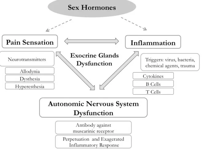
Autoimmunity is linked to chronic neuropathy in at least three different forms. The first and major outcome observed is the pain, in its different dysfunctional manifestations, as allodynia, dysesthesia, hypo or hyperestesia and hyperalgesia [9,16,17]. Second, chronic inflammation in the target organs generates a noxious stimulus that may persist in a further phase where the inflammatory process is already resolved [18,19]. Third, the central nervous system (CNS), mostly the autonomic nervous system dysfunction can induce or perpetuate an unbalanced inflammatory response in the target-innervated organs [3,20]. Therefore, an unified theory of the
8 Figure 1. A schematic model, showing the interrelationships among
the autonomic nervous system dysfunction, pathological pain and chronic inflammation in Sjögren’s syndrome (SS).
The chronic pain and persistent inflammation mediated by humoral factors and neurotransmitters associate autoimmunity and neuropathy in several target organs, including the hippocampus and the lacrimal functional unit (LFU) on SS [20,24-28]. The dissociation between signs and symptoms of dry eye and dry mouth delays and makes the diagnosis more difficult in SS but paradoxically, those individuals present a high association between pain sensation in different organs and dry eye and dry mouth [5,29-35].
9
This hypothesis predicts a spectrum of manifestations with two different poles of the SS disease. In one pole, the major characteristic is the chronic non-inflammatory pain with neuropathic features. In the opposite pole, primary SS present with inflammatory activity, including extra-glandular manifestations (EGM). However, these profiles can only be documented in long-term cohort studies [42]. In other words, we hypothesized that SS patients with Interferon-γ (IFN-γ)-inducible TKP activation could develop chronic pain and low EGM disease activity because the TKP promotes an immunosupressive and neuro sensitive effect. Therefore IFN-γ-inducible TKP could be the missing link between disease activity and neural manifestations in SS [43] (Figure 1).
2.a. SS and Neurological manifestations
10
XII and last but not least, dysautonomia, which is very common in SS, and can reach a frequency of 40% of SS patients, in combination or not with different neurological manifestations [49-52]. Moreover, CNS manifestations may cause focal syndromes (such as
11 Table 1. List of the main neurocognitive changes in Sjögren syndrome described in several study models
Description of the disorder Study model N Author, year
Major depressive and panic disorder Case series 2 Pelizza et al, 2010 [59]
Visual hallucinations Case report 1 Wong et al, 2014 [60]
Dementia Case report 1 Caselli et al, 1991 [61]
Migraine, neuropsychiatric disease, and focal acute neurological deficits Prospective 48 Escudero et al, 1995 [62] Cerebral manifestations (focal and diffuse) and spinal cord disease Case series 16 Alexander et al, 1982 [63] Peripheral nervous system abnormalities Cross-sectional 39 Barendregt et al, 2001 [46] Sensorimotor/sensory axonal polyneuropathy, spinal cord disease, and cognitive dysfunction Prospective 25 Lafitte et al, 2001 [64]
Peripheral neuropathy Cross-sectional 46 Gemignani et al, 1994 [65]
CNS disease, mostly non-focal dysfunction; PNS disease, mostly mild or severe sensory or sensory-motor
polyneuropathies Cohort 87 Govoni et al, 1999
[66]
Sensory ataxic neuropathy, painful sensory neuropathy without sensory ataxia, multiple mononeuropathy, multiple cranial neuropathy, trigeminal neuropathy, autonomic neuropathy, and radiculoneuropathy
Cross-sectional 92 Mori et al, 2005 [44] Motor neuropathy, sensory neuropathy, sensorimotor neuropathy, and small-diameter nerve fiber neuropathy Cross-sectional 62 Goransson et al, 2006 [48] CNS disorders: spinal cord involvement, brain involvement, and optic neuropathy. PNS involvement: symmetric
axonal sensorimotor polyneuropathy, cranial nerve involvement affecting trigeminal, facial, or cochlear nerves.
Retrospective 82 Delalande et al, 2004 [2] Complaints of painful distal paresthesias in the feet, abnormal sweating, and decreased pinprick sensation.
Small-fiber neuropathy Cohort 32 Lopate et al, 2006
[67]
Subcortical dementia Case report 1 Kawashima et al, 1993 [68]
PNS involvement: small fiber neuropathy; trigeminal, facial, or trochlear nerves involvement; multiple mononeuropathy; sensorimotor polyneuropathy; autonomic neuropathy; and myasthenia gravis. CNS involvement: headache; spinal cord involvement; seizures; motor and sensory deficit; movement disorders; neuromyelitis optica; aseptic meningitis. Cognitive dysfunction
Retrospective
cross-sectional case-control 93 Teixeira et al, 2013 [69]
Atypical neurologicmanifestations: pseudotumoral lesion; multiple mononeuropathy; progressive multiple
sclerosis; and myelitis along with progressive cognitive disorders Case series 4 Michel et al, 2011 [70]
Neuromyelitis optica Case series 2 Nitescu et al, 2011 [71]
Peripheral neuropathy: axonal sensorimotor polyneuropathy, pure sensory neuronopathy, mononeuropathy multiplex, and demyelinating polyradiculoneuropathy
Cross-sectional 102 Brito-Zeron et al, 2013 [72]
Multiple sclerosis Case report 1 Liu et al, 2014 [73]
Neuropathy axonal (pure sensory or sensorimotor) Cross-sectional 44 Pavlakis et al, 2011 [74]
CNS vasculitic involvement Case report 1 Hasiloglu et al, 2012 [75]
Neuromyelitis optica Retrospective 43 Qiao et al, 2015 [55]
12
13 This high variability of clinical manifestations of SS with different responses to treatment should be related to diverse and complex mechanisms of injury such as vasculitis and lymphocytic infiltration. Changes in the structure of the dorsal spinal cord and the dorsal ganglion roots, association with autonomic dysfunctions, decrease in the white matter, and loss of the gray matter of the hippocampal area were observed in SS patients [77-79].
2.b. SS and the mechanisms of neurological manifestations
The mechanisms triggering the neurological manifestations in SS are unclear. They involve genetic predisposition, environmental agents, trauma and posttraumatic stress, autoimmunity against the CNS and peripheral nervous systems (PNS), in addition to the neuroimmunendocrine network disruption [47,80-85]. More specifically, unknown causes trigger DNA demethylation, microRNA abnormal expression, unbalance of Interferon I (α and β) and II (γ), and anti-neuron autoantibodies production [55,83,86-88]. The 2-5 oligo-adenylate synthetase 1
(OAS1) gene defect leads to lower responsiveness to IFN-γ, and induces higher production of IFN-γ causing severe complications as neuropathy in SS [85].
The increase in corneal nerve thickness and higher number of antigen presenting cells in the cornea, nerve vasculitis, nonvascular encephalitis,
neuromyelitis, axonal and CNS degeneration are hypothesized to be responsible for those sensorial, autonomic, cognitive or behavioral neurological manifestations of SS [24,47,81,89].
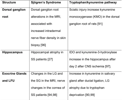
14 sensorial fibers, at the dorsal ganglion root, hippocampus, thalamus and LFU [2,12,13,27,39,77,90-95] (Table 2).
Table 2. Changes in the Nervous system and in the exocrine glands in Sjögren’s Syndrome (SS) and after the modulation of the
tryptophan/kynurenine pathway (TKP).
MRI: magnetic resonance image, SS: Sjögren’s syndrome, LG: lacrimal gland, SG: salivary gland, CNS: central nervous system, IDO: indoleamine 2,3-dioxygenase
2.c. The immune and the endocrine modulation of neurological findings
in SS
Structure Sjögren’s Syndrome Tryptophan/kynurenine pathway
Dorsal ganglion
root
Dorsal ganglion root
alterations in the MRI,
associated with
increased intradermal
nerve fiber density in skin
biopsy [96]
Sciatic injury increase kynurenine
monooxygenase (KMO) in the dorsal
ganglion root of rats [91]
Hippocampus Hippocampal atrophy in
SS patients [27]
IDO and kynurenine-3-hydroxylase
increase in the hippocampus after
day 2 after CNS ischemia [97].
Exocrine Glands
and LFU
Changes in the LG and
the SG in the MRI, nerve
changes in the cornea of
SS patients [94,98]
Increase in kynurenine in salivary
gland after ductal ligation, LG
atrophy due to tryptophan
15 The concept implicit in the neuroimmunendocrine network predicts that cellular and molecular communication among those three systems are
responsible for the homeostasis of the body organs; and a disruption in this network take part in the mechanism of the diseases [100-103]. Acetyl-choline (Ach), dopamine, glutamatergic and other neurotransmitters, are also secreted by lymphocytes [104]. In the other hand, the autonomic nervous system is capable of modulating lymphocytes proliferation in target organs like spleen, liver, kidney, and brain [23,105,106]. In addition, the sensory neurons are capable to secrete peptides with immunomodulatory properties as galanin, netrin-1, and somatostatin and promote or attenuate inflammatory responses [107,108].
Hormones, in particular the sex hormones, can modulate the inflammation and the pain, sensitizing the ionic receptors expressed in the neurons and epithelial cells, called transient receptor of potential (TRPs), and stimulating growth factors and cytokines expression in the target tissues as lacrimal and salivary glands (LG and SG), hippocampus and trigeminal ganglion of the CNS, and also on other target tissues[109-116]. Those mechanisms explain the sex hormones mediated amplification and perpetuation of the inflammatory process, the pain hypersensitivity and exocrine glands dryness manifestations in SS, where estrogen potentiate the pain and pro-inflammatory mediators and androgens work oppositely
16 These observations taken together indicate that hormones and neural pathways are involved in immune responses, resulting in variability in the pain sensation, inflammatory reaction, tissue integrity and functional disruption [5,23]. Those observations are in agreement with recent reports describing multiple comorbidities and extra glandular manifestations (EGM) in SS patients [2,92,122].
The loss or damage of the nerve fibers, dorsal root ganglionitis, nerve vasculitis and reduction of the CNS matter can be observed in image exams, as magnetic resonance image (MRI) or skin biopsies in SS; they are also implicated in the mechanisms of the diseases, and work as diagnostic markers of SS [24,77,78,96]. For example, the cognitive impairment on SS patients are associated with antibodies against NR2 subtype of the N-metyl Aspartate receptor (anti-NR2 antibodies) in the cerebrospinal fluid (CSF) mediating hippocampal gray matter atrophy, observed by MRI [27]. In
summary, the SS inflammatory activity in the PNS and CNS causes the signs and symptoms described above, and are modulated by hormones,
neurotransmitters and cytokines susceptible to the TKP interference [13,14,102].
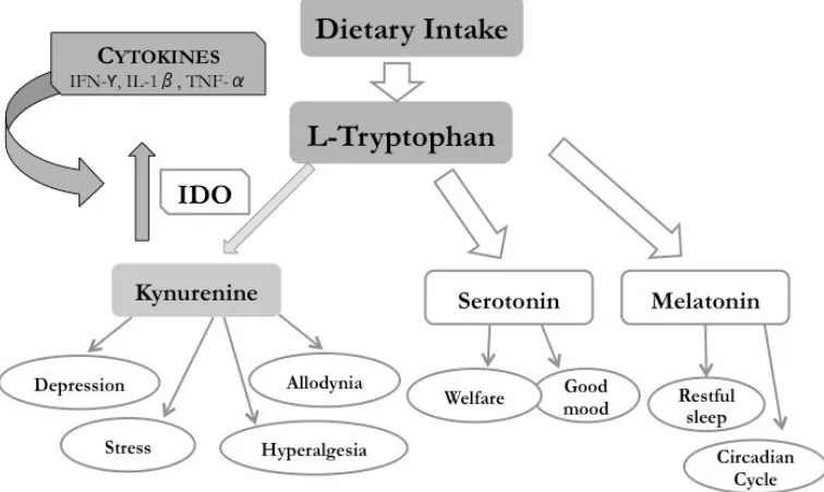
3. The tryptophan/kynurenine pathway (TKP)
17 tract control (satiety, secretion, and peristalsis) and tumor resistance.
Melatonin acts as a neurohormone in the circadian rhythm with also an anti-inflammatory, anti-angiogenic and anti-tumoral immunomodulatory effects [123] (Figure 2).
In the catabolic TKP, the tryptophan 2,3-dioxygenase (TDO) specifically metabolizes tryptophan in the liver and responds to hormonal regulations, such as cortisol and glucagon, and to the tryptophan
concentration [124] (Figure 2).
The IDO enzyme regulates both innate and adaptive immune
responses through the degradation of the essential amino acid tryptophan into kynurenine and other metabolites, which suppress effector T-cell function and promote the differentiation of regulatory T cells. The IDO also metabolizes serotonin, melatonin, and tryptophan until N-formil kynurenine and kynurenine in the lung, the brain, and blood, producing quinolinic acid and nicotinamide adenine nucleotide (NAD+) [125-127]. The TKP is responsible for 95-99% of the entire tryptophan catabolism [128] (Figure 2).
19 nuclear effect contributes to an enzyme “self-amplification” in an
IFN-dependent loop that may account for the phenotype of tolerance, attenuating or preventing immune reactions and mediating persistent pain in several conditions, including SS [129,130,132-134]. The IDO activity is induced by macrophages and by cytokines, like IFN-γ and TNF-α, by prostaglandins, by viral and bacterial infections and by lipopolysaccharides, by dendritic cells and the sub-products of kynurenine [135-139]. IDO overexpression has been
documented in patients with systemic lupus erythematosus (SLE) and SS, as well as in sepsis [15,36,140]. In patients who are positive for the IFN gene expression signature, Treg cell levels are elevated in combination with increased IDO activity, with tolerance and immune modulation[132]. Those regulatory T lymphocytes cells represent a diverse subclass of T cells, a protagonist in the maintenance of self-tolerance and immune modulation[141].
The TKP is catalyzed by the IDO enzyme and it can be induced by the cytokine IFN-γ, as observed in studies with mice where IFN-γ or IFN-γ
receptor knockout prevent kynurenine production [142]. The phenomenon is dependent on antigen-presenting cells (APCs) mentioned above, as observed in mice models with graft-versus host disease (GVHD) and induction of this TKP can extend the life-span and reduce the inflammation of the gut of the wild-type mice, but not of those IFN-γ receptor knockout mice [142].
20 and on the interaction between the T cells and other cells in the target tissues of inflammatory diseases, where the pretreatment IFN-γ can suppress the presentation of the auto antigens by those target cells [146-148].
The isoform IDO2, and also TDO1 are enzymes involved in the catabolism of the amino acid tryptophan and operate in similar ways in the immunomodulation, with variations in the target tissues, cells involved in the metabolic pathways in several conditions. However, further studies are necessary to clarify their diversity in diseases, including SS [149,150]. 4. The TKP and the neurologic manifestations
The relationship between tryptophan, serotonin, and depression has an extended history in psychiatry. The development of depressive symptoms is correlated with high levels of tryptophan metabolites in urine [37] and with a decrease of tryptophan in the blood and cerebrospinal fluid [151,152].The tryptophan transport across the blood-brain barrier, specific inflammation and damage caused by brain-reactive autoantibodies and immune complexes play a critical role in the regulation of tryptophan metabolism in the brain [123]. There is substantial evidence to suggest that in addition to serotonergic neurons, other cells such as astrocytes, dendritic cells, microglia, and macrophages also synthesize multiple neuroactive metabolites via the enzyme IDO and the TKP at CNS [153]. Evidences underlying the
mechanisms have come from clinical studies that examined the effects of IFN-α on the mood of cancer and hepatitis C virus-infected patients. In both, the development of depressive symptoms was associated with decreased
21 Tryptophan metabolites generated in the TKP have been associated with neurodegenerative diseases such as acquired immunodeficiency syndrome (AIDS)-related dementia, Alzheimer's and Huntington’s diseases, and neuropsychiatric diseases like bipolar disorder and schizophrenia [157,158]. The synthesis of kynurenine in the CNS is affected by dietary intake of
tryptophan and by the gut microbiota, and deprivation induces depression, cognitive dysfunction (attention, memory, and execution), among other neurologic dysfunctions through the glutamatergic receptors [14,40,159-162].
Tryptophan metabolism in astrocytes leads to the production of the kynurenic acid reported to take part in neuroprotective actions. On the other hand, microglia cells give rise to metabolites with reactive oxidative properties including hydroxykynurenine and 3-hydroxyanthranilic acid and quinolinic acid which also acts as an agonist at the glutamate N-methyl-D-aspartate (NMDA) receptor subtype and may contribute to excitotoxicity and neurotoxicity [163-165].
In healthy subjects, there is a well-adjusted system in the TKP by the action of kynurenines aminotransferases (KATs) towards kynurenic acid or to quinolinic acid by (KMO). Kynurenic acid, which is reported to promote neuroprotective and immunosuppressive actions in CNS, and plays a role as an NMDA antagonist, blocking this glutamatergic receptor. An enhanced IDO activity and a deviation to KMO downstream are observed by stimulation of cytokines such as IFN-γ [166]. KMO has a clear inflammatory and
22 neurologic diseases[123,157]. Likewise, 3-hidroxikynurenine has also
neurotoxic effect, probably associated with the conversion of reactive oxygen species and apoptosis [167-170]. [78]
The NR2 subtype NMDA receptor is ubiquitously distributed through the brain with a unusually high density in the hippocampus [171].
Hippocampus is a brain structure, linked to the autonomous nervous system, with critical importance for memory formation and learning, and is also affected in mood disorders and in SS [77,78,172,173]. Likewise, the NR2B subunit of NMDA receptor is widespread in dorsal root ganglion and may mediate peripheral sensitization and visceral pain [174]. Those receptors are critically involved in the initiation and maintenance of neuronal
hyperexcitability after noxious events, by C-fiber stimulation [175]. In the rodent model of peripheral nerve injury, tibial and peroneal nerves of one leg were sectioned, but the sural branch nerve was left intact. After seven days, it was observed that IDO1 was activated and the
kynurenine rose in the bloodstream, accompanied by the depressive behavior, confirmed by the extended time of immobility in the forced swim test and allodynia in the paw withdraw in response to mechanical stimulation with von Frey hair. Those findings were followed by an increase in the levels of KMO, quinolinic acid, and reduce the kynureneic acid in the contralateral
23 levels of tryptophan in patients with cancer receiving therapy with IL-2 and IFN-α, suggesting that those cytokines impact the levels of serotonin [155].
Studies observing the triggering of inflammation of the macaque CNS with poliovirus inoculation revealed that quinolinic acid, kynurenine, and other metabolites of the TKP accumulate in the spinal cord, CSF, but not as much in the bloodstream, in levels that were corresponding to the clinical
manifestations of functional damage [12,135]. Moreover, the in vitro conversion of L-tryptophan in kynurenine by fetal neuronal cells was dependent upon IFN-γ stimulation in the presence of macrophages in the culture [12].
Confirming the hippocampus as a target regulatory CNS organ in pain and depression, it was shown that chronic pain in rats exposed to social stress or paw arthritis increase the levels of IDO, and also increase the levels of kynurenine and decrease the levels of serotonin in the hippocampus [176]. This situation is similar to the observed in human plasma levels of IDO, kynurenine/tryptophan and serotonin/tryptophan levels in patients with back pain and depression, where the first two rise and the third one decreases, as revealed in the same paper. Moreover, the IDO1 knockout mice presented lower nociceptive and depressive behavior compared to the wild type, and this behavior was not attenuated in the wild type that received an intraperitoneal injection of the NSAID acetaminophen, suggesting that this behavior is not dependent of the inflammatory mechanisms alone. Furthermore, the authors found that IL-6 is overexpressed in rats with arthritis and the Jak2/Stat3 signaling pathway activated in the blood and the hippocampus of rats with depressive and nocioceptive behavior. The injection of IL-6 anti-serum
24 cells (a mice neuroblastoma cell line) in culture, the incubation with IL-6 induced increase in IDO1 mRNA and protein [176].
Therefore, persistent pain with allodynia and hyperalgesia has a central component (spinal and supraspinal cord), supporting the involvement of glutamatergicneurotransmission, associated with TKP signaling in the clinical manifestations and the role of the hippocampus as a criticalorgan in this process and the TKP in their physiopathology [9,16,28,176,177]. Those facts allow us to exam the relationship among TKP, neurologic manifestations and autoimmune diseases like SS.
4.a. The role of the hippocampus in the TKP of the neurologic
manifestations
Despite the agreement among clinical studies on changes in the TKP and SS, it is admissible that the lack of association between the symptoms of depression and fatigue in SS and the changes in the TKP is due to difficulties to monitor the changes in the CNS, more specifically in the hippocampus [43,132,178,179]
Not only SS patients but also individuals exposed to chronic stress present signs of hippocampal structure and NMDA signaling changes [27,180]. Animal models studies reveal that the hippocampus initially adapts to early, high and frequent stress, but the persistence of the aggression rise the levels of glutamate and disrupt the hormonal and neurotransmitters control, leading to a NMDA driven neuronal death and hippocampus atrophy [181,182].
25 synaptic connections and the expression of Nerve Growth Factor (NGF) in the hippocampus, supporting the hormone influence and the increased symptoms and pain sensitivity in the females with SS [111,114,181,183].
The cytokines, like IFN-γ, IL-1, TNF-α increase the kynurenine
expression, from the tryptophan by the IDO enzyme, deviating this amino acid form the production of serotonin in the CNS [13]. The resulted imbalance between the kynurenine metabolites and serotonin production in the
hippocampus induce depression, slow reactions and other cognitive disorders [184]. The target cells are microglial cells, astrocytes and other inflammatory cells present in the hippocampus and other CNS areas [19]. Once impacted by those cytokines, the cells reduce the glutamate re-uptake, increase the
glutamatergic signaling, reduce the capacity to produce serotonin, trigger the nociceptive and depressive behavior, induce cells death (mostly the
astrocytes) prolonging the inflammatory effect [13,19,28,39,185].
It is interesting to note that serotonin works as a modulator of the glutamate actions. The PNS sensory transmission has silent glutamate synapses that are activated by serotonin. Once those silent synapses are serotonin-activated, they amplify the peripheral nociceptive glutamate
26 Also, to demonstrate the differences among the TKP activity in the CNS and in other parts of the body, the systemic treatment with
dexamethasone to reduce the inflammation induced by lipopolysaccharide intraperitoneal injection (LPS) promoted a decrease of IDO enzymes in peripheral tissues (lung, spleen and liver) but an increase in the brain microglial cells and astrocytes [185,194]. Moreover, the use of systemic subcutaneous slow-release corticosteroid pellets in rats increased the levels of NR2 NMDA glutamatergic receptors mRNA in the hippocampus [195]. Those observations suggest that corticosteroid treatment, used to reduce chronic inflammation may potentiate the nociceptive and depressive behavior in the long-term.
Also, it was shown in HIV-infected patients, that quinolinic acid (the metabolite of the kynurenine, that mimics the glutamate in NMDA receptors), is several times higher in the brain, than in the cerebrospinal fluid or in the blood [196].
Taken together, these information locate the hippocampus as a primary responsible for nociception and mood control, also a site where the rise in TKP activity increases in response to inflammation, and its metabolites (i.e., quinolinic acid and glutamate) induce manifestation of pain and depressive behavior, which can be amplified by serotonin waking up of extra peripheral silent glutamatergic synapses. Moreover, in the chronic inflammations, like the SS, the levels of serotonin in the CNS are diminished by tryptophan
27 the hippocampus and the dorsal ganglion root [185,194,197]. These
observations support the possible mechanisms of SS neurologic manifestations and the TKP (Figure 3).
Figure 3. The tryptophan/kynurenine pathway (TKP) and its implications in the Sjögren’s syndrome physiopathology.
5. The TKP and the neuropathy in SS
Decades ago, reports revealed that the ingestion of
L-5-Hydroxytryptophan induced signs and symptoms similar to scleroderma, with high plasma levels of kynurenine [198]. Moreover, excessive doses of
tryptophan (upper than 1.2 g/day) triggered eosinophilia, severe muscular weakness and pain, and oral ulcers, with rise of the hepatic enzymes
28 Salivary gland (SG) ductal ligation in rats induces tissue suffering and atrophy and increase in the systemic levels of kynurenine (as measured in the hair) and was associated not just with salivary hypofunction but also with body weight loss, along of the six months of the experimental period. It indicates that higher levels of plasma kynurenine can reflect a peripheral organ suffering but also that SG damage is sufficient to impact the whole body metabolism as shown by the body weight loss compared to controls [99].
Those observations sound otherwise to the hypothesis for the neuropathic pain combined with less EGM profile for a subgroup of SS patients driven by the TKP, presented in session 2; and the anti-inflammatory and immune tolerance effects of the TKP introduced in session 3b, in models of GVHD, viral infection, tumors or chemical challenges. However, it is
necessary to understand the role of different triggers on each target organ, the individual capacity to metabolize and eliminate the side products of the TKP and those events along of the time course of the disease, including the influence of distinct therapeutic strategies.
As mentioned above, the tryptophan levels in the body are dependent on dietary intake, however, the influence in the serotonin conversion is limited to about 10% of the total intake, due to the hemato-encephalic barrier and other environmental stimuli, but also the individual metabolic conditions given by the genetic background [37,159-161,199-201]. For example, when female C57 ovariectomized mice ingest bisphenol A (BPA), an environmental contaminant with endocrine disruption capacity, it causes bowel inflammation and
29 environmental contaminants and the intestinal microbiota affect the TKP in the chronic inflammatory diseases [160].
The flow cytometric analysis of peripheral blood revealed a higher expression of IDO in dendritic cells of pSS patients, and in each of the subgroups, classified either by the presence of clinical or serological activity or none of those, compared to dendritic cells of healthy controls, and those findings were associated with immune regulation by IFN-α [202].
The measurement of the median expression of IDO in antigen
presenting cells (APCs) and in T cells were higher in peripheral blood cells of pSS patients than in controls, matched by age and sex, using also specific antibodies and flow cytometry, despite of the heterogeneity of the groups and the high internal variability of the results [203]. Therefore, the T cell mediated autoimmune activity present in autoimmune diseases, including SLE and SS have been associated with higher activity of the TKP, although their effects on auto antigen stimulation and the IFN-γ activity is unknown [202,203].
30 present in pSS men, confirming a higher activity of the IDO enzyme in the TKP [140,206]. Moreover, the higher levels of kynurenine were associated with higher levels of inflammatory markers in the serum, as erythrocyte
sedimentation rate, c-reactive protein, creatinine, IgA, β-2 microglobulin and positivity to anti-nuclear antibody. Higher levels of kynurenine was also associated with a lower proportion of individuals on corticosteroid usage but not with the frequency of neurologic manifestations in the pSS group [140,206]. In another recent study, polyneuropathy combines with more frequent
positivity for the autoantibodies anti-Ro (SSa) and anti-La (SSb) in pSS [42]. Taken together, these studies suggest that TKP higher activity is related to clinical and laboratory signs of systemic inflammation, which may disagree with our hypothesis but also can indicate that those studies documented a midway between the pain/neuropathic and the inflammatory poles of the disease [140,206,207]. Although the association of neurological or laboratory findings and kynurenine metabolites is evident in those studies, the
cause/effect relationship between the metabolites of the TKP and these manifestations is still unclear.
In another study, an association of fibromyalgia, and other
psychological symptoms, as anxiety, depression, insomnia, psychoticism and neuroticism with fatigue were observed in a large series of pSS patients with 106 cases, where 32 were fatigued and 74 non fatigued, identified by the
Functional Assessment of Chronic Illness Therapy-Fatigue (FACIT-F) scale,
31 other clinical or laboratorial association was identified, except for the number of individuals using hydro chloroquine, which were 50% among the pSS fatigue group and 28% in the pSS non-fatigue group. On the other hand, it was observed a higher expression of IDO-1 mRNA levels associated with plasma levels of IFN-γ [178].
Fatigue was associated with higher levels of TKP activity in SLE
patients, but only on those with clinical activity of the disease measured by the Systemic Lupus Erythematous Disease Activity Index (SLEDAI) with the score above five, which may confirms the possibility that the TKP is overexpressed when challenged by higher inflammatory activity [36]. Moreover, the levels of serum tryptophan are lower in SLE than in controls, scleroderma and pSS patients, who may reflect the higher activity and broad manifestations of the disease, compared to the other two conditions (i.e., pSS and SSc) that are more tissue-specific than SLE [179]. Experimental studies mimic the clinical findings of cognitive impairment, but not of depression, associated with microglia and astrocyte activation in the hippocampus of SLE mice models induced by the injection of anti-ribosomal antibodies in the CNS, compared to controls [208]. Unfortunately, the TKP was not investigated in that study.
32 hippocampus and dorsal ganglion root) after the inflammatory control. Those pitfalls must be taken into consideration in future studies addressing the present hypothesis of the association between SS neurologic features and TKP.
6. Therapy to modulate the TKP
The overexpression of IFN-γ, the induction of pro-inflammatorygenes, as TNF-α, interleukins, BAFF, promoting the activation of B cells and the rise of autoantibodies in the blood are involved in the physiopathology of SS [84,143,209-213]. Therefore, the therapeutic strategies to treat SS include immune modulators and biological therapy to refrain B and T cells activity and proliferation [214-217]. The limitations of those strategies open opportunities for new procedures and complementary therapies. Among the several
33 Table 3. List of studies showing potentials therapeutic strategies to modulate pain and inflammation targeting the therapeutic interventions in the kynurenine pathway
Description of the therapeutic interventions in the kynurenine pathway Study model Author, year
Administration of inhibitor of kynureninase (mNBA and oMBA) increased the content of kynurenine and KYNA in the brain, blood,
liver, and kidney following. There was a decrease in locomotor activity and protection of audiogenic seizures Experimental study in rats Chiarugi et al, 1995 [219] m-NBA administration significantly increased the concentration of kynurenine and kynurenate in the brain, blood, and in the liver.
This increase is associated with sedative and anticonvulsant actions, suggesting a functional antagonism of the excitatory amino acid receptors
Experimental study in rats Carpenedo et al, 1994 [220] Pellicciari et al, 1994 [221]
Administration of sulfur-containing amino acids, cysteine sulphinate, cysteate, homocysteine sulphinate and L-homocysteate reduced KYNA production and inhibited the activity of KAT I and / or KAT II
Experimental study in rats Kocki et al, 2003 [222] N(G)-nitro-L-arginine and its methyl ester impair brain synthesis of kynurenic acid, probably via NO-independent mechanism,
what could contribute, to the enhancement of neurotoxicity or seizures observed in some experimental designs based on their use
Experimental study in rats Luchowski et al, 2001 [223]
Activation of the PGC-1α1-PPARα/δ pathway increases skeletal muscle expression of kynurenine aminotransferases, thus enhancing the conversion of kynurenine into kynurenic acid. Reducing plasma kynurenine protects the brain from stress-induced changes associated with depression
Experimental study in rats Agudelo et al, 2014 [224]
KYNA was an important early mediator of leukocyte recruitment in an in vitro vascular flow model In vitro human study Barth et al, 2009 [225] In an EAE, IDO activity was increased in the spleen during the preclinical phase, and within the brain and spinal cord at the onset
of symptoms. Macrophages / activated microglia expressing IDO during EAE and IDO induction in microglia upon IFN-gamma treatment with synergistic effects of TNF-alpha. Inhibition of IDO by systemic administration of 1-Mt at clinical onset significantly exacerbated disease scores
Experimental study in rats Kwidzinski et al, 2005 [226]
The expression and activity of KMO significantly increased in the spinal cord in an EAE. The spinal cord content of 3-HK and quinolinic acid reached neurotoxic levels. Systemic administration of Ro 61-8048, a selective KMO inhibitor, reduced the increase of both 3-HK and quinolinic acid, and caused accumulation of KYNA
Experimental study in rats Chiarugi et al, 2001[227] QUIN levels were significantly elevated in the more caudal regions of the spinal cords of animals with EAE. The initial elevation in
QUIN occurred before the appearance of behavioral signs. Last, treatment with the glucocorticoid dexamethasone prevented both the signs of EAE and the elevation in spinal cord QUIN.
Experimental study in rats Flanagan et al, 1995 [228]
The NMDA receptor antagonist memantine to modify the neurological course of EAE. Significantly restored BBB integrity,
reduced symptoms, and limited inflammatory lesions Experimental study in rats Paul et al, 2002 [229]
In neuropathy, after CCI had an increase in the KMO mRNA levels in the spinal cord and the DRG) that were reduced by chronic administration of the microglial inhibitor minocycline. There was a decrease in the intensity of neuropathy. KMO inhibitors (Ro61-6048) administration potentiated the analgesic properties of morphine
34 Chronic pain induced depressive behavior and IDO1 upregulation in the bilateral hippocampus. Upregulation of IDO1 resulted in
the increased kynurenine/tryptophan ratio and decreased serotonin/tryptophan ratio in the bilateral hippocampus. IOD1 gene knockout or pharmacological inhibition of hippocampal IDO1 activity attenuated both nociceptive and depressive behavior
Experimental study in rats Kim H et al, 2012 [176]
35 Considering the broad spectrum of substrates, target tissues, and alternative ways of the TKP, the therapeutic strategies explore interference in different steps, observing one or more of the following outcomes: attenuate inflammation, reduce chronic pain or improve fatigue and depressive feelings in SS [132,203,218].
The traditional non-steroidal anti-inflammatory drugs (NSAID),
acetylsalicylic acid (ASA or aspirin) and sodium diclofenac were investigated [230,231]. In rats, the systemic intraperitoneal injection of tryptophan alone was able to increase the kynurenine levels in the blood and the liver between 20 and 120 min. However, after combined injection of subcutaneous diclofenac with intraperitoneal tryptophan, the concentration of kynurenine increased, not just in the plasma and the liver, but also in the spinal cord and the brain, with a remarkable increase in the kidney, after 60 and 120 min. Therefore,
diclofenac disturbs the renal clearance of TKP metabolites, which may amplify the anti-inflammatory and the excitatory stimuli on nociceptive NMDA
36 broadly used analgesic drug, have well known positive effects on dry eye symptoms and LG dysfunction, critical elements on the manifestations of SS [232-234].
In an in vitro study, using samples of T cells from 68 SS patients, the co-culture with mesenchymal stem umbilical cells revealed the suppression of proliferation and activation of these T circulating follicular helper cells,
associated with enhanced expression and enzymatic activity of IDO, measured by RT-PCR and HPLC, respectively [235]. Another study using human complementarity determining region 1 (hCDR1), a tolerogenic peptide complementary to the human anti-DNA monoclonal antibody has shown to reduce the expression of inflammatory cytokines, downregulate the
proliferation and activity of B cells, and increase the expression of
anti-inflammatory cytokines in rodent models of SLE [218]. In mature leukocytes of 16 SS individuals, in culture, it was shown that hCDR1 reduces the
expression of inflammatory cytokines, including IFN-γ, and increase the expression of anti-inflammatory cytokines, up-regulatingthe IDO gene expression. However, in the presence of 1mT, an IDO inhibitor, the effect of hCDR1 on the T cell regulator cytokine FOXP3 gene expression is reduced, suggesting that the immune modulatory effect of hCDR1 is partially
associated with its action over the IDO [218].
The suppression of the TKP by inhibiting the KMO can reduce the pain triggered by LPS injection in the dorsal ganglion root in rodents. It was
37 and decreased the pain and the protein expression of the following
inflammatory mediators IBA-1, IL-6, IL-1β and NOS2 [91].
Considering the strategy to overload the TKP to modulate the pain sensation, rats were subject to systemic administration of
L-4-chlorokynurenine [236]. The experiments revealed that L-4-L-4-chlorokynurenine, a NMDA/glutamate receptor antagonist, given by intraperitoneal injection reached the CNS and attenuated the hyperalgesia in four models of pain and behavior response (general behavioral, formalin plantar injection,
Carrageenan model and Chung neuropathy) compared to controls, MK-801 and gabapentin [236].
GVHD individuals, non-responders to corticosteroids, presented clinical improvement in the skin inflammation in a proportion of 12 out of the 20
patients with human chorionic gonadotropin (hCG) [237]. They also had a significant increase in the IDO mRNA expression in the PBMCs, and in the IL-10 expression in the blood serum [237]. The thought mechanism of action was to stimulate the IDO mediated immunotolerance, similar to the mother/fetus coexistence [130].
38 by removing the immune tolerance against those tumors. The drug, at doses of 200 mg/day, reduced the kynurenine plasma levels, indicating the reduction in the tryptophan degradation. The mean treatment time was 52 (from 7 to 284) days, and the daily doses ranged from 43 to 1400 mg. The side effects included fatigue, nausea, and pain, among others. However, no plasma changes on C-reactive protein or in the levels of the interleukins tested were observed [239]. Further conclusions are limited due to the small number and heterogeneity of the clinical cases.
Therefore, the observations of the present data on interventions in the TKP shows that it has a double direction avenue, where the inhibition at specific steps as KMO activity and quinolinic acid formation has beneficial effects on neuropathic pain and neurodegenerative disorders. On the other hand, enhancing the activity of IDO, what ultimately is capable of inhibiting pro-inflammatory cytokines and reducing the inflammatory process. How, and at which step those events can find a conciliatory mechanism to diminish the chronic pain and neurologic symptoms of SS patients but also prevent the chronic inflammatory reactions will be subject of further investigation. 7. Future perspectives
39 benefits of the TKP actions. Therefore, IDO and kynurenine metabolites higher expression in SS indicates so far a reactive process to modulate the large, redundant and competitive mechanism of inflammation induced by other pathways. Better ways to access the CNS and PNS organs by imaging analysis and to monitor the local activity of the TKP in SS involved organs, either glandular or not, would allow increase insights about the
physiopathology of this signaling pathway in SS, how it interferes with
exocrine secretion and the strategies to improve it. More effective treatments and enhanced quality of life for SS patients will be the potential benefits from this knowledge.
8. Conclusions
The glandular and EGM of SS are not exclusively inflammatory, but also involves a neuroimmunendocrine network, where the TKP takes part. The activity of the TKP is hard to track because of the delicate methods to trace the metabolites of this pathway in the CNS. A better understanding of this relationship between the SS physiopathology and the TKP in the CNS and the target tissues may help to clarify the discrepancies among signs and symptoms and the neurological manifestations. This knowledge can improve the therapy for SS.
Figure and Tables Legends
40 Table 1. Neurologic manifestations of Sjögren’s syndrome (SS) showing the study design, number of patients involved and clinical changes observed. Table 2. Central nervous system (CNS) structures altered in SS and sensitive to tryptophan/kynurenine pathway (TKP) modulation
Table 3. Potential therapeutic strategies to modulate pain and inflammation targeting the tryptophan/kynurenine pathway (TKP).
Figure 1. A schematic model, showing the interrelationships among the autonomic nervous system dysfunction, pathological pain and chronic inflammation in Sjögren’s syndrome (SS).
Figure 2. The tryptophan/kynurenine pathway (TKP) and their resulting metabolites.
41 Box 1. Summary of evidences linking Sjögren’s syndrome and the tryptophan /kynurenine pathway (TKP).
Association among chronic inflammation, pain and neuropathic
disorders
Triggered by Interferon
Modulated by sex hormones
Tryptophan deprivation induces dry eye
Sjögren’s syndrome and salivary gland aggression leads to increased
42
Back Matter
Supplementary Materials: not applicable
Acknowledgments: The authors would like to acknowledge the following
Author Contributions: Conceptualization: Fabíola Reis de Oliveira, Valeria Valim, Thiago Mattar Cunha and Eduardo Melani Rocha;
Methodology: Fabíola Reis de Oliveira, Valeria Valim and Eduardo
Melani Rocha; Formal Analysis: Marina Zilio Fantucci, Fabíola Reis de
Oliveira, Thiago Mattar Cunha and Eduardo Melani Rocha;
Investigation: Leidiane Adriano, Marina Zilio Fantucci, Fabíola Reis de
Oliveira, Thiago Mattar Cunha and Eduardo Melani Rocha; Resources:
Thiago Mattar Cunha, Eduardo Melani Rocha; Data Curation: Fabíola
Reis de Oliveira, Thiago Mattar Cunha, Valeria Valim and Eduardo
Melani Rocha; Writing – Original Draft Preparation: Fabíola Reis de
Oliveira and Eduardo Melani Rocha; Writing – Review & Editing:
Fabíola Reis de Oliveira, Valeria Valim, Leidiane Adriano, Marina Zilio
Fantucci, Thiago Mattar Cunha and Eduardo Melani Rocha; Project
Administration, Eduardo Melani Rocha; Funding Acquisition: Thiago
Mattar Cunha and Eduardo Melani Rocha.
43 9. References
1. Vitali, C.; Bombardieri, S.; Jonsson, R.; Moutsopoulos, H.; Alexander, E.; Carsons, S.; Daniels, T.; Fox, P.; Fox, R.; Kassan, S., et al. Classification criteria for Sjögren's syndrome: a revised version of the European criteria proposed by the American-European Consensus Group. Ann Rheum Dis 2002, 61, 554-558.
2. Delalande, S.; de Seze, J.; Fauchais, A.L.; Hachulla, E.; Stojkovic, T.; Ferriby, D.; Dubucquoi, S.; Pruvo, J.P.; Vermersch, P.; Hatron, P.Y. Neurologic manifestations in primary Sjogren syndrome: a study of 82 patients. Medicine 2004, 83, 280-291.
3. Humphreys-Beher, M.G.; Brayer, J.; Yamachika, S.; Peck, A.B.; Jonsson, R. An alternative perspective to the immune response in autoimmune exocrinopathy: induction of functional quiescence rather than destructive autoaggression. Scand J Immunol 1999, 49, 7-10. 4. Hayashi, T. Dysfunction of lacrimal and salivary glands in Sjögren's
syndrome: nonimmunologic injury in preinflammatory phase and mouse model. J Biomed Biotechnol 2011, 2011, 407031,
doi:10.1155/2011/407031.
5. van Bijsterveld, O.P.; Kruize, A.A.; Bleys, R.L. Central nervous system mechanisms in Sjogren's syndrome. Br J Ophthalmol 2003, 87, 128-130.
6. Seror, R.; Ravaud, P.; Bowman, S.J.; Baron, G.; Tzioufas, A.;
44 a consensus systemic disease activity index for primary Sjogren's syndrome. Ann Rheum Dis 2010, 69, 1103-1109,
doi:10.1136/ard.2009.110619.
7. Murube, J. The first definition of Sjogren's syndrome. Ocul Surf 2010, 8, 101-110.
8. Ramos-Casals, M.; Brito-Zeron, P.; Seror, R.; Bootsma, H.; Bowman, S.J.; Dorner, T.; Gottenberg, J.E.; Mariette, X.; Theander, E.;
Bombardieri, S., et al. Characterization of systemic disease in primary Sjogren's syndrome: EULAR-SS Task Force recommendations for articular, cutaneous, pulmonary and renal involvements. Rheumatology (Oxford, England) 2015, 54, 2230-2238,
doi:10.1093/rheumatology/kev200.
9. Koh, J.H.; Kwok, S.K.; Lee, J.; Son, C.N.; Kim, J.M.; Kim, H.O.; Park, S.H.; Sung, Y.K.; Choe, J.Y.; Lee, S.S., et al. Pain, xerostomia, and younger age are major determinants of fatigue in Korean patients with primary Sjogren's syndrome: a cohort study. Scand J Rheumatol 2017, 46, 49-55, doi:10.3109/03009742.2016.1153142.
10. Brito-Zeron, P.; Theander, E.; Baldini, C.; Seror, R.; Retamozo, S.; Quartuccio, L.; Bootsma, H.; Bowman, S.J.; Dorner, T.; Gottenberg, J.E., et al. Early diagnosis of primary Sjogren's syndrome: EULAR-SS task force clinical recommendations. Expert Rev Clin Immunol 2016, 12, 137-156, doi:10.1586/1744666X.2016.1109449.
45 hormones excess or deficiency in male Wistar rats. PLoS One 2018, 13, e0190355, doi:10.1371/journal.pone.0190355.
12. Saito, K.; Nowak, T.S., Jr.; Markey, S.P.; Heyes, M.P. Mechanism of delayed increases in kynurenine pathway metabolism in damaged brain regions following transient cerebral ischemia. J Neurochem 1993, 60, 180-192.
13. Muller, N.; Schwarz, M.J. The immune-mediated alteration of serotonin and glutamate: towards an integrated view of depression. Mol
Psychiatry 2007, 12, 988-1000, doi:10.1038/sj.mp.4002006.
14. Vecsei, L.; Szalardy, L.; Fulop, F.; Toldi, J. Kynurenines in the CNS: recent advances and new questions. Nature reviews 2013, 12, 64-82, doi:10.1038/nrd3793.
15. Filippini, P.; Del Papa, N.; Sambataro, D.; Del Bufalo, A.; Locatelli, F.; Rutella, S. Emerging concepts on inhibitors of indoleamine
2,3-dioxygenase in rheumatic diseases. Curr Med Chem 2012, 19, 5381-5393.
16. Rosenthal, P.; Borsook, D. The corneal pain system. Part I: the missing piece of the dry eye puzzle. Ocul Surf 2012, 10, 2-14,
doi:10.1016/j.jtos.2012.01.002.
17. Gur, A.; Oktayoglu, P. Central nervous system abnormalities in
fibromyalgia and chronic fatigue syndrome: new concepts in treatment. Curr Pharm Des 2008, 14, 1274-1294.
46 19. Fasick, V.; Spengler, R.N.; Samankan, S.; Nader, N.D.; Ignatowski, T.A.
The hippocampus and TNF: Common links between chronic pain and depression. Neurosci Biobehav Rev 2015, 53, 139-159,
doi:10.1016/j.neubiorev.2015.03.014.
20. Tracey, K.J. Physiology and immunology of the cholinergic antiinflammatory pathway. J Clin Invest 2007, 117, 289-296.
21. Lockwood, A.; Hope-Ross, M.; Chell, P. Neurotrophic keratopathy and diabetes mellitus. Eye 2006, 20, 837-839.
22. Partanen, J.; Niskanen, L.; Lehtinen, J.; Mervaala, E.; Siitonen, O.; Uusitupa, M. Natural history of peripheral neuropathy in patients with non-insulin-dependent diabetes mellitus. N Engl J Med 1995, 333, 89-94, doi:10.1056/NEJM199507133330203.
23. Pavlov, V.A.; Tracey, K.J. Neural regulation of immunity: molecular mechanisms and clinical translation. Nature neuroscience 2017, 20, 156-166, doi:10.1038/nn.4477.
24. Tuisku, I.S.; Konttinen, Y.T.; Konttinen, L.M.; Tervo, T.M. Alterations in corneal sensitivity and nerve morphology in patients with primary Sjogren's syndrome. Exp Eye Res 2008, 86, 879-885,
doi:10.1016/j.exer.2008.03.002.
25. Nguyen, D.H.; Vadlamudi, V.; Toshida, H.; Beuerman, R.W. Loss of parasympathetic innervation leads to sustained expression of
47 26. Stern, M.E.; Gao, J.; Siemasko, K.F.; Beuerman, R.W.; Pflugfelder,
S.C. The role of the lacrimal functional unit in the pathophysiology of dry eye. Exp Eye Res 2004, 78, 409-416.
27. Lauvsnes, M.B.; Beyer, M.K.; Kvaloy, J.T.; Greve, O.J.; Appenzeller, S.; Kvivik, I.; Harboe, E.; Tjensvoll, A.B.; Goransson, L.G.; Omdal, R. Association of hippocampal atrophy with cerebrospinal fluid antibodies against the NR2 subtype of the N-methyl-D-aspartate receptor in patients with systemic lupus erythematosus and patients with primary Sjogren's syndrome. Arthritis Rheumatol 2014, 66, 3387-3394, doi:10.1002/art.38852.
28. Levite, M. Glutamate receptor antibodies in neurological diseases: anti-AMPA-GluR3 antibodies, NR1 antibodies, anti-NMDA-NR2A/B antibodies, anti-mGluR1 antibodies or anti-mGluR5 antibodies are present in subpopulations of patients with either: epilepsy,
48 29. Hay, E.M.; Thomas, E.; Pal, B.; Hajeer, A.; Chambers, H.; Silman, A.J.
Weak association between subjective symptoms or and objective testing for dry eyes and dry mouth: results from a population based study. Ann Rheum Dis 1998, 57, 20-24.
30. Nichols, K.K.; Nichols, J.J.; Mitchell, G.L. The lack of association between signs and symptoms in patients with dry eye disease. Cornea 2004, 23, 762-770.
31. Vehof, J.; Kozareva, D.; Hysi, P.G.; Harris, J.; Nessa, A.; Williams, F.K.; Bennett, D.L.; McMahon, S.B.; Fahy, S.J.; Direk, K., et al. Relationship between dry eye symptoms and pain sensitivity. JAMA Ophthalmol 2013, 131, 1304-1308,
doi:10.1001/jamaophthalmol.2013.4399.
32. Galor, A.; Covington, D.; Levitt, A.E.; McManus, K.T.; Seiden, B.; Felix, E.R.; Kalangara, J.; Feuer, W.; Patin, D.J.; Martin, E.R., et al.
Neuropathic Ocular Pain due to Dry Eye Is Associated With Multiple Comorbid Chronic Pain Syndromes. The journal of pain : official journal of the American Pain Society 2016, 17, 310-318,
doi:10.1016/j.jpain.2015.10.019.
33. Alves, M.; Reinach, P.S.; Paula, J.S.; Vellasco e Cruz, A.A.; Bachette, L.; Faustino, J.; Aranha, F.P.; Vigorito, A.; de Souza, C.A.; Rocha, E.M. Comparison of diagnostic tests in distinct well-defined conditions related to dry eye disease. PLoS One 2014, 9, e97921,
doi:10.1371/journal.pone.0097921.
49 of dry eye in Sjögren's syndrome patients]. Arq Bras Oftalmol 2008, 71, 547-552, doi:S0004-27492008000400015 [pii].
35. Seror, R.; Gottenberg, J.E.; Devauchelle-Pensec, V.; Dubost, J.J.; Le Guern, V.; Hayem, G.; Fauchais, A.L.; Goeb, V.; Hachulla, E.; Hatron, P.Y., et al. European League Against Rheumatism Sjogren's
Syndrome Disease Activity Index and European League Against Rheumatism Sjogren's Syndrome Patient-Reported Index: a complete picture of primary Sjogren's syndrome patients. Arthritis Care Res (Hoboken) 2013, 65, 1358-1364, doi:10.1002/acr.21991.
36. Akesson, K.; Pettersson, S.; Stahl, S.; Surowiec, I.; Hedenstrom, M.; Eketjall, S.; Trygg, J.; Jakobsson, P.J.; Gunnarsson, I.; Svenungsson, E., et al. Kynurenine pathway is altered in patients with SLE and associated with severe fatigue. Lupus Sci Med 2018, 5, e000254, doi:10.1136/lupus-2017-000254.
37. Curzon, G.; Bridges, P.K. Tryptophan metabolism in depression. J Neurol Neurosurg Psychiatry 1970, 33, 698-704.
38. Heyes, M.P.; Saito, K.; Crowley, J.S.; Davis, L.E.; Demitrack, M.A.; Der, M.; Dilling, L.A.; Elia, J.; Kruesi, M.J.; Lackner, A., et al. Quinolinic acid and kynurenine pathway metabolism in inflammatory and
50 40. Remus, J.L.; Dantzer, R. Inflammation Models of Depression in
Rodents: Relevance to Psychotropic Drug Discovery. Int J Neuropsychopharmacol 2016, 19, doi:10.1093/ijnp/pyw028. 41. Bortolato, B.; Berk, M.; Maes, M.; McIntyre, R.S.; Carvalho, A.F.
Fibromyalgia and Bipolar Disorder: Emerging Epidemiological Associations and Shared Pathophysiology. Curr Mol Med 2016, 16, 119-136.
42. Ter Borg, E.J.; Kelder, J.C. Development of new extra-glandular manifestations or associated auto-immune diseases after establishing the diagnosis of primary Sjogren's syndrome : A long-term study of the Antonius Nieuwegein Sjogren (ANS) cohort. Rheumatology
international 2017, 37, 1153-1158, doi:10.1007/s00296-017-3715-4. 43. Valim, V.; Sardemberg, W.M.; Brun, J.G.; Zandonade, E.; Balarini,
G.M.; Tanure, L.V.; Ferreira, G.V.; Serrano, Έ.V.; Tonini, J.F.V.; Brokstad, K.A., et al. Interferon-gamma-inducible kynurenines inflammation pathway: the missing link between disease activity and symptoms in Sjögren's syndrome Annals of the Rheumatic Diseases 2017, 76, 1102.
44. Mori, K.; Iijima, M.; Koike, H.; Hattori, N.; Tanaka, F.; Watanabe, H.; Katsuno, M.; Fujita, A.; Aiba, I.; Ogata, A., et al. The wide spectrum of clinical manifestations in Sjogren's syndrome-associated neuropathy. Brain 2005, 128, 2518-2534, doi:10.1093/brain/awh605.
51 46. Barendregt, P.J.; van den Bent, M.J.; van Raaij-van den Aarssen, V.J.; van den Meiracker, A.H.; Vecht, C.J.; van der Heijde, G.L.; Markusse, H.M. Involvement of the peripheral nervous system in primary
Sjogren's syndrome. Ann Rheum Dis 2001, 60, 876-881. 47. Chai, J.; Logigian, E.L. Neurological manifestations of primary
Sjogren's syndrome. Curr Opin Neurol 2010, 23, 509-513, doi:10.1097/WCO.0b013e32833de6ab.
48. Goransson, L.G.; Herigstad, A.; Tjensvoll, A.B.; Harboe, E.; Mellgren, S.I.; Omdal, R. Peripheral neuropathy in primary sjogren syndrome: a population-based study. Arch Neurol 2006, 63, 1612-1615.
49. Indart, S.; Hugon, J.; Guillausseau, P.J.; Gilbert, A.; Dumurgier, J.; Paquet, C.; Sene, D. Impact of pain on cognitive functions in primary Sjogren syndrome with small fiber neuropathy: 10 cases and a literature review. Medicine 2017, 96, e6384,
doi:10.1097/MD.0000000000006384.
50. Terkelsen, A.J.; Karlsson, P.; Lauria, G.; Freeman, R.; Finnerup, N.B.; Jensen, T.S. The diagnostic challenge of small fibre neuropathy:
clinical presentations, evaluations, and causes. Lancet Neurol 2017, 16, 934-944, doi:10.1016/S1474-4422(17)30329-0.
51. Kocer, B.; Tezcan, M.E.; Batur, H.Z.; Haznedaroglu, S.; Goker, B.; Irkec, C.; Cetinkaya, R. Cognition, depression, fatigue, and quality of life in primary Sjogren's syndrome: correlations. Brain Behav 2016, 6, e00586, doi:10.1002/brb3.586.
52 is associated with significant cognitive dysfunction. Int J Rheum Dis 2016, 19, 981-988, doi:10.1111/1756-185X.12912.
53. Milin, M.; Cornec, D.; Chastaing, M.; Griner, V.; Berrouiguet, S.; Nowak, E.; Marhadour, T.; Saraux, A.; Devauchelle-Pensec, V. Sicca
symptoms are associated with similar fatigue, anxiety, depression, and quality-of-life impairments in patients with and without primary
Sjogren's syndrome. Joint Bone Spine 2016, 83, 681-685, doi:10.1016/j.jbspin.2015.10.005.
54. Imrich, R.; Alevizos, I.; Bebris, L.; Goldstein, D.S.; Holmes, C.S.; Illei, G.G.; Nikolov, N.P. Predominant Glandular Cholinergic Dysautonomia in Patients With Primary Sjogren's Syndrome. Arthritis Rheumatol 2015, 67, 1345-1352, doi:10.1002/art.39044.
55. Qiao, L.; Wang, Q.; Fei, Y.; Zhang, W.; Xu, Y.; Zhang, Y.; Zhao, Y.; Zeng, X.; Zhang, F. The Clinical Characteristics of Primary Sjogren's Syndrome With Neuromyelitis Optica Spectrum Disorder in China: A STROBE-Compliant Article. Medicine 2015, 94, e1145,
doi:10.1097/MD.0000000000001145.
56. Abu-Amero, K.K.; Helwa, I.; Al-Muammar, A.; Strickland, S.; Hauser, M.A.; Allingham, R.R.; Liu, Y. Screening of the Seed Region of MIR184 in Keratoconus Patients from Saudi Arabia. BioMed research
international 2015, 2015, 604508, doi:10.1155/2015/604508.
53 58. Carvalho, D.C.; Tironi, T.S.; Freitas, D.S.; Kleinpaul, R.; Talim, N.C.;
Lana-Peixoto, M.A. Sjogren syndrome and neuromyelitis optica spectrum disorder co-exist in a common autoimmune milieu. Arquivos de neuro-psiquiatria 2014, 72, 619-624.
59. Pelizza, L.; Bonacini, F.; Ferrari, A. Psychiatric disorder as clinical presentation of primary Sjogren's syndrome: two case reports. Ann Gen Psychiatry 2010, 9, 12, doi:10.1186/1744-859X-9-12.
60. Wong, J.K.; Nortley, R.; Andrews, T.; D'Cruz, D. Psychiatric manifestations of primary Sjogren's syndrome: a case report and literature review. BMJ Case Rep 2014, 2014, doi:10.1136/bcr-2012-008038.
61. Caselli, R.J.; Scheithauer, B.W.; Bowles, C.A.; Trenerry, M.R.; Meyer, F.B.; Smigielski, J.S.; Rodriguez, M. The treatable dementia of
Sjogren's syndrome. Ann Neurol 1991, 30, 98-101, doi:10.1002/ana.410300117.
62. Escudero, D.; Latorre, P.; Codina, M.; Coll-Canti, J.; Coll, J. Central nervous system disease in Sjogren's syndrome. Ann Med Interne (Paris) 1995, 146, 239-242.
63. Alexander, E.L.; Provost, T.T.; Stevens, M.B.; Alexander, G.E. Neurologic complications of primary Sjogren's syndrome. Medicine (Baltimore) 1982, 61, 247-257.
54 65. Gemignani, F.; Marbini, A.; Pavesi, G.; Di Vittorio, S.; Manganelli, P.;
Cenacchi, G.; Mancia, D. Peripheral neuropathy associated with primary Sjogren's syndrome. J Neurol Neurosurg Psychiatry 1994, 57, 983-986.
66. Govoni, M.; Bajocchi, G.; Rizzo, N.; Tola, M.R.; Caniatti, L.; Tugnoli, V.; Colamussi, P.; Trotta, F. Neurological involvement in primary Sjogren's syndrome: clinical and instrumental evaluation in a cohort of Italian patients. Clin Rheumatol 1999, 18, 299-303.
67. Lopate, G.; Pestronk, A.; Al-Lozi, M.; Lynch, T.; Florence, J.; Miller, T.; Levine, T.; Rampy, T.; Beson, B.; Ramneantu, I. Peripheral neuropathy in an outpatient cohort of patients with Sjogren's syndrome. Muscle Nerve 2006, 33, 672-676, doi:10.1002/mus.20514.
68. Kawashima, N.; Shindo, R.; Kohno, M. Primary Sjogren's syndrome with subcortical dementia. Intern Med 1993, 32, 561-564.
69. Teixeira, F.; Moreira, I.; Silva, A.M.; Vasconcelos, C.; Farinha, F.; Santos, E. Neurological involvement in Primary Sjogren Syndrome. Acta Reumatol Port 2013, 38, 29-36.
70. Michel, L.; Toulgoat, F.; Desal, H.; Laplaud, D.A.; Magot, A.; Hamidou, M.; Wiertlewski, S. Atypical neurologic complications in patients with primary Sjogren's syndrome: report of 4 cases. Semin Arthritis Rheum 2011, 40, 338-342, doi:10.1016/j.semarthrit.2010.06.005.
55 72. Brito-Zeron, P.; Akasbi, M.; Bosch, X.; Bove, A.; Perez-De-Lis, M.;
Diaz-Lagares, C.; Retamozo, S.; Gandia, M.; Perez-Alvarez, R.; Soto-Cardenas, M.J., et al. Classification and characterisation of peripheral neuropathies in 102 patients with primary Sjogren's syndrome. Clin Exp Rheumatol 2013, 31, 103-110.
73. Liu, J.Y.; Zhao, T.; Zhou, C.K. Central nervous system involvement in primary Sjogren`s syndrome manifesting as multiple sclerosis.
Neurosciences (Riyadh) 2014, 19, 134-137.
74. Pavlakis, P.P.; Alexopoulos, H.; Kosmidis, M.L.; Stamboulis, E.; Routsias, J.G.; Tzartos, S.J.; Tzioufas, A.G.; Moutsopoulos, H.M.; Dalakas, M.C. Peripheral neuropathies in Sjogren syndrome: a new reappraisal. J Neurol Neurosurg Psychiatry 2011, 82, 798-802, doi:10.1136/jnnp.2010.222109.
75. Hasiloglu, Z.I.; Albayram, S.; Tasmali, K.; Erer, B.; Selcuk, H.; Islak, C. A case of primary Sjogren's syndrome presenting primarily with central nervous system vasculitic involvement. Rheumatol Int 2012, 32, 805-807, doi:10.1007/s00296-011-1824-z.
76. Koh, J.H.; Kwok, S.K.; Lee, J.; Park, S.H. Autonomic dysfunction in primary Sjogren's syndrome: a prospective cohort analysis of 154 Korean patients. Korean J Intern Med 2017, 32, 165-173,
doi:10.3904/kjim.2015.219.
77. Yoshida, T.; Sueyoshi, T.; Suwazono, S.; Kinjo, M.; Nodera, H. Detection of atrophy of dorsal root ganglion with 3-T magnetic
56 Sjogren's syndrome. Eur J Neurol 2018, 25, e78-e79,
doi:10.1111/ene.13647.
78. Lauvsnes, M.B.; Beyer, M.K.; Appenzeller, S.; Greve, O.J.; Harboe, E.; Goransson, L.G.; Tjensvoll, A.B.; Omdal, R. Loss of cerebral white matter in primary Sjogren's syndrome: a controlled volumetric magnetic resonance imaging study. Eur J Neurol 2014, 21, 1324-1329,
doi:10.1111/ene.12486.
79. Mori, K.; Koike, H.; Misu, K.; Hattori, N.; Ichimura, M.; Sobue, G. Spinal cord magnetic resonance imaging demonstrates sensory neuronal involvement and clinical severity in neuronopathy associated with
Sjogren's syndrome. J Neurol Neurosurg Psychiatry 2001, 71, 488-492. 80. Sullivan, D.A.; Wickham, L.A.; Rocha, E.M.; Krenzer, K.L.; Sullivan,
B.D.; Steagall, R.; Cermak, J.M.; Dana, M.R.; Ullman, M.D.; Sato, E.H., et al. Androgens and dry eye in Sjogren's syndrome. Annals of the New York Academy of Sciences 1999, 876, 312-324.
81. Pavlakis, P.P.; Alexopoulos, H.; Kosmidis, M.L.; Mamali, I.; Moutsopoulos, H.M.; Tzioufas, A.G.; Dalakas, M.C. Peripheral neuropathies in Sjogren's syndrome: a critical update on clinical
features and pathogenetic mechanisms. Journal of autoimmunity 2012, 39, 27-33, doi:10.1016/j.jaut.2012.01.003.
82. Roberts, A.L.; Malspeis, S.; Kubzansky, L.D.; Feldman, C.H.; Chang, S.C.; Koenen, K.C.; Costenbader, K.H. Association of Trauma and Posttraumatic Stress Disorder With Incident Systemic Lupus
57 83. Konsta, O.D.; Thabet, Y.; Le Dantec, C.; Brooks, W.H.; Tzioufas, A.G.;
Pers, J.O.; Renaudineau, Y. The contribution of epigenetics in Sjogren's Syndrome. Frontiers in genetics 2014, 5, 71,
doi:10.3389/fgene.2014.00071.
84. Garcia-Carrasco, M.; Fuentes-Alexandro, S.; Escarcega, R.O.; Salgado, G.; Riebeling, C.; Cervera, R. Pathophysiology of Sjogren's syndrome. Arch Med Res 2006, 37, 921-932.
85. Li, H.; Reksten, T.R.; Ice, J.A.; Kelly, J.A.; Adrianto, I.; Rasmussen, A.; Wang, S.; He, B.; Grundahl, K.M.; Glenn, S.B., et al. Identification of a Sjogren's syndrome susceptibility locus at OAS1 that influences isoform switching, protein expression, and responsiveness to type I interferons. PLoS Genet 2017, 13, e1006820,
doi:10.1371/journal.pgen.1006820.
86. Owada, K.; Uchihara, T.; Ishida, K.; Mizusawa, H.; Watabiki, S.; Tsuchiya, K. Motor weakness and cerebellar ataxia in Sjogren syndrome--identification of antineuronal antibody: a case report. Journal of the neurological sciences 2002, 197, 79-84.
87. Rosler, D.H.; Conway, M.D.; Anaya, J.M.; Molina, J.F.; Carr, R.F.; Gharavi, A.E.; Wilson, W.A. Ischemic optic neuropathy and high-level anticardiolipin antibodies in primary Sjogren's syndrome. Lupus 1995, 4, 155-157.
88. Rassi, D.M.; De Paiva, C.S.; Dias, L.C.; Modulo, C.M.; Adriano, L.; Fantucci, M.Z.; Rocha, E.M. MicroRNAs in ocular surface and dry eye diseases: Good, bad and healing situations. Ocul Surf 2017,
58 89. Josephs, K.A.; Rubino, F.A.; Dickson, D.W. Nonvasculitic autoimmune
inflammatory meningoencephalitis. Neuropathology 2004, 24, 149-152. 90. Imada, T.; Nakamura, S.; Hisamura, R.; Izuta, Y.; Jin, K.; Ito, M.;
Kitamura, N.; Tanaka, K.F.; Mimura, M.; Shibuya, I., et al. Serotonin hormonally regulates lacrimal gland secretory function via the serotonin type 3a receptor. Scientific reports 2017, 7, 6965, doi:10.1038/s41598-017-06022-4.
91. Rojewska, E.; Piotrowska, A.; Makuch, W.; Przewlocka, B.; Mika, J. Pharmacological kynurenine 3-monooxygenase enzyme inhibition significantly reduces neuropathic pain in a rat model.
Neuropharmacology 2016, 102, 80-91, doi:10.1016/j.neuropharm.2015.10.040.
92. Akpek, E.K.; Mathews, P.; Hahn, S.; Hessen, M.; Kim, J.; Grader-Beck, T.; Birnbaum, J.; Baer, A.N. Ocular and systemic morbidity in a
longitudinal cohort of Sjögren's syndrome. Ophthalmology 2015, 122, 56-61, doi:10.1016/j.ophtha.2014.07.026.
93. Belmonte, C.; Nichols, J.J.; Cox, S.M.; Brock, J.A.; Begley, C.G.; Bereiter, D.A.; Dartt, D.A.; Galor, A.; Hamrah, P.; Ivanusic, J.J., et al. TFOS DEWS II pain and sensation report. Ocul Surf 2017, 15, 404-437, doi:10.1016/j.jtos.2017.05.002.