Chapter 4
CONTENTS
Page
METHODS USED TO STUDY MENTAL DISORDERS. . . . Biochemistry . . . .. . . . Anatomy and Activity . . . . SCHIZOPHRENIA . . . . Biochemistry . . . . Anatomy and Activity . . . . Other Factors . . . . Synthesis . . . . MOOD DISORDERS . . . . Biochemistry . . . .. . . . Anatomy and Activity . . . . Other Factors . . . . Synthesis . . . . ANXIETY DISORDERS . . . . Obsessive-Compulsive Disorder . . . . Panic Disorder . . . . Synthesis . . . . IMPLICATIONS FOR TREATMENT . . . . SUMMARY AND CONCLUSIONS . . . . CHAPTER 4 REFERENCES . . . . 71 72 74 77 77 79 81 82 82 82 85 86 88 88 88 90 92 92 93 94 Boxes Box Page
4-A. Cloning Dopamine Receptors . . . 75
4-B. Serotonin and Suicide . . . 83
Figures Figure Page 4-1. Neurons . . . 72
4-2. The Synapse . . . 73
4-3. Brain Structures Involved in Mental Disorders . . . 76
4-4. MRI Scan of an Individual With Schizophrenia . . . 79
4-5. PET Scan of an Individual With Schizophrenia . . . 80
4-6. PET Scans of an Individual With Bipolar Disorder . . . 86
4-7. PET Scan of an Individual With Obsessive-Compulsive Disorder . . . 89
4-8. PET Scan of an Individual With Panic Disorder . . . 92
Tables Table Page 4-1. Techniques for Imaging the Brain . . . 77
Chapter 4
Mental Disorders and the Brain
Studying the factors that play a role in mental disorders is like putting together a jigsaw puzzle. The pieces of the puzzle are bits of information about the workings of the human brain. This chapter considers the chemistry, structure, and function of the human brain in mental disorders. Another key piece in the puzzle is the heritability of these disorders, which is discussed in chapter 5.
The nature and amount of information available about the biology of mental disorders reflects the course of neuroscience research over the years. During the 1960s and 1970s there were advances in the methods used to study the chemistry of the brain and a resulting increase in knowledge about brain pharmacology and biochemistry. Many scientists therefore focused their work on the roles of natural chemicals and pharmaceuticals in mental disorders. The following decade, the 1980s, saw advances in molecular biology and imaging technologies, which in turn led to study of brain anatomy and activity and the molecules involved. The pace and extent of research into the biological components of mental disorders mirror these developments, with the body of knowledge concerning the chemistry of the brain being much larger than the growing database about other factors. Currently, some of the most active research involves techniques that enable investiga-tors to study the activity of the brain in living subjects. These advances and the expectation of future discoveries have infused researchers in the area of mental disorders with optimism that further studies will pay off in a greater knowledge of the brain, a better understanding of disorders, and the development of new treatments for them.
Scientists examine the activity of the brain to determine its normal functioning and to see whether biological factors are associated with a given mental disorder. When a factor is identified, an important distinction must be made as to whether it is correlated with the disorder or in fact causes it. A correlated factor is one that is linked to the disorder and may result in some of its symptoms. For example, a perturbation of the chemical functioning of an area of the brain may be correlated with symptoms characteristic of a disorder. Understand-ing the perturbation can explain how the symptoms occur-that is, what the biological underpinnings
are-but it does not explain what caused the chemical disturbance. Thus, a correlated factor—in this case the chemical perturbation-is secondary to the underlying cause of the disorder. The consistent association of either a causative or correlative factor with a disorder can provide a biological marker to aid in the diagnosis of the disorder, which in turn can be critical to research and treatment. The identifica-tion of factors that are associated with a disorder can also provide an understanding of the mechanisms underlying symptoms; this is crucial to the develop-ment of rational therapeutic interventions. Most basic of all is the identification of specific causes of mental disorders. To date, research into the biology of the mental disorders considered here has identi-fied several factors that are associated with their symptoms; there is much less evidence regarding the causes of these disorders.
To solve the puzzle of what causes and contrib-utes to mental disorders, all of the pieces have to be studied and fit together. It is important to note that not all of them will necessarily be biological. Although beyond the scope of this report, psycho-logical and social factors also contribute. Thus, when a biological factor is identified, research must point out how it interacts with psychological and social factors that may produce, modify, or deter-mine how mental disorders are expressed. For example, it may be that biological factors create a predisposition to certain disorders. The psychologi-cal and social experiences of an individual, such as exposure to stress or a negative life event, may then shape the likelihood that that factor will manifest itself as the clinical condition.
METHODS USED TO STUDY
MENTAL DISORDERS
To understand the involvement of biological factors in mental disorders, researchers conduct experiments in animals, analyze biological samples from patients, and study patients’ biochemistry, brain anatomy, behavior, and mental activities. In general, basic mechanisms of the brain’s physiol-ogy, chemistry, and anatomy are studied in either animal models that approximate aspects of a disor-der or in tissue samples from living persons and brain samples from deceased ones. Patient
72 ● The Biology of Mental Disorders
tions are examined to learn more about symptoms and characteristics associated with disorders. Under-standing mental disorders depends on connecting information from these diverse observations. Ulti-mately, the most comprehensive information is derived from studies and techniques that permit direct measurements in humans, both those with and those without mental disorders. Although it is very difficult to study the working brain in humans, new techniques enable investigators to observe some physiological processes in living subjects. Refine-ment of these techniques, and the developRefine-ment of additional ones, will most likely enhance the under-standing of mental disorders.
It is often difficult to put disparate biological pieces together into a unified hypothesis about the biological underpinnings of a mental disorder. Many times, results from studies contradict each other or are inconsistent, which further complicates this process. A number of factors contribute to these contradictions and inconsistencies. A better under-standing of the workings of the healthy brain is essential to understanding what might go wrong in mental disorders. As a result, there is still much to be learned. Also, some older research techniques pro-vide only crude measures of brain activity, produc-ing less precise findproduc-ings. Finally, the difficulty of distinguishing specific mental disorders may result in a heterogeneous research population, which can then produce difficult-to-interpret results. To some
extent, the explosion in neuroscience research in recent years and the development of new, sophisti-cated techniques and methodologies for more pre-cise, complex analysis have reduced, and will continue to alleviate, many of these problems.
Biochemistry
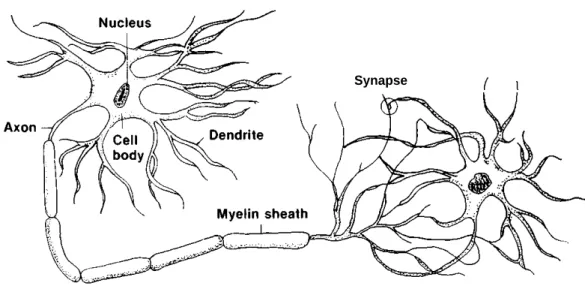
Study of the biochemistry of the brain involves examining the many chemicals involved in commun-ication and processing of information in the brain. Neurotransmitters are chemicals released by nerve cells, or neurons, to communicate with each other. Neurons are the cells that process information in the brain. A neuron consists of a cell body with long extensions, much like the branches of a tree, called dendrites (figure 4-l). Also projecting out of the cell body is a single fiber called the axon, which can extend a great distance (figure 4-l). When a neuron is activated, it releases a neurotransmitter into the synapse, the gap between two neurons (figure 4-2). The molecules of the neurotransmitter move across the synapse and attach themselves, or bind, to proteins, called receptors, in the outer wall of an adjacent cell (figure 4-2). Usually, the axon terminal is the part of the cell that releases neurotransmitters into the synapse, and the dendrites and cell body are the areas of the neuron which contain receptors that form synapses with the axons of other neurons.
Once the neurotransmitter has activated a recep-tor, it unbinds from the receptor. It then has to be Figure 4-l—Neurons
Synapse
Two neurons in synaptic contact.
Chapter Mental Disorders and the Brain ● 7 3
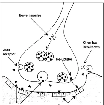
Figure 4-2—The Synapse
Nerve impulse
Auto- breakdown
receptor
Neurotransmitters Receptors Receiving cell The synapse and associated structures.
SOURCE: Office of Technology Assessment, 1992.
removed from the synapse so the synapse will be available for a new message. This is done either by the neurotransmitter’s being taken backup into the neuron that released it (a process called reuptake) or by it being broken down chemically into compounds called-metabolizes (figure 4-2).
For each neurotransmitter in the brain, there are specific receptors to which it can attach. Binding by the neurotransmitter activates the receptor, which can have different effects, depending on the recep-tor. Receptors can be linked to a variety of biochem-ical and cellular mechanisms that are turned on or off by the activation of the receptor. A neuron can have thousands of receptors for many different neuro-transmitters. Some neurotransmitters activate neu-rons (excitatory neurotransmitters), while others decrease the activity of neurons (inhibitory neuro-transmitters).
When a neuron is activated, changes occur in its membrane, resulting in a shift in the balance of ions (electrically charged molecules) between the inside and outside of the neuron. This change in ionic balance triggers an electrical impulse inside the neuron. The electrical impulse travels from the cell body, down the axon, to the axon terminal. At the
axon terminal, the impulse causes the release of neurotransmitter from the neuron into the synapse.
Sometimes a receptor for one neurotransmitter can affect a receptor for another neurotransmitter. In such cases, the receptors are biochemically coupled: The activation of one modulates the functioning of the other, either increasing or decreasing its activity. A neuron can also have receptors for the neurotrans-mitter it releases; these are usually located near the site where the neurotransmitter is released into the synapse (figure 4-2). Such receptors are acted on by the neuron’s own neurotransmitter to regulate the release of the neurotransmitter. Thus, these autorecep-tors, as they are called, act as a feedback mechanism to regulate a neuron’s activity. The activity of a neuron will be determined by the cumulative activity of all its various receptors.
While receptors are specific for a neurotransmit-ter, there may be a variety of receptor subtypes, linked to different cellular mechanisms, that all respond to the same neurotransmitter. In this way, one neurotransmitter can have diverse effects in various areas of the brain. Also, the number of receptors in the brain is not static. In response to increased production of a neurotransmitter, the number of receptors for that neurotransmitter will decrease; conversely, depletion of a neurotransmit-ter will result in an increase in the number of receptors for that neurotransmitter. This mechanism allows the brain to compensate for changes in neurotransmitter levels. Such receptor changes are important in therapeutics; some drugs mimic neuro-transmitters by stimulating increases or decreases in receptor numbers. In some cases, these changes may be directly related to the drug’s therapeutic effect.
Many chemicals have been identified as neuro-transmitters, among them acetylcholine, the cat-echolamines (norepinephrine, epinephrine, dopam-ine), serotonin, various amino acids, and peptides, including certain hormones. Various chemicals in the brain other than neurotransmitters and their receptors are necessary for brain function. They may be associated with the biochemical mechanisms activated by neurotransmitter-receptor interactions, involved with the production and breakdown of neurotransmitters, or responsible for carrying out metabolic activity.
Abnormalities in any of these chemicals, their receptors, or the cellular mechanisms that are turned on or off by the receptors could contribute to mental
74 . The Biology of Mental Disorders
disorders. For example, there may be too much or too little of a neurotransmitter, or the receptors for a neurotransmitter may not function properly. Mecha-nisms activated by receptors maybe defective, or the systems responsible for deactivating neurotransmit-ters maybe faulty. Also, breakdowns in the chemical systems responsible for the normal functioning of cells in the nervous system may play a role in mental disorders. Such alterations in neurotransmitter sys-tems have been implicated in the symptoms of certain mental disorders (see later discussions).
Scientists use a variety of tools and methods to study these factors. Biochemical assays are available to measure receptor number and activity, concentra-tions of neurotransmitters, and many other biochem-ical parameters of brain function. The majority of these assays are used with tissue from animals or from patients (i.e., postmortem brain samples or tissue samples from living patients). For example, information about concentrations of neurotransmitters is derived from measuring these compounds or their metabolizes in samples of blood or cerebrospinal fluid (i.e., fluid inside and surrounding the brain and spinal cord). Nevertheless, such samples provide only an indirect measure of what is occurrin g in the brain. The inability to observe and measure the chemical activity of the brain directly has hampered investigators’ understanding of how these processes may go awry in mental disorders. One new tech-nique that enables scientists to study biochemistry in the living brain is positron emission tomography (PET) (see later discussion). In particular, it can be used to assay some biochemical measures, such as distribution and number of receptors, in living human subjects.
The last decade has also seen the application of molecular biological techniques to study the brain. Genetic information about the brain and its compo-nents is studied and manipulated to understand the cellular and molecular workings of the brain. While these new techniques are just beginning to have an impact on the study of mental disorders, they have already provided valuable information about recep-tor subtypes (box 4-A) and other aspects of the biochemistry of the brain.
Information about underlying biochemical abnor-malities is also often derived from studying the actions of therapeutically effective drugs (i.e., psy-chopharmacology). In fact, many initial advances in understanding the biochemistry of mental disorders
came from studies of drug actions in the brain. If a drug is found to be effective in treating a disorder, examination of that drug’s chemical action in the brain may lead to the discovery of an intrinsic pathology. For example, the finding that effective antidepressant drugs act on catecholamines led to the study of these neurotransmitters in depression (see later discussion). Conversely, drug develop-ment may be guided by previously acquired knowl-edge about a disorder, which directs research efforts to create compounds that will act on an already identified pathology. If a specific neurotransmitter system is identified as being aberrant in a disorder, drugs can be designed to interact with some aspect of that system, such as the receptors, to try to reverse the abnormality.
Anatomy and Activity
Abnormalities in the structure of the brain or in its activity in specific locations can contribute to mental disorders. In the brain, neurons that share the same anatomical region, and to varying degrees the same function, are assembled into groups called nuclei. The brain is made up of hundreds of nuclei. Some consist of neurons that produce many different neurotransmitters, while others are predominantly of one type. Axons extending from nuclei convey information between and among them. Thus, the brain comprises many nuclei, which are connected by pathways of axons that contain various neuro-transmitters. Information is conveyed and processed via networks made up of interconnected nuclei.
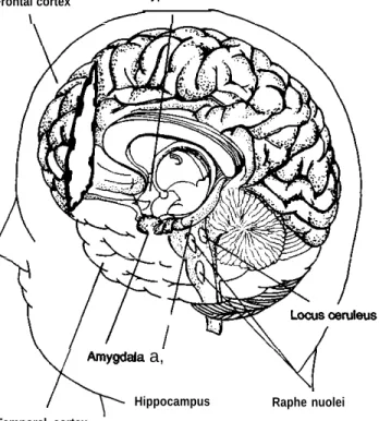
Some networks of nuclei are particularly relevant to mental disorders (figure 4-3). In general, these are networks that control cognitive (i.e., perception, recognition, reasoning, judgment, imagination), be-havioral, and emotional functions. Disruptions of these areas are likely to be involved in the thinking and mood disturbances characteristic of severe mental disorders. The cerebral cortex (the portion of the brain that is critical in decisionmaking) is important in this regard, especially the frontal lobes, which are considered to be the seat of higher-order. . thinkmg and which enables humans to reason abstractly. The limbic system, a network of struc-tures (e.g., hippocampus, amygdala, parts of the temporal lobe of the cortex) located in the upper part of the brain (figure 4-3) and involved in control of emotional behavior, is also important in mental disorders. Additional areas of the brain implicated in mental disorders are the basal ganglia, a group of
Chapter 4--Mental Disorders and the Brain ● 75
Box 4-A-Cloning Dopamine Receptors
Advances in the ability to manipulate and express genetic information provide an important new means of studying the brain. One area in which the tools of the molecular biologist have contributed significantly is the identification of receptor subtypes for neurotransmitters, These techniques have permitted the cloning of genes for specific receptors and have provided a detailed characterization of the receptor’s three-dimensional structure. Not only is this information important for understanding better how the brain works, but it also aids the development of drugs specifically designed to act only on certain receptors. This specificity can increase the efficacy of a drug
while decreasing the side effects it causes. The recent identification of several receptor subtypes for the
neuro-transmitter dopamine is an example of the contribution molecular biology is making to understanding the brain. Previous to the use of molecular biological techniques, two dopamine receptors had been characterized, based on the ability of various drugs to bind to them. For example, drugs that are effective in treating schizophrenia (called
typical antipsychotic drugs) ail bind to the same dopamine receptor--the D2 receptor. In addition, another receptor
that binds dopamine, but not typical antipsychotic drugs, was identified and called the D1 receptor. Other evidence,
derived using pharmacological techniques, suggested that there might be additional dopamine receptors, but it was not until the gene for each of the dopamine receptor subtypes was identified that their existence was confirmed.
Currently, six dopamine receptor subtypes have been identified and cloned using molecular biological techniques. Although all of these receptors are acted on by dopamine, they all have slightly different molecular structures. In addition, there are some differences in their location in the brain, the cellular mechanisms that they
turn on when they are activated, and their ability to bind typical antipsychotic drugs. Both the Dl and D5 receptors
are linked to the same cellular mechanism, are located in the hippocampus and cortex, and do not readily bind typical antipsychotic drugs. They differ in their ability to bind dopamine and dopamine-like drugs. There are two types of
D2 receptors: Both bind typical antipsychotic drugs, and both are found in parts of the limbic system, basal ganglia,
and cortex. They differ in that each is linked to a different cellular mechanism, and one is a dopamine
autoreceptor— a receptor that lies on the dopamine neuron itself, regulating the cell’s activity (see text). The D3 and
D4 receptors are found predominantly in the limbic system. While it is unclear what cellular mechanisms are
activated by these receptors, the D3 receptor is thought to be an autoreceptor. Neither binds typical antipsychotic
drugs as effectively as D2 receptors, and the D4 receptor readily binds a new atypical antipsychotic drug, clozapine
(see text).
The identification of these dopamine receptor subtypes has provided new insights into how more efficacious antipsychotic medications can be designed. Since clozapine is a highly effective antipsychotic drug but produces
fewer side effects than typical antipsychotics, it is thought that its mixture of strong binding to D4 receptors and weak
binding to D2 receptors accounts for its action. Currently there is great interest in understanding the various
dopamine receptor subtypes to determine their role in schizophrenia and how drugs can be designed to target them.
The complexity of the dopamine receptor system indicates the many ways that a single neurotransmitter can have myriad effects in the brain. Molecular biological techniques provide an important tool for clarifying these basic brain mechanisms, providing new information about how they maybe disturbed in mental disorders and leading the way for the development of more efficacious medications.
SOURCES: E. Kandel, J. Schwartz and T. Jessell (eds.), Principles of Neuroscience (New York NY: Elsevier Science publishing, 1991); P. Sokoloff, B. Giros, M. Martres, et al., ‘‘Molecular Cloning and Characterization of a Novel Dopamin e Receptor (D3) as a Target for Neuroleptics,” Nature 347: 146-151, 1990; R Sunahara, H. Guarn, B. O’Dowd, et al., ‘‘Cloning of the Gene for a Human Dopamine D5 Receptor With Higher Affinity for Dop amine Than Dl,’ Nature 350:614-619, 1991; H. van Tol, J. Bunzow, H. Guan, et al., “Cloning of the Gene for a Human Dopamine D4 Receptor With High Affinity for the Antipsychotic Clozapine, ’ Nature
350:610-614, 1991.
nuclei just below the cerebral cortex, some of which flight response); and the raphe nuclei, also found in
coordinate movement and others of which are part of the brainstem (figure 4-3), made up of serotonin
the limbic system; the hypothalamus, a collection of neurons that regulate sleep, are involved with
nuclei at the base of the brain (figure 4-3) that behavior and mood, and are connected to the limbic
regulate hormones and behaviors such as eating, system. It must be kept in mind, however, that if any
. .
drinking, and sex; the locus ceruleus, a nucleus in the of these, or other brain structures, are impaired in a
brainstem (figure 4-3) made up of norepinephrine mental disorder, it is unlikely that only the function
neurons that are intimately involved in the body’s of that structure will be affected. Since the brain is
Chapter Mental Disorders and the Brain ● 7 3
Figure 4-2—The Synapse
Nerve impulse
Auto- breakdown
receptor
Neurotransmitters Receptors Receiving cell The synapse and associated structures.
SOURCE: Office of Technology Assessment, 1992.
removed from the synapse so the synapse will be available for a new message. This is done either by the neurotransmitter’s being taken backup into the neuron that released it (a process called reuptake) or by it being broken down chemically into compounds called-metabolizes (figure 4-2).
For each neurotransmitter in the brain, there are specific receptors to which it can attach. Binding by the neurotransmitter activates the receptor, which can have different effects, depending on the recep-tor. Receptors can be linked to a variety of biochem-ical and cellular mechanisms that are turned on or off by the activation of the receptor. A neuron can have thousands of receptors for many different neuro-transmitters. Some neurotransmitters activate neu-rons (excitatory neurotransmitters), while others decrease the activity of neurons (inhibitory neuro-transmitters).
When a neuron is activated, changes occur in its membrane, resulting in a shift in the balance of ions (electrically charged molecules) between the inside and outside of the neuron. This change in ionic balance triggers an electrical impulse inside the neuron. The electrical impulse travels from the cell body, down the axon, to the axon terminal. At the
axon terminal, the impulse causes the release of neurotransmitter from the neuron into the synapse.
Sometimes a receptor for one neurotransmitter can affect a receptor for another neurotransmitter. In such cases, the receptors are biochemically coupled: The activation of one modulates the functioning of the other, either increasing or decreasing its activity. A neuron can also have receptors for the neurotrans-mitter it releases; these are usually located near the site where the neurotransmitter is released into the synapse (figure 4-2). Such receptors are acted on by the neuron’s own neurotransmitter to regulate the release of the neurotransmitter. Thus, these autorecep-tors, as they are called, act as a feedback mechanism to regulate a neuron’s activity. The activity of a neuron will be determined by the cumulative activity of all its various receptors.
While receptors are specific for a neurotransmit-ter, there may be a variety of receptor subtypes, linked to different cellular mechanisms, that all respond to the same neurotransmitter. In this way, one neurotransmitter can have diverse effects in various areas of the brain. Also, the number of receptors in the brain is not static. In response to increased production of a neurotransmitter, the number of receptors for that neurotransmitter will decrease; conversely, depletion of a neurotransmit-ter will result in an increase in the number of receptors for that neurotransmitter. This mechanism allows the brain to compensate for changes in neurotransmitter levels. Such receptor changes are important in therapeutics; some drugs mimic neuro-transmitters by stimulating increases or decreases in receptor numbers. In some cases, these changes may be directly related to the drug’s therapeutic effect.
Many chemicals have been identified as neuro-transmitters, among them acetylcholine, the cat-echolamines (norepinephrine, epinephrine, dopam-ine), serotonin, various amino acids, and peptides, including certain hormones. Various chemicals in the brain other than neurotransmitters and their receptors are necessary for brain function. They may be associated with the biochemical mechanisms activated by neurotransmitter-receptor interactions, involved with the production and breakdown of neurotransmitters, or responsible for carrying out metabolic activity.
Abnormalities in any of these chemicals, their receptors, or the cellular mechanisms that are turned on or off by the receptors could contribute to mental
76 ● The Biology of Mental Disorders
Figure 4-3—Brain Structures Involved in Mental Disorders
Frontal cortex Hypothalamus
\ I
a,
Hippocampus Raphe nuolei Temporal cortex
Three-dimensional drawing of the human brain.
SOURCE: Adapted from Lewis E. Calver, University of Texas,
Southwest-ern Medical Center, Dallas, TX, 1992.
functional impairment in part of a network can create a disturbance throughout the network.
Structural changes associated with mental disor-ders can include anatomical abnormalities in the structure of the brain or irregularities in the individ-ual cells within a region of the brain. The classical techniques for gathering information about brain structure-the macroscopic and microscopic post-mortem examination of normal brains and brains from individuals who have had mental disorders— have been augmented with a number of newer techniques and machines that make possible the study of the structure of the brain in living persons (table 4-l). Computerized axial tomography (CAT), which uses computers to combine a series of x-rays, provides clearer pictures of the brain than x-rays alone. Remarkably clear and detailed images of brain structure are obtained using magnetic reso-nance imaging (MRI) scans, which detect molecular
changes in the brain that occur when an individual is exposed to a strong magnetic field. Abnormalities that can be detected by either CAT or MRI scans include structural brain abnormalities, changes in the volume of brain tissue, and enlargement of the cerebral ventricles.1
Decreases in the volume of brain tissue and enlargement of the cerebral ventri-cles indicate either atrophy or underdevelopment of a brain region.
The activity of the brain can also be studied to determine damage or malfunctioning in a region of the brain. Neuropsychologica1 testing seeks to deter-mine brain damage by measuring deficits in a person’s performance on various tasks. For example, deficits on tests that measure language performance imply damage to the regions of the brain that subserve language skills, while poor performance on certain types of puzzles indicates abnormalities in regions devoted to various kinds of cognitive and sensory information processing. While neuropsy-chologica1 testing is helpful in identifying areas of brain pathology, the measures used are indirect, and exact locations of involved regions can only be inferred. However, when combined with more direct methods of looking at the brain, they provide a powerful tool for studying brain function and anatomy.
Measurement of electrical activity in the brain using the electroencephalograph (EEG) provides a more direct indication of brain function, and com-bining the EEG with computer analysis provides an even more detailed measure. Electrical activity can be measured while subjects are resting or engaging in some sort of sensory or cognitive task. By examining the electrical patterns of the brain, investigators can observe changes in normal brain responses and where they occur. A shortcoming of these measures is that they reflect the cumulative activity of broad areas of the brain, usually near the surface. This makes it more difficult to locate areas of possible pathology.
PET scanning and single-photon-emission com-puted tomography (SPECT) are imaging techniques that reveal brain activity. They do so by creating computerized images of the distribution of radioac-tively labeled materials in the brain. Researchers administer labeled materials to a subject either by injection or inhalation. Subsequent distribution of The cerebral ventricles are spaces in the brain that are filled with fluid.
Chapter 4-Mental Disorders and the Brain ● 77
Table 4-l—Techniques for Imaging the Brain
Technique How it works What it images
Computerized axial Computer construction of x-ray tomography (CAT) images
Magnetic resonance Images molecular changes in imaging (MRI) brain cells when exposed to a
strong magnetic field
Computer analysis of Creates maps of brain electrical electroencephalogram activity by computer analysis of
(EEG) EEG
Single-photon-emission Creates images of the distribu-computed tomography tion of radioactively labeled sub-(SPECT) stances in the brain following
either injection into the blood or inhalation
Positron emission Creates images of the distribu-tomography (PET) tion of radioactively labeled sub-stances following injection into the blood
Structure
Structure and activity (when used in conjunction with a magnetically active substance)
Activity
Activity (regional cerebral blood flow)
Activity (regional cerebral blood flow, glucose utilization) and neurochemical activity (receptor number and distri-bution)
SOURCE: Office of Technology Assessment, 1992.
these materials reflects the activity of the brain. Recently, MRI scanning has also been adapted for this function. PET, SPECT, and MRI enable research-ers to measure the utilization of glucose or the amount of blood flowing in a region of the brain (i.e., regional cerebral blood flow). Both glucose utiliza-tion and regional cerebral blood flow are indicators of brain activity: The more active a region is, the more blood will flow through it and the more glucose it will use. Abnormal activity levels in specific brain regions, in the whole brain, or in the normal asymmetry of activity between the two sides of the brain can be discerned with these techniques. Also, as previously mentioned, PET scanning can be done following the injection of labeled drugs that attach to specific receptors, making it possible to visualize the number and distribution of receptor populations. As with EEG measures, these scanning techniques can be done either while the subject is at rest or doing a task. While the imaging techniques now being used to study mental disorders will undoubtedly be refined, they provide for the first time a window through which to view the human brain at work.
SCHIZOPHRENIA
The symptoms of schizophrenia reflect a broad range of cognitive and emotional dysfunctions that are commonly categorized as either positive or negative symptoms (see ch. 3). Positive symptoms include hallucinations and delusions, paranoid psy-chosis, as well as bizarre behaviors and thought
disorder. Negative symptoms include emotional flattening (i.e., flat affect), loss of motivation, general loss of interest, and social withdrawal. In some cases, specific alterations in the biochemistry or anatomy of the brain have been associated with either positive or negative symptoms. As with other severe mental disorders, there are many clues available regarding schizophrenia, and a number of hypotheses have been put forward in an attempt to unify this information into an explanation of the underlying pathology.
Biochemistry
Dopamine
The most prominent and enduring theory regard-ing the biochemistry of schizophrenia concerns the role of the neurotransmitter dopamine. This theory is based on two sets of observations about drug action. First, drugs that increase dopamine activity, such as amphetamine, L-dopa, cocaine, and meth-ylphenidate, sometimes induce a paranoid psychosis in normal individuals that is similar to some aspects of schizophrenia. The same drugs, when adminis-tered to patients with schizophrenia, sometimes induce a transitory worsening of symptoms, particu-larly increasing psychosis and disturbance of thought. Second, and in contrast, drugs that block certain dopamine receptors often ease the symp-toms, in particular the positive sympsymp-toms, of schizo-phrenia. In general, there is a close correlation between how well these drugs block dopamine receptors and their antipsychotic effect. The
thera-78 ● The Biology of Mental Disorders
peutic effectiveness of dopamine-blocking drugs and the ability of dopamine-enhancing drugs to worsen the symptoms of schizophrenia provide evidence for the role of excessive concentrations or transmission of dopamine in at least some aspects of the disorder. Nevertheless, studies that have tried to measure dopamine activity in schizophrenia are less conclusive (81).
The concentrations of certain chemicals (e.g., homovanillic acid, a metabolize of dopamine) can be measured by scientists to provide information about dopamine activity. A number of investigators have measured these chemicals in the tissue and fluids of persons with and without schizophrenia. If higher concentrations of dopamine are associated with schizophrenia, then higher concentrations of these chemicals would be expected in persons with schizophrenia. The results are inconclusive. A re-view of this research (12) shows that in some studies higher dopamine concentrations were found in persons with schizophrenia; in others no such association was found. Conflicting results have also been found in studies of dopamine receptors, al-though one subtype of dop amine receptor (the D2
receptor) may be increased in schizophrenia (12). Thus, regardless of the index of dopamine activity examined in studies, results are variable. Data supporting the dopamine-excess hypothesis are usu-ally contradicted by data that fail to find differences between persons with schizophrenia and controls. Part of this variability is undoubtedly due to the imprecision of some of the measures used. The use of newer, more sophisticated techniques may re-solve some of these contradictions. For example, as previously described, there are a number of dopa-mine receptor subtypes (see box 4-A). As the ability to study these receptor subtypes improves, altera-tions in a specific receptor population may become evident. Part of the variability in results may also be due to differences in the characteristics of the patients with schizophrenia studied. Factors such as how long an individual has had the disorder and his or her age when it first appeared can affect findings. There also is some evidence that dopamine activity may be associated with specific symptoms of schizophrenia. For example, when changes reflect-ing increased dopamine function are observed, they often occur in patients with prominent positive symptoms, particularly psychosis (12). Other stud-ies support the proposition that dopamine deficiency
is associated with the negative symptoms of schizo-phrenia. For example, a correlation between low levels of dopamine chemicals in cerebrospinal fluid and negative symptoms has been reported (59). In addition, dopamine-blocking drugs used to treat the positive symptoms of schizophrenia produce behav-iors suggestive of the negative symptoms in animals and humans free of mental disorders (81,88). Thus, the contradictions between dopamine excess and dopamine deficiency as explanations for schizophre-nia can be reconciled by proposing that each is associated with different symptoms of the disease— that is, positive and negative symptoms, respectively (12,60,88)-and that each involves different neural networks (see later discussion). However, these hypotheses are controversial and it is possible that one or both are incorrect. Nonetheless, most scien-tists agree that dopamine plays some role in schizo-phrenia.
Other Neurotransmitters
Alterations in the amount or function of other neurotransmitters in persons with schizophrenia have also been examined by researchers. Both increased and decreased levels of serotonin have been postulated as being associated with schizophre-nia. The decreased-level hypothesis was based on the effects of LSD (lysergic acid diethylamide), which blocks serotonin activity in the brain and causes effects that are similar to some of the positive symptoms (e.g., hallucinations) seen inpatients with schizophrenia (21). These observations were not supported by other data; in particular, they were contradicted by evidence that some drugs effective against schizophrenia reduce serotonin activity. Also, as with the dop amine hypothesis, measures of serotonin in the blood, cerebrospinal fluid, and postmortem brain tissue do not provide clear an-swers. Both increased and decreased measures of serotonin and its metabolizes in blood and cerebro-spinal fluid have been observed, and results from measures in postmortem brain tissue have been inconclusive (53). Thus, while there is some indica-tion that serotonin activity may be altered in schizophrenia, the exact nature of its involvement is unclear.
Both an excess and a deficiency in the activity of the neurotransmitter norepinephrine have been asso-ciated with schizophrenia, although data indicate that an excess of norepinephrine is more likely to produce symptoms (80,90). Increased
Chapter Mental Disorders and the Brain . 79
rine has been observed in the cerebrospinal fluid and blood of patients with schizophrenia (21,82). Since dopamine and norepinephrine have complex interac-tions, namely, that disturbances in one affect the other, it is unclear whether any observed increase in norepinephrine is primary to schizophrenia or sec-ondary to changes in the dop amine system (53).
These neurotransmitters interact with and modu-late the activity of many other neurotransmitter and neuropeptide systems; awareness of this fact has led to the study of these chemicals in schizophrenia. The opiate peptides are thought to affect dopamine neurons, and naloxone, a drug that blocks opiate receptors, may have antipsychotic properties (53). In addition, there have been findings relating other peptides to schizophrenia (53). Since alterations in such neuropeptides could facilitate, inhibit, or other-wise alter the pattern of activity of other nerve cells, further study of the status of peptides in schizophre-nia is warranted.
Finally, a more specific neurotransmitter hypoth-esis involves the action of the drug phencyclidine (PCP) (31). PCP can produce symptoms that resem-ble both the positive and the negative symptoms of schizophrenia and can exacerbate these symptoms in people with the disorder. It is thought that PCP produces these dual effects by acting at different sites in the brain (21). PCP inhibits the activity of the excitatory neurotransmitter glutamate by interfering with the receptor for glutamate. From that observa-tion, it has been speculated that inhibition of the glutamate receptor in the hippocampus results in the negative symptoms associated with schizophrenia (21). PCP also weakly blocks the reuptake of dopamine, and in other areas of the limbic system there is some evidence that PCP inhibition of glutamate secondarily causes an increase in dopa-mine activity. These effects on dopadopa-mine could result in the positive symptoms of schizophrenia. In both instances, there is implicit involvement of glutamate in the onset of symptoms of schizophre-nia. While there is currently little experimental evidence to support this hypothesis, it represents a new avenue of investigation in schizophrenia re-search.
Thus, while it is likely that dopamine plays some role in schizophrenia, the involvement of other neurotransmitters is unclear. Since brain neurotrans-mitter systems interact with each other, it is often difficult to isolate a cause-effect relationship. The
modulatory action that neurotransmitters exert sug-gests that there may be complex interactions in schizophrenia between dopamine systems and these other brain chemicals.
Anatomy and Activity
Alterations in a number of brain structures, notably the frontal cortex and limbic system, have been implicated in schizophrenia. In particular, ventricular enlargement and evidence of changes in the size of various brain regions have been observed in imaging studies and postmortem examinations (figure 4-4). Limbic structures, such as the hip-pocampus and parts of the temporal lobes, are most affected. However, the specificity of these findings for schizophrenia has been questioned because they also occur in normal aging and in a variety of other neurological and psychiatric conditions (10, 21).
Attempts to replicate these findings have yielded contradictory results. In some cases, evidence of changes in the volume of the frontal cortex and temporal lobes has been observed, but the data are not conclusive and additional studies are needed to confirm the results (10). Some PET studies have examined brain metabolism in schizophrenia and have observed decreased activity in the frontal cortex and limbic structures, as well as increased
Figure 4-4—MRI Scan of an Individual With Schizophrenia
Brain structure shown of individual who does not have schizophre-nia (top) and a person who does (bottom). The ventricles are enlarged in schizophrenia (black areas).
SOURCE: W. Carpenter, Maryland Psychiatric Research Center, and H. I_Oats Associates, Inc.
80 ● The Biology of Mental Disorders
Figure 4-5-PET Scan of an Individual With Schizophrenia
Brain activity in an individual who does not have schizophre-nia (right) and a person who does (left). The frontal shows more activity in schizophrenia (white areas).
SOURGE: W. Carpenter, Maryland Psychiatric Research Center, and H. Associates, Inc.
activity in the basal ganglia (10,21) (figure 4-5). In general, these studies indicate that decreased frontal cortex activity is associated with the negative symptoms of schizophrenia. This coincides with data from animal studies, which indicate that dam-age to the frontal lobes produces behaviors similar to the negative symptoms.
EEG studies that show a higher incidence of abnormal electrical activity in the brain of patients with schizophrenia than in normal subjects provide further evidence of the involvement of the frontal cortex. These abnormalities often appear in the temporal lobe as well. Impairments on neuropsy-chological tests, such as problem-solving and atten-tion deficits, also indicate that these structures are affected in schizophrenia (34,39). For example, 65 percent of patients with schizophrenia often have difficulty visually following a moving object, com-pared to 8 percent of subjects who do not have schizophrenia (21) (see ch. 5). It is thought that defects in maintaining attention, as a result of a dysfunction in the frontal lobes and limbic system, contribute to these visual task defects.
These data have led to a number of hypotheses that attempt to unify the information regarding brain structures implicated in schizophrenia. In particular, these theories assign important functions to the limbic system and frontal cortex. The limbic system
is integral to motivation, gratification, memory, and many other emotions and thought-processes whose disturbance is associated with psychosis and the other positive symptoms of schizophrenia. The frontal cortex has been implicated in the negative symptoms of schizophrenia. The proposed theories differ as to whether the two sets of dysfunctions should be viewed as dependent or independent and in the specific structures presumed to be involved.
One theory postulates that they are dependent and that an insult during the development of the brain that affected the functioning of the frontal cortex (see later discussion) can lead to negative symptoms (84). Since the frontal cortex and limbic system are interconnected, with the frontal cortex inhibiting activity in the limbic system, this theory posits that the dysfunction of the frontal cortex reduces the inhibition on the Iimbic system, leading to the positive symptoms.
Another theory suggests that positive and nega-tive symptoms are mediated by two different net-works within the frontal cortex and limbic system and that dysfunction in one of these networks is separate from dysfunction in the other (9). This coincides with data from PET studies which show that negative symptoms are only correlated with decreased activity in the frontal cortex.
A third theory implicates a disruption in the activity of the basal ganglia and its interaction with other brain structures (16,17). This theory is based on three experimental findings: First, there is increased metabolic activity in the basal ganglia of patients with schizophrenia; second, patients with schizophrenia have difficulty performing visual attention tasks, which are mediated by areas of the cortex; and third, animals with lesions in dopamine-containing areas of the brain that project to the basal ganglia display some of the behavioral impairments seen in schizophrenia. This evidence has led to the hypothesis that schizophrenia is related to impaired activity in a network composed of the basal ganglia, certain cortical areas, and other brain structures and that the impairment is secondary to decreased dopamine activity.
These hypotheses attempt to find a basis for the disturbed behaviors observed in schizophrenia; they are overlapping and not always mutually exclusive. The available data point to networks involving certain limbic structures in psychosis and networks involving the frontal cortex in negative symptoms.
Chapter 4-Mental Disorders and the Brain ● 81
The precise interaction between these networks and the possible involvement of other brain structures and areas still need to be clarified. Based on these theories, predictions (which can themselves be tested with additional studies) can be made about the involvement of various areas of the brain in schizop-hrenia.
Other Factors
Immune and Viral Factors
Viral theories for the cause of schizophrenia are derived from reports that a number of viral and immune indices, such as the number and function of immune system cells, are deviant inpatients with the disorder (13,32,83). Also, there is some epidemio-logical evidence to support a viral hypothesis. Schizophrenia may have a north-to-south prevalence gradient, may be endemic in a few areas (e.g., northern Sweden), and occurs somewhat more often in persons born in the winter. It has also been observed that fetuses in the second trimester of gestation during an influenza epidemic have an increased risk of developing schizophrenia as adults. However, it has been difficult to conduct definitive studies, since any potential marker of an immune or viral process associated with schizophrenia is appli-cable in only some cases and is subject to interpreta-tion as secondary to condiinterpreta-tions associated with the disease (e.g., crowding of hospitalized patients, exposure of individuals living in low socioeconomic circumstances, and poor health habits).
Developmental Factors
The observed changes in brain volume of persons with schizophrenia, and the cellular alterations that accompany these changes, are thought to be irrevers-ible but not progressive (54). Current information suggests that the magnitude and nature of anatomi-cal and morphologianatomi-cal changes in schizophrenia are present at the onset of the disorder and do not vary over the lifetime of an individual. A possibility is that these abnormalities reflect changes that oc-curred very early in life or in utero, either as the result of some specific damage or a pathological alteration in the normal development of the brain.
Evidence that developmental factors may play a role derives from the observation that infants born after a complicated pregnancy or labor are at increased risk for developing schizophrenia as adults (41,42). One mechanism by which gestational
or birth complications may alter brain development is diminished oxygen supply (i.e., hypoxia). This theory is attractive for two reasons. First, many pregnancy and birth complications are associated with temporary hypoxia. Second, limbic structures, especially the hippocampus, are among the most sensitive areas in the developing brain to the adverse consequences of hypoxia. Also, subtle deviations in neurological and psychological functioning have been observed from infancy in children who are at high risk of developing schizophrenia (11). Al-though far from established, it is possible that early, adverse gestational influences on the developing brain create a risk of both birth complications and, later, schizophrenia. A corollary to this proposition is that a pathological influence operating early in gestation alters the development of the brain to create subsequent vulnerability to schizophrenia. An intriguing possible instance of this proposition is the relationship, discussed earlier, between pregnancy during influenza epidemics and the development of schizophrenia.
What specific alterations in the brain may result from such insults is unknown; however, it is clear that subtle deviations in the development of the brain could create dysfunctions associated with specific behaviors. Furthermore, it is possible that such subtle brain abnormalities are not manifest until much later in life, when new demands are placed on the brain systems during adolescence and adulthood. Postmortem findings of abnormalities in the number and organization of some nerve cells (1,37) suggest that the developmental process of cell migration, by which the cells in the brain become organized into the normal pattern of neuronal networks, may have gone awry in schizophrenia. Altered cell migration could be a genetic result, might be caused by gestational insults, or might involve an interaction of both. Another process that might be involved is the pruning of nerve cells that occurs as the brain develops (18). During early development, the brain has more neurons than it needs; the fine-tuning necessary for efficient functioning involves elimi-nating certain nerve cells and many of the synapses connecting cells. Failure to prune nerve cells and synapses adequately, or an error in selecting which ones to prune, could underlie dysfunctions that lead to the manifestation of the symptoms of schizophre-nia. Whatever developmental processes play a role in schizophrenia is still an open question.
82 ● The Biology of Mental Disorders
Synthesis
Although various alterations in the biochemistry, anatomy, and activity of the brain have been observed in schizophrenia, there are several impor-tant points regarding these data (21). The high variability among patients on any one of the biological factors and the lack of agreement among studies about many of them suggest that schizophre-nia is a heterogeneous disorder, with patients exhibiting different clusters of symptoms. This variability could also reflect the effects of other factors, such as psychosocial variables, on the biological components. The fact that some of the biological abnormalities observed in schizophrenia are also seen in other conditions calls into question their specificity to, and their role in, schizophrenia. Despite the equivocal nature of the research findings, conclusions can still be drawn from them. Dopamine plays a role in at least some of the symptoms of schizophrenia; however, the character-istics of that involvement are unclear, and dopa-mine’s precise relationship to the positive and negative symptoms of schizophrenia remains to be elucidated. The role of other neurotransmitters in schizophrenia, and if and how they interact with dopamine systems, needs to be clarified. Given the cognitive and emotional functions governed by the frontal cortex and limbic system, it is not surprising that alterations in these regions have been implicated in schizophrenia. Abnormal functioning of the frontal lobes has been one of the most consistent findings in schizophrenia, and, while less well documented, there does seem to be a relationship between decreased frontal cortex activity and nega-tive symptoms. Posinega-tive symptoms appear to be associated with increased metabolic activity in the limbic system.
While completed studies furnish valuable infor-mation regarding what might be wrong in the brain of a person with schizophrenia, the question of why schizophrenia occurs remains unanswered. The role of abnormal brain development or an injury to the brain, either during development or early in life, is an important avenue of investigation. As with other factors associated with schizophrenia, the precise mechanisms that may be involved are subject to speculation. The interaction of such a precipitating event with genetic factors (see ch. 5) is another plausible cause.
MOOD DISORDERS
Mood disorders include major depression and bipolar disorder. As the name indicates, major depression is marked by a deep depression that can be unremitting. Bipolar disorders are characterized by periods of depression alternating with manic episodes. Sadness is a normal human emotion in response to various life events, but depression that has no known cause or unremitting depression that interferes with normal activity is pathological. Available data regarding the role of biological factors in major depression and bipolar disorder often overlap. This reflects the fact that these disorders may be closely linked. The depressed state may be mediated by the same brain regions in both conditions. However, different brain mechanisms may be involved in the manic state and the swing from depression to mania that is characteristic of bipolar disorder.
Biochemistry
Neurotransmitter Systems
Prominent hypotheses concerning depression have focused on altered function of the group of neuro-transmitters called monoamine (i.e., norepineph-rine, epinephnorepineph-rine, serotonin, dopamine), particularly norepinephrine (NE) and serotonin (25,51,70). Evi-dence that monoamines are involved comes from the knowledge of the mechanism of action of the two classes of clinically effective antidepressant medica-tions—tricyclic antidepressants and monoamine ox-idase inhibitors (MAOIS). Tricyclic antidepressants block the reuptake of neurotransmitters, and MAOIs block the action of monoamine oxidase, the enzyme involved in the chemical breakdown of the monoa-mine transmitters once they are released into the synapse. The net effect of both of these types of drugs is to prolong the activity of these neurotrans-mitters in the synapse.
In the 1960s, the clinical observation that patients who were taking a norepinephrine-blocking drug for high blood pressure developed depression led to the hypothesis that depression was the result of low concentrations of monoamine, in particular NE (25). Some experiments have shown that patients with bipolar disorder have decreased NE metabolizes during depression and increased amounts during mania, which supports the NE imbalance hypothe-sis; other studies, however, have shown that patients
Chapter 4--Mental Disorders and the Brain ● 83
Box 4-B—Serotonin and Suicide
More than 30,000 Americans commit suicide each year, making it the eighth leading cause of death in the Nation. It is the second leading cause of death among adolescents. Changes in a number of indices of serotonin activity are correlated with suicide attempts and suicide completions. Suicidal behavior is associated with decreased concentrations of serotonin and its metabolizes in cerebrospinal fluid and the brain. Among successful suicides, decreased concentrations are usually found in the brainstem, where the raphe nuclei, the major serotonin-containing nuclei in the brain, are located. Also, increased numbers of serotonin receptors have been observed in the brains of suicide victims, usually in the frontal cortex. Since certain frontal cortex neurons receive connections from those of the raphe nuclei, it is possible that the receptor increase is a compensatory response to decreased serotonin activity in the raphe neurons. A decrease in the number of serotonin autoreceptors in suicide victims has been reported in some studies. Finally, suicide attempters, as compared to nonattempters, show decreased release of the hormone prolactin following administration of a serotonin-stimulating drug. This blunted prolactin response is indicative of a low level of serotonin activity. These data indicate that a net decrease in serotonin activity in the brain is associated with suicidal behavior.
These data do not mean that decreased serotonin causes a person to commit suicide. First of all, not every suicide victim exhibits decreased serotonin. For example, serotonin metabolize concentrations are not reduced in individuals with bipolar disorder who attempt suicide, compared to individuals with bipolar disorder who do not attempt suicide. Also, there is some evidence that among suicide attempters, measures of decreased serotonin activity correlate with the lethality of the method us@ that is, the more violent the attempted suicide method (e.g., cutting arteries v. drug overdose), the more depressed the serotonin activity. In addition, some of these same measures of serotonin activity, such as low levels of serotonin metabolizes in the cerebrospinal fluid and blunted response to prolactin, can be observed in individuals who exhibit impulsive and aggressive behavior. This suggests that, rather than causing suicide, decreased serotonin activity is correlated with a behavioral predisposition that can lead to suicide. If individuals who are burdened with feelings of despondency also have depressed serotonin activity, the propensity for aggression may be directed internally, tragically resulting in a successful suicide attempt.
SOURCES: E.F. Coccaro and J.L. AstiIl, ‘‘Central Serotonergic Function in Parasuicide,’ Progress in Neuro-Psychop harmacology &
Biological Psychiatry 14:663-674, 1990; K.Y. Little and D.L. Sparks, “Brain Markers and Suicide: Can a Relationship Be Found?” Journal of Forensic Sciences 35:1393-1403, 1990; J.J. Mann, V. Arango, M.D. Underwood, et al., “Neurochemical correlates of
suicidal Behavior Involv ement of Serotonergic and Non-Serotonergic Systems,” Pharmacology and Toxicology 3:37-60, 1990;
J.J. Mann, V. Arango, and M.D. Underwood, ‘ ‘Serotonin and Behavior, ‘‘Annals of the New York Academy of Sciences 60&476-484, 1990; L.C. Ricci and M.M. Wellman, ‘‘Monoamine s: Biochemical Markers of Suicide?” Journal of Clinical Psychology
46:106-116, 1990.
with major depression exhibit increased concentra- addition, decreased dopamine metabolizes have been
tions of NE metabolizes (51,70).
As more information has been gathered, it has become clear that the chemistry of depression is more complex and that other neurotransmitters, especially serotonin, maybe involved. For example, fluoxetine, a highly effective antidepressant drug that is widely prescribed, acts exclusively on sero-tonin.
Although the results from studies of serotonin metabolizes are not conclusive, there is some indica-tion that concentraindica-tions of serotonin are decreased in mood disorders (25). Also, low concentrations of serotonin metabolizes have been observed in people who commit suicide (box 4-B). As a result of these findings, it has been hypothesized that decreased serotonin plays a role in mood disorders (25,46). In
observed in some, but not all, studies of depression. One fact that argues against the monoamine imbalance hypothesis is the time lag between administration of antidepressant medications and their clinical effect. These medications increase neurotransmitter levels almost immediately upon administration, but their therapeutic effects often do not appear until 2 or 3 weeks after initiation of drug therapy. This time lag has led to the suggestion that the receptors for monoamines may be involved— specifically, that the clinical effects of these drugs are due to reductions in the number of receptors to compensate for the drug-induced increased levels of monoamine neurotransmitters. It would take weeks for such compensatory changes to come about.
One hypothesis related to drug-induced changes in receptors involves a receptor for NE (25). There
84 ● The Biology of Mental Disorders
are a number of NE receptor subtypes. One is an autoreceptor that decreases the release of NE into the synapse when it is activated. It has been hypothe-sized that these NE autoreceptors are overly active in depression. The therapeutic effects of antidepres-sant drugs are the result of exposing the autorecep-tors to higher concentrations of neurotransmitter, which decreases their number, ultimately increasing the activity of the NE-containing neurons. While this hypothesis would explain the delayed appearance of clinical effects of antidepressant dregs, studies of the NE autoreceptor in depression have found no specific evidence of an abnormality to date.
Currently, no clear evidence links abnormal serotonin receptor activity in the brain to depression (51). Increased receptors for serotonin have been observed in the brains of suicide victims (box 4-B), but there are conflicting results from studies that have examined serotonin receptors in depressed persons (25). Changes in serotonin receptor activity have been measured indirectly in mood disorders, notably bipolar disorder, using blood platelets (a type of cell found in the blood) which also contain serotonin and are used to investigate mechanisms related to serotonin and its receptors (46). Both increased numbers of a subtype of serotonin receptor and decreased sites for the reuptake of serotonin into the platelets have been seen in the platelets of persons with depression (46). However, it is unclear how these changes relate to serotonin activity in the brain of persons with major depression and bipolar disorder.
While there is sufficient evidence to support the notion that abnormalities in monoamine systems are an important component of depression, the data currently available do not provide consistent evi-dence either for altered neurotransmitter levels or for disruption of normal receptor activity. This has led to a dysregulation hypothesis, which states that there is a more general perturbation in the mechanisms that regulate the activity of the monoamine neuro-transmitters and that clinically effective drugs re-store efficient regulation (70). Linked to this hypoth-esis is the fact that the mono amine systems interact with each other-the activity of one affects and modulates the activity of another. Based on available data, it has been proposed that decreased activity within the NE-serotonin component of the system is associated with depression, while increased activity of the NE-dopamine component tends to promote mania (25).
Complicating the picture is the fact that other neurotransmitters have been implicated in bipolar disorder. Based on evidence that agents which activate acetylcholine systems can induce depres-sion and that agents which block such activity have some ability to alleviate depression (51), it has been postulated that increased acetylcholine activity in-duces depression and decreased activity inin-duces mania (15,25,30,70). Furthermore, a number of investigators have proposed that the salient mecha-nism may be the balance between NE and tylcholine systems, with a predominance of ace-tylcholine activity associated with depression and a predominance of NE activity associated with mania (25,28,51,70). A role for the inhibitory neurotrans-mitter gamma-aminobutyric acid (GABA) has also been put forth, based on the paradoxical finding that increased GABA activity has both an antidepressant and an antimanic effect (25,40,51). Given that GABA is a ubiquitous inhibitory neurotransmitter, it is possible that increasing its activity can result in the modulation of a number of other neurotransmit-ter systems, which could explain its broad range of effects. Finally, there is evidence that some neu-ropeptides may also be involved in mood disorders (6,23,25). In particular, decreased levels of somato-statin and increased levels of corticotropin-releasing factor are associated with depression.
Thus, a number of neurotransmitters have been implicated in mood disorders, with NE and serotonin being the most prominent. Lithium is the most effective drug for the treatment of mania and for controlling the mood swings between depression and mania that characterize bipolar disorder. It is not known how lithium affects the activity of neurons, but like many other chemicals that are important to normal brain functioning, lithium is an ion—that is, a molecule that has an electrical charge. It is thought that, whatever its action on neurons, lithium has many different effects in the brain (8). It increases serotonin activity, decreases acetylcholine activity, affects the activity of both norepinephrine and dopamine, and inhibits some of the intracellular mechanisms that are initiated by activation of receptors. Since lithium has such a broad range of actions that can affect the neurotransmitter systems implicated in mood disorders, its therapeutic effect may be due to its capacity to correct the neurotrans-mitter abnormalities associated with mania and depression, and to prevent the changes in
neuro-Chapter 4-Mental Disorders and the Brain ● 85
chemical balance that are thought to be responsible for the mood swings of bipolar disorder.
Neuroendocrine Systems
Abnormalities in hormone regulation are common in depression (7,25,70,71), perhaps because regula-tion of hormones and the glands that secrete them is under the control of the same neurotransmitters in the brain that are thought to be dysfunctional in depression. In particular, the activities of the pitui-tary, adrenal, and pineal glands are affected. Many of the symptoms associated with depression (e.g., changes in appetite, sleep, and sex drive) may be related to these hormonal changes, which means that the hormonal abnormalities may be secondary to the neurotransmitter alterations of the disorder (7). Nonetheless, there is great interest in studying these changes in hopes of discovering a biological marker and developing diagnostic tests for depression.
A number of hormones have been studied to determine if depressed persons consistently exhibit abnormal concentrations of them or show an abnor-mal release of them in response to some sort of pharmacological challenge. Current information in-dicates that while some of these hormones are altered in depression, variability in baselines and pharmacological response, as well as the possibility of changes due to other causes, makes them unrelia-ble markers for depression (7,70,71).
Depressed persons often have elevated concentra-tions of the hormone cortisol (7,25,70,71), which results from increased concentrations of corticotropin-releasing factor. In healthy individuals, administra-tion of the drug dexamethasone suppresses the concentration of cortisol in the blood. The dexametha-sone-suppression test (DST), developed as a test of hormone functioning, has been studied as a possible diagnostic tool in depression. Approximately 40 to 50 percent of persons diagnosed with major depres-sion have an abnormal DST in that they do not suppress cortisol in response to dexamethasone (70). In very severe cases, particularly psychotically depressed patients, the percentage ranges from 60 to 80. It is not known why only some patients show an abnormal DST. Whether this reflects variability in response to DST or a subpopulation of depressed patients has yet to be resolved. Therefore, there are several problems with using the DST as a diagnostic tool (7,25,70,71). Aside from the fact that not all depressed patients show an abnormal DST, a num-ber of other clinical (e.g., Alzheimer’s disease,
anorexia nervosa) and nonclinical conditions (e.g., fasting, ingestion of caffeine) can result in abnormal DSTs. Although its effectiveness as a diagnostic tool is limited, the test maybe useful in predicting which patients are likely to relapse following cessation of drug therapy: If DST results remain abnormal during therapy, the patient is more likely to relapse once the antidepressant is withdrawn (7).
Another aspect of endocrine function related to depression is the association between depression and reproduction-related events in women (2). Hormonal alterations related to menstruation, preg-nancy, childbirth, and menopause can affect neuro-transmitters that regulate mood and behavior. The evidence regarding the relationship between these hormonal fluctuations and the occurrence of depres-sion is mixed. Nevertheless, the clear association of mood alterations with these reproductive events in some women suggests an area for additional investi-gation. Understanding these biochemical interac-tions could provide new insights into the pathology of depression.
Anatomy and Activity
It is unclear which areas of the brain may be involved in mood disorders. The data regarding anatomical defects and activity in the brain of persons with mood disorders are equivocal. Given that few studies have been conducted and that their results have varied, it is impossible to come to any conclusions regarding relationships between the data and the cause and symptoms of mood disorders. Overall, the data suggest an association between mood disorders and abnormalities of large regions of the brain, especially the frontal and temporal lobes; they also imply an abnormal difference between the left and right sides of the brain. Normally, the left and right sides of the brain are involved in different, although overlapping, functions. For example, the left is usually more specialized for language and logical “thinking, while the right is more involved with spatial processing. As a result of these different functions, the two halves of the brain often exhibit different levels of activity. In mood disorders, some of the normal differences in activity level between the right and left sides of the brain appear to be altered.
Few postmortem studies have investigated ana-tomical alterations in the brains of persons with mood disorders, although a number of studies have