Thyroid vascularity and blood flow are not dependent on
serum thyroid hormone levels: studies
in vivo
by color flow
doppler sonography
Fausto Bogazzi, Luigi Bartalena, Sandra Brogioni, Alessandro Burelli, Luca Manetti, Maria Laura Tanda, Maurizio Gasperi and Enio Martino
Dipartimento di Endocrinologia e Metabolismo, Ortopedia e Traumatologia, Medicina del Lavoro, University of Pisa, Pisa, Italy
(Correspondence should be addressed to E Martino, Dipartimento di Endocrinologia e Metabolismo, Ortopedia e Traumatologia, Medicina del Lavoro, Universita` di Pisa, Ospedale di Cisanello, Via Paradisa, 2, 56122 Pisa, Italy)
Abstract
Objective: Thyroid blood flow is greatly enhanced in untreated Graves’ disease, but it is not known whether it is due to thyroid hormone excess or to thyroid hyperstimulation by TSH-receptor antibody. To address this issuein vivopatients with different thyroid disorders were submitted to color flow doppler sonography (CFDS).
Subjects and methods: We investigated 24 normal subjects, and 78 patients with untreated hyperthy-roidism (49 with Graves’ hyperthyhyperthy-roidism, 24 with toxic adenoma, and 5 patients with TSH-secreting pituitary adenoma (TSHoma)), 19 patients with thyrotoxicosis (7 with thyrotoxicosis factitia, and 12 with subacute thyroiditis), 37 euthyroid patients with goitrous Hashimoto’s thyroiditis, and 21 untreated hypothyroid patients with Hashimoto’s thyroiditis.
Results: Normal subjects had CFDS pattern 0 (absent or minimal intraparenchimal spots) and mean intraparenchimal peak systolic velocity (PSV) of 4.861.2 cm/s. Patients with spontaneous hyperthy-roidism due to Graves’ disease, TSHoma, and toxic adenoma had significantly increased PSV (P< 0.0001, P= 0.0004, P< 0.0001 respectively vs controls) and CFDS pattern. Patients with Graves’ disease had CFDS pattern II (mild increase of color flow doppler signal) in 10 (20%) and pattern III (marked increase) in 39 cases (80%). Mean PSV was 1563 cm/s. Patients with toxic adenoma had CFDS pattern I (presence of parenchymal blood flow with patchy uneven distribution) in 2 (8%), pattern II in 16 (70%) and pattern III in 5 (22%). Mean PSV was 1162.4 cm/s. Patients with TSHoma showed CFDS pattern I in one case (20%) and pattern II in 4 (80%). Mean PSV was 14.864.2 cm/s. Patients with thyrotoxicosis had normal PSV (4.261.1 cm/s in subacute thyroiditis, 460.8 cm/s in thyrotoxicosis factitia,P= not significant vs controls) and CFDS pattern 0. Untreated euthyroid patients with goitrous Hashimoto’s thyroiditis had CFDS pattern 0, and mean PSV (4.360.9 cm/s; P= not significant vs controls). Untreated hypothyroid patients with goitrous Hashimoto’s thyroiditis had CFDS pattern I in 14 cases (67%), pattern II in 4 (19%) and pattern 0 in 3 (14%) and mean PSV (5.661.4 cm/s) was higher than that of controls (P= 0.026).
Conclusions: An increase in both intrathyroidal vascularity and blood velocity was observed in patients with spontaneous hyperthyroidism but not in thyrotoxicosis due to either ingestion of thyroid hormones or to a thyroidal destructive process. The slightly increased vascularity and blood velocity observed in patients with hypothyroid Hashimoto’s thyroiditis suggests that thyroid stimulation by either TSH-receptor antibody or TSH is responsible for the increased thyroid blood flow.
European Journal of Endocrinology140452–456
Introduction
Hyperthyroidism is associated with hemodynamic changes including high output state, increased heart rate and cardiac contractility, and decreased peripheral resistance (1, 2), that are related both to direct cardio-stimulatory effects of thyroid hormone and to increased peripheral oxygen consumption (3, 4). While cardiac parameters are invariably increased, as assessed by the shortening of systolic and diastolic time-intervals, the
increase of systolic and diastolic function and the reduction in the left ventricular ejection fraction (3– 6), regional blood flow distribution is not uniform in hyperthyroidism. For example, blood flow in skeletal muscle, skin, cerebral, and coronary arteries is increased, whereas hepatic blood flow is unchanged (7–9).
Untreated Graves’ patients have a hypervascular gland responsible for a thyroid bruit and thrill. The availability of color flow doppler sonography (CFDS), a
rapid, informative, and non-invasive technique, has recently allowed direct measurement of intrathyroidal blood flow (10–17). Using CFDS it has been shown that intrathyroidal blood flow, which can be quantitated by measurement of peak systolic velocity (PSV), is greatly increased in patients with untreated Graves’ hyperthy-roidism (10, 11). In hyperthyhyperthy-roidism hemodynamic changes are thought to be due to serum thyroid hormone excess. However, in Graves’ patients it is not well understood whether thyroidal hypervascularity is related to thyroid hormone excess or to the excessive stimulation of the thyroid gland by TSH-receptor antibody (TRAb). To address this problem, in the present study we have evaluated the intrathyroidal blood flow and PSV by CFDS in patients with various thyroid disorders.
Materials and subjects
We enrolled in this study 155 consecutive patients seen at our Institution in the period 1995–1997. The main clinical and biochemical features of the subjects are described in Table 1.
Conventional and CFD sonography
CFDS was performed using an ALU-4 ESAOTE color doppler system (Esaote Biomedicals, Genua, Italy) with a 7.5 MHz linear electronic transducer. Each patient was first submitted to gray scale sonography; subsequently color flow doppler information was gained. Thyroid
volume was measured by ultrasound and calculated by the ellipsoid model (width×length×thickness×0.52 for each lobe) as previously described (11). CFDS images were obtained in the transverse and longitudinal planes and color gain was adjusted to a level not associated with relevant artifacts with the probe repetition frequency at 750 Hz. Color flow imaging was depicted by red and blue spots that indicated the direction of flow toward (red) and away (blue) from the transducer. For quantitative doppler evaluation, the peak systolic velocity (the doppler waveform recorded at the point with the highest frequency shift) was used. Measure-ments were performed at the level of the intrathyroid arteries, with a sampling volume of 2 mm. Multiple sampling (n= 5) was performed to account for varia-tions, and the mean value was expressed. The coefficient of variation of PSV was 8%. The angle correction was not employed due to the tortuosity of small intrathy-roidal vessels and their incomplete visualization. PSV values and CDFS pattern were recorded by one of us (F B) who did not know the patients’ diagnoses. The possible CFDS patterns were as follows: pattern 0: absent intraparenchymal (or nodular) vascularity or minimal spots; pattern I: presence of parenchymal (or nodular) blood flow with patchy uneven distribution; pattern II: mild increase of color flow doppler signal with patchy distribution (for nodules: mainly peri-pheral); pattern III: markedly increased color flow doppler signal with diffuse homogeneous distribution, including the so-called ‘thyroid inferno’ (10) (for nodules: intranodular and peripheral vascularity).
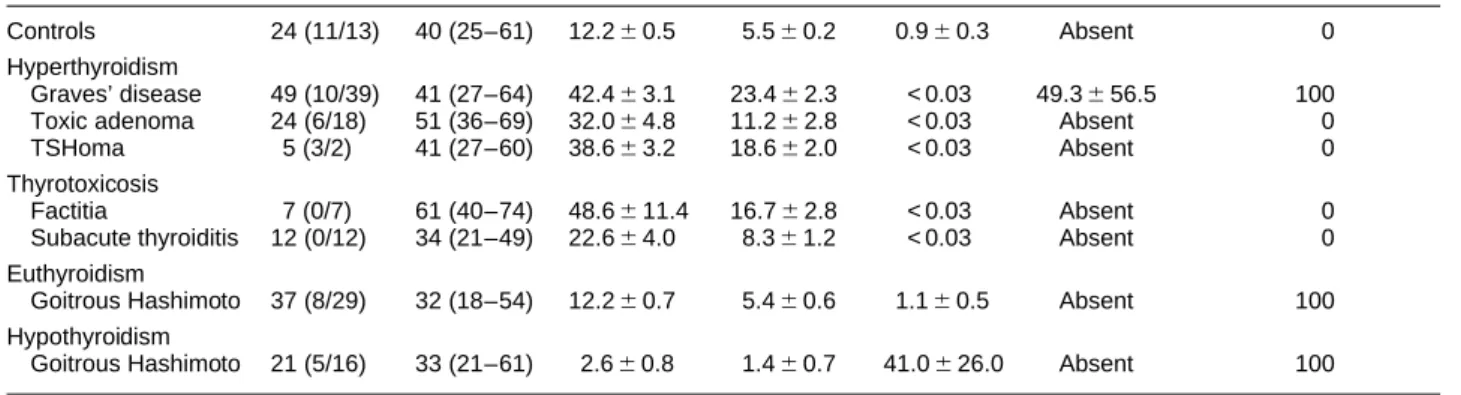
Table 1 Clinical and biochemical features of the subjects.
Mean age
(range) Serum FT4 Serum FT3 Serum TSH TRAb Positive AbTg/AbTPO
Subjects n(M/F) (years) (pmol/l) (pmol/l) (mU/l) (U/l) (percent)
Controls 24 (11/13) 40 (25–61) 12.260.5 5.560.2 0.960.3 Absent 0
Hyperthyroidism
Graves’ disease 49 (10/39) 41 (27–64) 42.463.1 23.462.3 < 0.03 49.3656.5 100
Toxic adenoma 24 (6/18) 51 (36–69) 32.064.8 11.262.8 < 0.03 Absent 0
TSHoma 5 (3/2) 41 (27–60) 38.663.2 18.662.0 < 0.03 Absent 0
Thyrotoxicosis
Factitia 7 (0/7) 61 (40–74) 48.6611.4 16.762.8 < 0.03 Absent 0
Subacute thyroiditis 12 (0/12) 34 (21–49) 22.664.0 8.361.2 < 0.03 Absent 0
Euthyroidism
Goitrous Hashimoto 37 (8/29) 32 (18–54) 12.260.7 5.460.6 1.160.5 Absent 100
Hypothyroidism
Goitrous Hashimoto 21 (5/16) 33 (21–61) 2.660.8 1.460.7 41.0626.0 Absent 100
Normal subjects were recruited among medical personnel of our Institution. Patients with toxic adenoma had a single hot thyroid nodule at scintiscan with absent radioiodine uptake in the extranodular tissue. Patients with thyrotoxicosis factitia had low/suppressed thyroidal 24-h radioiodine uptake values (361%), normal urinary iodine excretion, and undetectable serum Tg (18). All patients admitted surreptitious thyroid hormone pill intake. Five patients were previously described in detail (16). Patients with TSH-secreting pituitary adenoma (TSHoma) had elevated serum FT4and FT3and inappropriately high serum TSH concentration with absent TSH response to i.v. thyrotropin-releasing hormone stimulation and anasubunit/TSH molar ratio greater than 1. All patients underwent adenomectomy: four patients had a pure TSHoma; in one patient immunohistochemistry revealed a mixed neoplasm with 4% growth hormone-producing cells.
Assays
Serum free thyroxine (FT4), free tri-iodothyronine (FT3) (Technogenetics, Milan, Italy), thyrotropin (TSH) (Del-phia hTSH ultra Kit; Pharmacia, Turku, Finland), thyroglobulin (Tg) (Sorin Biomedica, Saluggia, Italy), TRAb (TRAK assay; Henning, Berlin, Germany), anti-Tg (Serodia, Tokyo, Japan), and anti-thyroperoxidase (TPO) (Sorin Biomedica) were determined by commercial kits. Normal values in our laboratory were as follows: FT4, 8.3–20.5 pmol/l; FT3, 3.8–8.4 pmol/l; TSH, 0.4–3.7 mU/l; Tg, <3–30 mg/l; anti-Tg, negative; anti-TPO, negative; and TRAb, <5 U/l. Urinary iodine excretion was measured using an autoanalyzer apparatus (Technicon, Rome, Italy). The median urinary excretion of iodine in our area is 110mg/l.
Statistics
Data are expressed as means6S.D. Comparison of parameters among the study groups was performed by the Mann–Whitney U test.
Results
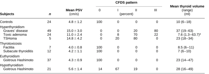
The results are summarized in Table 2 and in Fig. 1. Patients with hyperthyroidism had higher PSV values than controls (P#0.004), patients with thyrotoxicosis (P#0.004), and hypothyroid and euthyroid patients with goitrous Hashimoto’s thyroiditis (P#0.0005).
Patients with thyrotoxicosis had PSV values which did not differ from those observed in controls and euthyroid patients with goitrous Hashimoto’s thyroiditis. Hypothyroid patients with goitrous Hashimoto’s thyroiditis had higher PSV values than euthy-roid patients with goitrous Hashimoto’s thyeuthy-roiditis (P= 0.0004), controls (P= 0.026) and patients with thyrotoxicosis ((P#0.01).
Discussion
The results of the present study confirmed that CFDS is a useful tool to investigate intrathyroidal blood flow and clearly demonstrated that thyroid vascularity and blood flow are mainly dependent on the activation of TSH receptor by TSH, TRAb, or by gain of function mutations of TSH receptor (19), while thyrotoxicosisper sedoes not cause hemodynamic changes in intrathyroidal vessels. Increased PSV values were observed only in conditions associated with elevated TSH values (mainly central hyperthyroidism due to TSHoma and, to a lesser extent, untreated hypothyroidism due to goitrous Hashimoto’s thyroiditis), with the presence of the abnormal TSH-receptor stimulator (TRAb, in untreated Graves’ hyperthyroidism) or with gain of function mutations of TSH receptor (toxic adenoma), irrespective of serum thyroid hormone levels (elevated in TSHoma, toxic adenoma and Graves’ disease, low in Hashimoto’s hypothyroid patients). The fact that the increase in PSV values was modest in hypothyroid patients, and marked in the other two groups, in spite of very high TSH values in the former group, is in keeping with the concept that a responsive (and not damaged) thyroid gland is also required to achieve a substantial increase in thyroid vascularity and blood flow. Autoimmunity per seis unlikely to be the cause of thyroid hypervascu-larity. In fact, autoimmunity is present in patients with Graves’ disease and in patients with Hashimoto’s thyroiditis, but hypervascularity is observed only in the presence of TRAb or of increased serum TSH values. Thyroid volume might play some role. However, patients with increased serum TSH levels (TSHoma and hypothyroid patients) had similar thyroid volume (Table 2) but PSV values significantly differed in the two groups (Fig. 1). The importance of thyroid stimulators is supported by recent reports (20, 21) showing that TSH can drive thyroidal angiogenesis in
Table 2 Sonography and color doppler characteristics of the subjects.
CFDS pattern
Mean thyroid volume
Mean PSV 0 I II III (range)
Subjects n (cm/s) (percent) (ml) Controls 24 4.861.2 100 0 0 0 10 (6–18) Hyperthyroidism Graves’ disease 49 15.063.0 0 0 20 80 37 (19–63) Toxic adenoma 24 11.062.4 0 8 70 22 7.6 (1.3–63.7)* TSHoma 5 14.864.2 0 20 80 0 23 (18–34) Thyrotoxicosis Factitia 7 4.060.8 100 0 0 0 8.5 (6–11) Subacute thyroiditis 12 4.261.1 100 0 0 0 7 (6–10) Euthyroidism Goitrous Hashimoto 37 4.360.9 100 0 0 0 23 (14–47) Hypothyroidism Goitrous Hashimoto 21 5.661.4 14 67 19 0 28 (16–49)
vitro: this effect might be mediated by the increased expression of growth factors of the vasculotropins and the tyrosine kinase receptor families, which are then released by thyroid follicular cells and in turn act on endothelial cells. It is conceivable that similar effects might be exerted by TRAb. On the other hand, the ineffectiveness of elevated serum thyroid hormones to increase intrathyroidal blood flow might be explained by the recent findings of Diekmanet al.(22), who reported that human endothelial cells contain thyroid hormone receptor, but do not produce endothelin in response to thyroid hormone stimulation. Endothelin is a 21 amino acid peptide which is both a vasoconstrictor and a smooth-muscle mitogen, and might have a role in modulating regional blood flow (23, 24).
Our data showed that some hypothyroid patients had CFDS pattern II similar to patients with Graves’ disease or TSHoma. However, PSV values were only slightly elevated in hypothyroid patients, but greatly increased in the hyperthyroid patients. Thus, CFDS pattern and PSV values are not always superimposable: CFDS pattern II might be found in association, for example, either with a 6 cm/s PSV (hypothyroid patients with Hashimoto’s thyroiditis) or with 15 cm/s PSV (Graves’ hyperthyroidism or TSHoma). For these reasons, we would like to suggest that PSV represents a better index of thyroid function than the CFDS pattern. High PSV values are probably indicative of hyperthyroidism, since they reflect a sustained stimulation on a thyroid which is capable of producing and secreting excess thyroid hormone; this does not occur in goitrous Hashimoto’s hypothyroidism, in which the sustained stimulation of TSH leads only to a moderate increase in thyroid vascularity and blood velocity, due to the damage of thyroid tissue.
Our data support the concept that spontaneous hyperthyroidism (i.e. a stimulation of the thyroid gland) and not thyrotoxicosis (i.e. a serum increase of thyroid hormone levels) is associated with increased
intrathyroidal vascularity. This might be diffuse as in Graves’ disease or circumscribed as in toxic adenoma. Thyrotoxicosis due to destructive process of thyroid gland (subacute thyroiditis or type II amiodarone-induced thyrotoxicosis (17)) or to exogenous ingestion of thyroid hormons (16) are not associated with an increased PSV or CFDS pattern.
Because it is easy, non-invasive and rapid, CFDS and, even more, PSV can provide prompt information on thyroid status. For the interpretation of CFDS pattern and PSV values the examiner must keep in mind that thyroid vascularization depends on thyroid stimulation by TSH or TRAb.
Acknowledgements
We thank Professor Aldo Pinchera for his continuous encouragement and advice. This work was partially supported by Grants from the University of Pisa (Fondi d’Ateneo) to E M and L B.
References
1 DeGroot WJ & Leonard JJ. Hyperthyroidism as a high cardiac output state.American Heart Journal197079265–272. 2 Ladenson PW. Recognition and management of cardiovascular
disease related to thyroid dysfunction. American Journal of Medicine199088638–641.
3 Klein I. Thyroid hormone and the cardiovascular system.
American Journal of Medicine199088631–637.
4 Polikar R, Burger AG, Scherrer U & Nicod P. The thyroid and the heart.Circulation1993871435–1441.
5 Cohen MV, Schulman IC, Spenillo A & Surks MI. Effects of thyroid hormone on left ventricular function in patients treated for thyrotoxicosis.American Journal of Cardiology19814833–38. 6 Woeber KA. Thyrotoxicosis and the heart.New England Journal of
Medicine199232794–98.
7 Meyers JD, Brannon ES & Holland BC. Correlative study of the cardiac output and hepatic circulation in hyperthyroidism.
Journal of Clinical Investigation1950291069–1072.
8 Rowe GG, Huston JH & Weinstein AB. Hemodynamics of thyrotoxicosis with special reference to coronary blood flow and
Figure 1 PSV distribution in the study groups.
The horizontal line represents the mean value. Mean PSV in patients with elevated serum TSH or TRAb levels (untreated Graves’ dis-ease (TDG), TSHoma, untreated hypothyroid-ism due to goitrous Hashimoto’s thyroiditis (HT hypo)) or with toxic adenoma (TA) was significantly higher than that found in the control group, in patients with thyrotoxicosis factitia (TF) or subacute thyroiditis (SAT) and euthyroid Hashimoto’s thyroiditis (HT euthyr). For statistical differences see text. Numbers refer to the number of patients having the same individual PSV value.
myocardial oxygen metabolism. Journal of Clinical Investigation
195635372–379.
9 Kontos HA, Shapiro W & Mauck HP Mechanism of certain abnormalities of the circulation to the limbs in thyrotoxicosis.
Journal of Clinical Investigation196544947–951.
10 Ralls PW, Mayekawa DS, Lee KP, Colletti PM, Radin DR, Bosnell WD et al. Color-flow doppler sonography in Graves’ disease: ‘thyroid inferno’.American Journal of Radiology1988150781– 784.
11 Fobbe F, Finke R, Rechenstein E, Schleusener H & Wolf K-J. Appearance of thyroid diseases using colour-coded duplex sonography.European Journal of Radiology1989929–31. 12 Shimamoto K, Endo T, Ishigaki T, Sakuma S & Makino N. Thyroid
nodules: evaluation with color doppler ultrasonography.Journal of Ultrasound Medicine199312673–678.
13 Lagalla R, Caruso G, Novara V & Cardinale AE. Analisi flussimetrica nelle malattie tiroidee: ipotesi di integrazione con lo studio qualitativo con color-doppler.Radiologia Medica199385
606–610 (In Italian).
14 Vitti P, Rago T, Mazzeo S, Brogioni S, Lampis M, De Liperi Aet al.
Thyroid blood flow evaluation by color-flow doppler sonography distinguishes Graves’ disease from Hashimoto’s thyroiditis.Journal of Endocrinological Investigation199518857–862.
15 Castagnone D, Rivolta R, Rescalli S, Baldini MI, Tozzi R & Cantalamessa L. Color doppler sonography in Graves’ disease: value in assessing activity of disease and predicting outcome.
American Journal of Radiology1996166203–207.
16 Bogazzi F, Bartalena L, Vitti P, Rago T, Brogioni S & Martino E. Color flow doppler sonography in thyrotoxicosis factitia.Journal of Endocrinological Investigation199619603–606.
17 Bogazzi F, Bartalena L, Brogioni S, Mazzeo S, Vitti P, Burelli Aet al.
Color flow doppler sonography rapidly differentiates type I and
type II amiodarone-induced thyrotoxicosis.Thyroid19977541– 545.
18 Mariotti S, Martino E, Cupini C, Lari R, Giani C, Baschieri Let al.
Low serum thyroglobulin as a clue to the diagnosis of thyrotoxicosis factitia. New England Journal of Medicine 1982
307410–412.
19 Tonacchera M, Van Sande J, Parma J, Duprez L, Cetani F, Costagliola Set al.TSH receptor and disease.Clinical Endocrinology
199644621–633.
20 Tseng YC, Lahiri S, Jackson S, Burman KD & Wartofski L. Endothelin binding to receptors and endothelin production by human thyroid follicular cells: effects of transforming growth factor-beta and thyrotropin.Journal of Clinical Endocrinology and Metabolism199376156–161.
21 Viglietto G, Manzo G, Romano A, Paoletti I, Mauriello V, Bruni P
et al.TSH and iodine exert opposite effects on thyroid vasculature by modulation of the expression of VEGF and PLGF in rat thyroid glandin vivoand in rat cultured thyrocytesin vitro.Journal of Endocrinological Investigation199619(Suppl 6) 49.
22 Diekman MJM, Bakker O & Wiersinga WM. Human endothelial cells in vitro contain a1-T3 receptors but do not respond to T3 with endothelin-1 production. Journal of Endocrinological Investigation199619(Suppl 6) 9.
23 Vanhovitte PM & Shimokawa H Endothelium-derived relaxing factor and coronary vasospasm.Circulation1989801–9. 24 Plouet J & Bayard F. Regulations of vasculotropin/vascular
endothelial growth factor bioavailability. Hormone Research
19944214–19.
Received 4 November 1998 Accepted 11 January 1999