저작자표시-비영리-변경금지 2.0 대한민국 이용자는 아래의 조건을 따르는 경우에 한하여 자유롭게 l 이 저작물을 복제, 배포, 전송, 전시, 공연 및 방송할 수 있습니다. 다음과 같은 조건을 따라야 합니다: l 귀하는, 이 저작물의 재이용이나 배포의 경우, 이 저작물에 적용된 이용허락조건 을 명확하게 나타내어야 합니다. l 저작권자로부터 별도의 허가를 받으면 이러한 조건들은 적용되지 않습니다. 저작권법에 따른 이용자의 권리는 위의 내용에 의하여 영향을 받지 않습니다. 이것은 이용허락규약(Legal Code)을 이해하기 쉽게 요약한 것입니다. Disclaimer 저작자표시. 귀하는 원저작자를 표시하여야 합니다. 비영리. 귀하는 이 저작물을 영리 목적으로 이용할 수 없습니다. 변경금지. 귀하는 이 저작물을 개작, 변형 또는 가공할 수 없습니다.
Adipose tissue-derived stem cells-
hydrogel scaffold injection for
prevention of esophageal stricture after
endoscopic submucosal dissection in a
porcine model
Hyunsoo Chung
Department of Medicine
The Graduate School, Yonsei University
[UCI]I804:11046-000000523501
[UCI]I804:11046-000000523501
Adipose tissue-derived stem cells-
hydrogel scaffold injection for
prevention of esophageal stricture after
endoscopic submucosal dissection in a
porcine model
Directed by Professor Yong Chan Lee
Doctoral Dissertation
submitted to the Department of Medicine
the Graduate School of Yonsei University
in partial fulfillment of the requirements for the degree
of Doctor of Philosophy
Hyunsoo Chung
This certifies that the Doctoral
Dissertation of Hyunsoo Chung is
approved.
The Graduate School
Yonsei University
ACKNOWLEDGEMENTS
I would like to express my sincere gratitude to my advisor Prof.
Yong Chan Lee for the continuous support of my Ph.D study
and related research, for his patience, motivation, and
immense knowledge. I could not have imagined having a
better advisor and mentor for my Ph.D study.
Besides my advisor, I would like to thank the rest of my thesis
committee for their insightful comments and encouragement,
but also for the hard question which incented me to widen my
research from various perspectives.
Last but not the least, I would like to thank my family: my
parents, Yoonjean, Seoyoung and Seohyun for supporting me
spiritually throughout writing this thesis and my life in general.
<TABLE OF CONTENTS>
ABSTRACT
··· 1
I. INTRODUCTION
··· 3
II. MATERIALS AND METHODS
··· 5
1.
in vitro examination··· 5
A. Confirmation of synthesis of the hydrogel conjugate ··· 5
B. Material characterization of the hydrogel ··· 6
C
biocompatibility and paracrine effects of the hydrogel ··· 7
2. Mouse experiment ··· 8
A. Confirmation of injectability, in situ self-crosslinkability of the
hydrogel ··· 8
B. Cell transplantation test ··· 8
C. Histological analysis ··· 9
3. Porcine experiment ··· 9
A. Animals and experimental design ··· 9
B. Semi-circumferential ESD and intralesional injection of hADSCs
and hADSC-gel (hydrogel) complex ··· 9
C. Evaluation of the degree of esophageal stricture after ESD ··· 9
D. Histological analysis ··· 10
III. RESULTS
··· 12
1.
in vitro examination··· 12
2. Mouse experiment ··· 22
3. Porcine experiment ··· 25
IV. DISCUSSION
··· 33
V. CONCLUSION
··· 36
REFERENCES
··· 37
APPENDICES
··· 40
LIST OF FIGURES
Figure 1. Study design and Mucosal constriction rate ··· 11
Figure 2. ascidian-inspired, biocompatible, injectable and in
situ self-crosslinkable hydrogel conjugate ··· 15
Figure 3. Material characterization of the hydrogel ··· 18
Figure 4. in vitro examination of biocompatibility and
paracrine effects of the hydrogel. ··· 22
Figure 5. Mouse experiment ··· 25
Figure 6. Porcine experiment: Serial endoscopic findings by
group ··· 27
Figure 7. Porcine experiment: mucosal contracture rate ···· 28
1
ABSTRACT
Adipose tissue-derived stem cells-hydrogel scaffold injection for
prevention of esophageal stricture after endoscopic submucosal
dissection in a porcine model
Hyunsoo Chung
Department of Medicine
The Graduate School, Yonsei University
(Directed by Professor
Yong Chan Lee)
Esophageal stricture after extensive esophageal endoscopic submucosal
dissection (ESD) deteriorates the quality of life, prevention of
postoperative strictures has recently been an issue of interest. We
hypothesized that adipose tissue-derived stem cells (ADSCs) -hydrogel
scaffold mixture can prevent the esophageal stricture. In our study, a
biocompatible, injectable and in situ self-crosslinkable hydrogel was
designed from a highly auto-oxidative chemical moiety inspired by marine
ascidian. First,
in vitro test was performed for checking material
characterization, biocompatibility and paracrine effects of the hydrogel.
The ADSCs encapsulated in the in vivo-crosslinked hydrogel which has
similar or slightly higher mechanical property compared to the hydrogel
formed by in vivo-mimetic crosslinking would exhibit better paracrine
effects. For the next step, in vivo test was performed in mouse model and
the hydrogel was successfully crosslinked and stably adhered to the
surface of subcutaneous tissues which was confirmed when the injected
region was surgically opened after 1-day post-injection. Histological
analysis also showed that the hydrogel was tightly integrated onto the
2
tissue and we could conclude that the hydrogel and its crosslinking
processes were highly biocompatible even in an
in vivo environment.
Finally, porcine experiment was performed to determine whether it is
effective in an environment similar to the actual clinical situation. Fifteen
female domestic pigs weighing 20-25kg were used and they were
randomly assigned to a control group (n = 5), ADSCs group (n= 5) or
ADSCs-gel group (n=5). On 7, 14, and 21 days after the procedure, the
degree of stricture was evaluated by endoscopic and fluoroscopic
examination. After evaluation, 2 pigs on day 7, 1 pig on day 14 and 2 pigs
on day 21 were sacrificed and histologic evaluation was performed. The
mean (± SD) rate of mucosal contraction was 79.5% ±2.0%, 62.8% ± 1.7%,
and 37.9% ±2.9% in control, ADSCs group, and ADSCs-gel group,
respectively (P < 0.05). Contractions are significantly milder in ADSCs
group, and ADSCs-gel group, compared to control (P < 0.05 and P< 0.01)
and milder in ADSCs-gel group, compared to ADSCs group (P < 0.05).
This effect can imply that ADSCs could be effective to reduce esophageal
stricture and this effect can be augmented in ADSCs-Gel mixture. In
conclusion, our study demonstrated that ADSCs mixture with
ascidian-inspired, biocompatible, injectable and in situ self-crosslinkable hydrogel
was significantly reduced the post-ESD esophageal stricture by
augmenting the paracrine effects of ADSCs and thus may suggest the
possibility of using ADSCs-gel mixture to prevent or reduce the
esophageal stricture after ESD.
Key words: adipose tissue-derived stem cell, esophageal stricture,
hydrogel
3
Adipose tissue-derived stem cells-hydrogel scaffold injection for
prevention of esophageal stricture after endoscopic submucosal
dissection in a porcine model
Hyunsoo Chung
Department of Medicine
The Graduate School, Yonsei University
(Directed by Professor
Yong Chan Lee)
I. INTRODUCTION
1. Esophageal stricture as a complication from endoscopic resection for superficial esophageal cancer.
Esophageal cancer is the eighth-most common cancer and the sixth most common cause of cancer death worldwide.1 Recently, the early diagnosis rate of esophageal cancer is increased according to the development of diagnostic endoscopy and accordingly, endoscopic resection (i.e. endoscopic submucosal dissection (ESD)) for superficial esophageal cancer is increasingly performed. There was no difference in long-term survival between ESD and surgery group in superficial esophageal cancer patients.2 However, esophageal stricture may occur after ESD. If mucosal defects occur above 75% of the esophageal lumen, the risk of esophageal stricture increases to 66-88% and in particular, in patients with mucosal defects in 100% of the lumen, an average of 33.5 endoscopic balloon dilatation procedures were required to treat esophageal
4
stricure.3,4 To prevent stenosis after ESD, topical or oral injection of steroids is generally performed.5 Although these methods are effective, adverse events such as perforation, mediastinal abscess, and steroid-induced side effects are a concern.
2. Adipose-derived stem cells in esophageal stricture
Adipose-derived stem cells (ADSCs) have been studied in regenerative medicine because they have the potential to differentiate to lineages of mesenchymal tissues, including bone, cartilage, fat, tendon, muscle, and marrow stroma.6 More recently, many studies have demonstrated that ADSCs possess inflammatory effects by secreting various growth factors and anti-inflammatory molecules7.
For esophageal stricture after ESD, topical injections of ADSCs or repeated oral administration of the conditioned medium from ADSCs in animal model showed that the rate of esophageal stenosis has been reduced, but there is a need to maximize the duration of action of ADSCs and to reduce the number of administration.8,9
3. Scaffolds, the stable and effective way to deliver ADSCs
To overcome the above-mentioned limitation, various scaffolds have been developed to stably deliver therapeutic cells to diseased sites with improved efficiency.10-12 Among those, hydrogel systems have received more attention due to their unique properties. A hydrogel, which is a complex 3D polymer network filled with abundant water, has the softness, flexibility, and structural similarity to extracellular matrix of tissues.13,14 Due to these advantages, the hydrogel system can be used as carriers to provide an ideal environment for cells within to maintain and function well, even minimizing loss of the cells to be delivered. Thus, it can be applied to diverse biomedical applications including cell therapy.15 An important issue in practical and clinical
5
applications using these hydrogel systems is injectability. In this case, injectability means that an aqueous solution of gel component (pre-gel solution) with delivery materials such as cells and drugs can be administrated using a syringe with a small-diameter needle and be gelled inside the body under mild conditions such as pH, temperature, and chemical reactions.16 Especially, the injectability should be more critical for endoscopy which consists of long and narrow tubing with very small gauge needle.17,18 However, an appropriate viscosity of pre-gel solution can be helpful for accurate and local injection, since a watery liquid would diffuse out from desired injection sites. In this study, we used a bio-inspired and in situ self-crosslinkable hydrogel composed of hyaluronic acid conjugated with pyrogallol group for endoscopic delivery of ADSCs into damaged tissues after semi-circumferential ESD (75-80% of th e circumference). In this hydrogel system, hyaluronic acid has a role as the biocompatible and biodegradable polymer backbone for providing a cell-compatible environment, and pyrogallol group functions as the in situ self-crosslinkable crosslinker via complex oxidative processes within in vivo environment. This hydrogel can culture the encapsulated ADSCs without any cytotoxicity, maintaining their own functions including paracrine effects. Furthermore, it can be easily injected to animal tissues using a syringe, and in situ crosslink while maintaining its own shape and injected volume.
II. MATERIALS AND METHODS
1. in vitro examination
A. Confirmation of synthesis of the hydrogel conjugate
The synthesis of the hydrogel was carried out in the same manner reported previously by our group.19 Sodium hyaluronate (HA; Molecular weight 200 kDa, Lifecore Biomedical, IL, USA) was dissolved in triple-distilled water (TDW) at a concentration of 10 mg/ml.
1-(3-Dimethylaminopropyl)-3-6
ethylcarbodiimide hydrochloride (EDC; Thermo Fisher Scientific, Waltham, MA, USA) was added to the HA solution at 1.5 molar ratio to HA and stirred for 15 min. Then, N-hydroxysuccinimide (NHS; Sigma-Aldrich, St. Louis, MO, USA) was added to the HA/EDC solution at an equal molar ratio to HA. After reacting for 15 min, 5-hydroxydopamine was added a little at a time into the solution at an equal molar ratio to HA and reacted for 24 hours at room temperature, while maintaining the pH level around 4.5 using 0.5 M NaOH solution. Then, unreacted reactants and byproducts were removed by dialysis using a Cellu Sep T2 dialysis membrane with a MW cut-off of 6-8 kDa (Membrane Filtration Products Inc., Seguin, TX, USA) in acidic PBS (pH 4.3) and TDW. The synthesized hydrogel was then lyophilized and stored at 4 ℃ until use. The successful conjugation of 5-hydroxydopamine to HA was confirmed using 1H-NMR (300 MHz, Bruker, Billerica, MA, USA) and UV-vis light spectrophotometer (JASCO Corporation, Tokyo, Japan). Degree of substitution (DS) of the hydrogel was measured using 1H-NMR by calculating the integral area ratio of the peak of aromatic protons of PG in 5-hydroxydopamine to that of the methyl groups of the HA backbone.
B. Material characterization of the hydrogel
To induce in vivo-mimetic crosslinking of the hydrogel, the lyophilized conjugate was dissolved in PBS and evenly mixed with different concentrations of horseradish peroxidase (HRP; Sigma-Aldrich) solution. The final concentration of hydrogel was adjusted to 2 wt%, and that of HRP was set to predetermined concentrations (0.06, 0.6, and 6 U/ml, respectively). For in vivo crosslinking, the pre-gel solution was prepared by dissolving lyophilized conjugate in PBS at a concentration of 2 wt% and injected directly into desired tissues in vivo. The rheological analyses were performed using a rheometer (MCR 102, Anton Paar, Ashland, VA, USA). Viscoelastic properties of the hydrogel were examined by measuring storage (G) and loss
7
modulus (G) in a frequency sweep mode at a frequency range of 0.1 to 1 Hz. The elastic modulus of the hydrogel was determined by calculating the average of storage moduli of each hydrogel at 1 Hz (n = 3-4). The elasticity (tan δ) of the hydrogels was determined by calculating the ratio of the loss modulus to storage modulus (G/G) at 1 Hz (n = 3–4). The images of scanning electron microscope (SEM) were obtained using a field emission scanning electron microscope (FE-SEM; JEOL-7001F, JEOL Ltd., Tokyo, Japan). The samples for SEM were prepared by lyophilizing the hydrogels formed by each condition and coating it with platinum for 1 min.
C. biocompatibility and paracrine effects of the hydrogel
To evaluate the biocompatibility of the hydrogel in vitro, human adipose-derived stem cells (hADSCs; StemPro® Human Adipose-Derived Stem Cells, Invitrogen, Carlsbad, CA, USA) were encapsulated in the hydrogels (1.0 × 10⁶ cells per 100 μL of hydrogel), and stained using Live/Dead viability/cytotoxicity kit (Invitrogen) following the manufacturer’s protocol at days 1, 3, 7, and 14 for in culture. Then, the stained hADSCs were observed using a fluorescence microscope (IX73, Olympus, Tokyo, Japan), and the ratio of viable cells (green) to dead cells (red) was quantified by manually counting the number of cells in each condition from the acquired images (n =10). hADSCs were cultured using MesenPRO-RSTM medium (Invitrogen). For investigation on paracrine effects of stem cells encapsulated in hydrogel, hADSCs (8.0 × 10⁵ cells per 80 μL of hydrogel) were encapsulated in the hydrogels formed via two different in vivo-mimetic crosslinking using 0.6 and 6 U/ml of HRP. Each hydrogel containing hADSCs was incubated with MesenPRO-RSTM medium, and the medium used for culture was collected at pre-determined time points (2, 4, 7, 10, 14 days after encapsulation). The retrieved medium was used for quantifying the factors released from the encapsulated cells (n = 3-4), and each factor (VEGF and IL-10) was measured
8
using a human VEGF ELISA kit and human IL-10 ELISA kit (R&D Systems, Minneapolis, MN, USA) following the manufacturer’s protocol.
2. Mouse experiment
A. Confirmation of injectability, in situ self-crosslinkability of the hydrogel
To confirm injectability of the hydrogel through a syringe with the needle narrower than clinically-used endoscopic needle, the pre-gel solution prepared by dissolving the conjugate at a concentration of 2 wt% was loaded into a syringe with 26-gauge needle, and then it was injected into subcutaneous tissues of mouse. The injected region was observed apparently at immediately and 1 day after injection. Then, the mouse was sacrificed, and the injected region was surgically opened for observing the in vivo-crosslinked hydrogel. Subsequently, the hydrogel with adjacent tissues was removed together and used for further rheological and histological analyses.
B. Cell transplantation test
To preliminarily test cell transplantation, hADSCs were labeled using Qtracker 655 cell labeling kit (Invitrogen) following the manufacturer’s protocol. The labeled hADSCs (1.0 × 10⁶ cells per 100 μL of hydrogel) were resuspended in the pre-gel solution (2 wt% of hydrogel solution) and injected into subcutaneous tissues in mice using a syringe with 26-gauge needle. The formed hydrogel with cells and adjacent tissues was retrieved together at predetermined time points (day 7 and 14) and fixed with 10% (v/v) formalin (Sigma-Aldrich) The fluorescent Qdot inside the cells was observed using a confocal microscope (LSM 880, Carl Zeiss, Jena, Germany), and the number of Qdot per unit area was quantified by image-based analysis.
9
After 1, 3, and 7 days of injection, the mice were sacrificed, and the tissues with crosslinked hydrogels were retrieved for histological analysis. The tissues were fixed with 10% (v/v) formalin and processed with serially diluted sucrose solutions (15 and 30%) for cryoprotection (Sigma-Aldrich). The prepared tissue-embedded OCT blocks were sectioned at 6-μm thickness and stained with hematoxylin (Sigma-Aldrich) and eosin Y (Samchun Chemicals, Seoul, Korea) (H&E) for confirming successful in vivo crosslinking and Toluidine Blue O (Sigma-Aldrich) for evaluating biocompatibility in vivo.
3. Porcine experiment
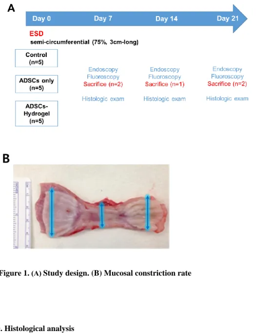
A. Animals and experimental design (Figure 1A)
This study was approved by the Yonsei University Institutional Animal Care and Use Committee (2019-0129) and conducted in a dedicated animal facility (Seoul, Korea). Fifteen female domestic pigs weighing 20-25kg were used and were fasted for 24 h before the procedure, but water intake was allowed ad libitum. They were randomly assigned to a control group (n = 5), ADSCs-only group (ADSC group) (n= 5) or ADSCs-gel (hydrogel) mixture group (ADSC-gel group) (n=5). They were premedicated by intramuscular administration of atropine sulfate at 0.05 mg/kg. They were then anesthetized with 15 mg/kg ketamine hydrochloride and 3 mg/ g xylazine hydrochloride and then anesthetized with an intramuscular injection of 1-3mg/kg alfaxalone. After endotracheal intubation, isoflurane and nitrous oxide gas were used for maintenance of anesthesia during the procedure, under mechanical ventilation.
B. Semi-circumferential ESD and intralesional injection of hADSCs and hADSC-gel (hydrogel) complex
A single-channel esophagogastroduodenoscope (GIF-Q260; Olympus, Tokyo, Japan) with a transparent attachment hood fitted to the tip ((D-201-11304; Olympus, Tokyo, Japan). Semi-circumferential (75-80% of the circumference)
10
ESD was performed in the esophagus between 30 cm and 35 cm from the dental arch with DualKnife (KD-650Q; Olympus, Tokyo, Japan). En bloc resections were performed sequentially until a 5-cm semi-circumferential resection had been achieved. An electrosurgical generator (ForceTriad, Medtronic, Minneapolis, MN, USA) was set to the pure cut mode (30 W) or forced coagulation mode (35 W). All ESD procedures were performed by 1 experienced endoscopist (HC) who had performed more than 1000 ESD procedures in humans, including the esophagus and stomach. Immediately after ESD, the labeled hADSCs (1.0 × 10⁶ cells per 100 μL of hydrogel) were resuspended in the PBS (A group) and the pre-gel solution (2 wt% of hydrogel solution) (AG group) and injected into residual submucosal tissues using a syringe with 23-gauge needle. We injected 0.2-0.5 mL of the solution into each of 8-10 spots, the total amount being 2 mL. For postoperative care, all pigs were given liquid from the next day after ESD and then semi-solids the following days.
C. Evaluation of the degree of esophageal stricture after ESD
On 7, 14, and 21 days after the procedure, the degree of stricture was evaluated by endoscopic and fluoroscopic examination. After evaluation, 2 pigs on day 7, 1 pig on day 14 and 2 pigs on day 21 were sacrificed by intravenous injection of KCl in each group and macroscopic and histological evaluations were conducted. The resected esophagus was immediately placed on a corkboard and fixed with pins. The degree of stricture at the lesion site was expressed as the lateral mucosal constriction rate calculated by the following formula, as described previously. 8 Mucosal constriction rate (%) = [1 - (length of short axis at site of maximal constriction)/ (length of short axis at a normal mucosal site on upper side + length of short axis at a normal mucosal site on a lower side)/2] x 100 (Figure 1B)
11
Figure 1. (A) Study design. (B) Mucosal constriction rate
D. Histological analysis
Esophagi were fixed in 10% formaldehyde saline solution, embedded in paraffin, and cut into 5-mm sections. Tissue sections were stained with hematoxylin and eosin, and Masson’s trichrome to examine the accumulation of collagen fibers. To track ADSCs in the resected esophagus, fluorescence microscopic evaluation was also performed. Tissue sections were stained with anti–a-smooth muscle
12
actin antibody (mouse, monoclonal antibody, Abcam, Cambridge, UK) for 60 minutes, anti-myeloperoxidase antibody (rabbit, polyclonal antibody, Abcam, Cambridge, UK) for 40 minutes, anti–Ki-67 antibody (rabbit, monoclonal antibody, Invitrogen, Carlsbad, CA, US) for 60 minutes at room temperature, and anti-VEGF antibody (rabbit, polyclonal antibody, Abcam, Cambridge, UK).
4. Statistical Analysis
All quantitative data are expressed as mean ± standard deviation (SD). Statistical analysis was performed to determine statistical significance by calculating p values < 0.05, 0.01 or 0.001 using Students’ t-test (GraphPad Software, CA, La Jolla, USA). All data were expressed as median (interquartile range) or mean ± standard deviation (SD). Normally distributed variables were compared by using a t test, and non–normally distributed variables were compared by use of the Mann-Whitney U test.
III. RESULTS
1. in vitro examination
A. Confirmation of synthesis of the ascidian-inspired, biocompatible, injectable and in situ self-crosslinkable hydrogel conjugate
The hydrogel was designed from a highly auto-oxidative chemical moiety (pyrogallol; PG, red) inspired by TOPA-containing polypeptides in a marine ascidian (Figure. 2A). Also, hyaluronic acid (HA, blue), one of the most widely used extracellular matrix components in biomedical application, was utilized as a polymer backbone of this hydrogel system due to its great biocompatibility and biodegradability.13,20 The bioinspired polymer was synthesized by chemically
13
conjugating 5-hydroxydopamine containing PG group to the HA backbone via a carbodiimide coupling reaction using 1-ethyl-3-(3-dimethylaminopropyl)carbodiimide (EDC) and N-hydroxysuccinimide (NHS). The conjugation of 5-hydroxydopamine to carboxyl group in HA backbone was confirmed by 1H nuclear magnetic resonance (NMR) spectroscopy. The presence of proton peaks at ~ 6.4 ppm indicating aromatic protons in the PG groups demonstrated successful synthesis of hydrogel polymer (Figure 2B). The degree of substitution (DS) of hydrogel was also measured using 1H-NMR by calculating the integral area ratio of the peak of aromatic protons in the PG groups to that of the methyl groups of the HA backbone. The DS was determined to be approximately 8%. Furthermore, the absorbance spectrum and its peak at 278 nm using ultraviolet-visible (UV-vis) spectroscopy also showed the presence of galloyl group within the polymer (Figure 2C).
15
Figure 2. ascidian-inspired, biocompatible, injectable and in situ self-crosslinkable hydrogel conjugate
(A) ascidian-inspired, biocompatible, injectable and in situ self-crosslinkable hydrogel conjugate (B) The presence of proton peaks at ~ 6.4 ppm indicating aromatic protons in the PG groups demonstrated successful synthesis of hydrogel polymer (C) The absorbance spectrum and its peak at 278 nm using ultraviolet-visible (UV-vis) spectroscopy showed the presence of galloyl group within the polymer
B. Material characterization of the hydrogel
The hydrogel could be formed via oxidative crosslinking within in vivo oxidative environment (Figure 3A). We used a peroxidase enzyme (horseradish peroxidase, HRP) for in vivo-mimetic gelation since the enzyme was considered as one of the key factors for complex oxidative processes in vivo.21-24 The stability of the formed hydrogel was confirmed by a consistently higher storage modulus (G') than loss modulus (G̍̎̍ ̍̎̍'') which were measured by the frequency sweep mode using a rheometer in the range from 0.1 to 1 Hz (Figure. 3B).
For the analysis of the physical and rheological properties of the hydrogel, the storage and loss moduli at three different concentrations of HRP were measured using the rheometer. While The average storage modulus of hydrogel (at 1 Hz) formed with 0.06 U/ml of HRP was similar to that of the hydrogel without any enzyme, the modulus of those with 0.6 and 6 U/ml of HRP were significantly increased (Figure 3C). Of that, the hydrogel formed by using 6 U/ml of HRP even exhibited a similar level of physical property of the hydrogel made within an in vivo oxidative condition. Interestingly, several previous studies had shown that the concentration of HRP for enzymatic crosslinking mainly affects the gelation rate rather than the physical properties of the formed hydrogels, but the concentration of HRP comparatively influenced the physical properties of hydrogel.25,26 We assumed that peroxidase could be directly involved in and
16
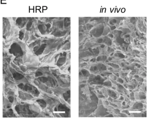
change the path of reaction of the galloyl groups in hydrogel, and this phenomenon could also happen in an in vivo oxidative environment.27,28 Contrastively, hydrogels formed with different concentrations of HRP exhibited a reverse trend in the elasticity (tan δ, G″/G′), which decreased as the concentration of HRP was higher due to the mechanical reinforcement and stiffening by peroxidase (Figure 3D). Based on these results, hydrogel crosslinked in the condition of 0.6 and 6 U/ml of HRP appeared to highly resemble the hydrogel formed within an in vivo condition in terms of physical and rheological properties. Furthermore, the internal structures of HRP-crosslinked and in vivo-crosslinked hydrogels seemed to be similar according to the images obtained from a scanning electron microscope (SEM) (Figure 3E). Thus, these crosslinking conditions to use 0.6 and 6 U/ml of HRP were chosen for the subsequent in vitro experiments.
18
Figure 3.Material characterization of the hydrogel
(A) oxidative crosslinking within in vivo oxidative environment (B) The stability of the formed hydrogel was confirmed by a consistently higher storage modulus (G') than loss modulus (G̍̎̍ ̍̎̍ '') (C) the modulus of those with 0.6 and 6 U/ml of HRP were significantly increased. (D) The hydrogels formed with different concentrations of HRP exhibited a reverse trend in the elasticity (tan δ, G″/G′) (E) Scanning electron microscope (SEM) image: the internal structures of HRP-crosslinked and in vivo-crosslinked hydrogels seemed to be similar.
C. in vitro examination of biocompatibility and paracrine effects of the hydrogel
First, we examined the cytotoxicity of the hydrogels in vitro, which is an important factor for stem cell transplantation and long-term maintenance in vivo. Cellular viability was assessed through Live/Dead assay, and the human adipose-derived stem cells (hADSCs) encapsulated in the hydrogel crosslinked by in vivo -mimetic crosslinking using HRP exhibited good viabilities at early time points
19
(Day 1 and 3), which means the oxidative crosslinking of hydrogel does not negatively affect the viability of the encapsulated cells (Figure 4A, 4B). Furthermore, the encapsulated hADSCs were highly viable even until day 14 (about 90%), which shows that stem cells could be effectively delivered to in vivo environments by utilizing the hydrogel system and successfully maintain their conditions for a long time.
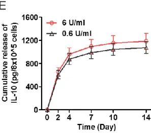
Next, we checked the paracrine effect of the stem cells encapsulated in the hydrogel, which was expected to contribute to therapeutic effects at transplanted regions (Figure 4C). Adipose-derived stem cells (ADSCs) are well-known for their ability to secrete various growth factors and cytokines for regeneration and immune modulation.29-31 We examined the paracrine effects of the 3D-encapsulated ADSCs by measuring two well-known proteins released from the hADSCs encapsulated in hydrogel which were formed by two conditions of in vivo-mimetic crosslinking. Vascular endothelial growth factor (VEGF), one of the representative growth factors important to regenerative process, was released from the hADSCs encapsulated in hydrogel over 14 days after seeding although the amount gradually decreased (Figure 4D). Furthermore, interleukin 10 (IL-10), one of the typical anti-inflammatory cytokines, was also secreted from the hADSCs encapsulated in hydrogel, although the released amount of IL-10 also decreased over time (Figure 4E). Interestingly, the released amount of both proteins from the hADSCs encapsulated in hydrogel crosslinked using 6 U/ml of HRP were slightly higher than that from cells encapsulated in hydrogel crosslinked using 0.6 U/ml of HRP. We assumed that the mechanically stiffer environment provided by the hydrogel with 6 U/ml of HRP might facilitate the encapsulated cells to release larger amount of paracrine factors than the relatively softer environment provided by the hydrogel with 0.6 U/ml of HRP.32,33 In this respect, the hADSC encapsulated in the in vivo-crosslinked hydrogel which has similar or slightly higher mechanical property compared to the hydrogel formed by in vivo-mimetic crosslinking would exhibit better paracrine effects.
20
The injectability of hydrogels is critical in terms of practical and clinical availability.16 It would especially be a key factor when the hydrogel is applied as an injecting solution for an endoscopic device that consists of a tubing and needle with a very narrow gauge.
22
Figure 4.in vitro examination of biocompatibility and paracrine effects of
the hydrogel.
(A) & (B) Cellular viability was assessed through Live/Dead assay. hADSCs encapsulated in the hydrogel crosslinked by in vivo-mimetic crosslinking using HRP exhibited good viabilities at early time points (Day 1 and 3). (C) The scheme of the paracrine effect of the stem cells encapsulated in the hydrogel. (D) Vascular endothelial growth factor (VEGF) was released from the hADSCs encapsulated in hydrogel over 14 days after seeding although the amount gradually decreased. (E) Interleukin 10 (IL-10) also secreted from the hADSCs encapsulated in hydrogel.
2. Mouse experiment
A. Confirmation of injectability, in situ self-crosslinkability of the hydrogel
For investigation of the injectability and in situ self-crosslinking of hydrogel, we used a mouse model. The pre-gel solution was prepared by dissolving the lyophilized hydrogel with phosphate-buffered saline (PBS) at a concentration of 2 wt%. Then the liquid was subcutaneously injected using a 1 ml syringe with
23
26-gauge needle. Although the solution was slightly viscous, it was easily released through the 26-gauge needle which is narrower than clinically used endoscopic needles such as 22- to 25-gauge needles. The injected solution did not instantly spread all over the surrounding subcutaneous regions due to its viscosity and maintained its own shape, making the injected region look swollen (Figure 5A). Then, the injected pre-gel solution would be crosslinked by in vivo oxidative processes, maintaining its original shape and volume even 1 day after injection. Interestingly, the location of the formed hydrogel did not change at all, which might have resulted from the tissue-adhesiveness of pyrogallol group and its intermediates during oxidative processes.19 In fact, the hydrogel was successfully crosslinked in the in vivo environment, and stably adhered to the surface of subcutaneous tissues which was confirmed when the injected region was surgically opened after 1-day post-injection (Figure 5B).
B. Cell transplantation test
Stem cell delivery test using hydrogel was conducted. The Qtracker 655 (red-fluorescent Qdot 655; QD655)-labeled hADSCs were loaded into hydrogel pre-gel solution and injected into subcutaneous tissues in mice. Subsequently, the hydrogels formed within the tissues were removed and fixed at 7 and 14-day post-injection. Then, the fluorescent Qdots inside the delivered cells were observed using a confocal microscope, and an image-based quantification was performed using the acquired images. After 7 days of injection, a considerable amount of hADSC existed in the hydrogel, and quite a few hADSCs still remained in the hydrogel even after 14 days of transplantation. While the number of cells per unit area did slightly decreased, it was not statistically significant. (Figure 5C, 5D).
C. Histological analyses of mice model
Histological analysis also showed that the hydrogel was tightly integrated onto the tissue, which was shown in the hematoxylin & eosin (H&E) staining image (Figure 5E). Then, the biocompatibility of hydrogel was examined in vivo, which is an integral factor for further biomedical applications. For assessing the in vivo
24
biocompatibility of hydrogel, the pre-gel solutions were subcutaneously injected into mice, and then the crosslinked hydrogels and the adjacent tissue were removed altogether at the predetermined time points for histological analysis. Toluidine blue staining showed that the hydrogels did not induce problematic inflammatory responses in surrounding tissue at both early and late time points (Day 3, 7) (Figure 5F, 5G).
25
Figure 5.Mouse experiment
(A) The injected solution did not instantly spread all over the surrounding subcutaneous regions due to its viscosity. (B) The hydrogel was successfully crosslinked and stably adhered to the surface of subcutaneous tissues after 1-day post-injection. (C) & (D) After 7 days of injection, a considerable amount of hADSC existed in the hydrogel, and quite a few hADSCs still remained in the hydrogel even after 14 days of transplantation. (E) hematoxylin & eosin (H&E) staining image. The hydrogel was tightly integrated onto the tissue. (F) & (G) Toluidine blue staining images. The hydrogels did not induce problematic inflammatory responses in surrounding tissue at both early and late time points (Day 3, 7)
3. Porcine experiment
Semi-circumferential ESD was performed safely in all pigs but pig 14 (ADSC group) was expired unexpectedly on day 6. Necropsy was performed and esophageal perforation with severe mediastinal inflammation was noted.
A. Macroscopic appearance
Serial endoscopic findings were compared among groups. On day 7, extensive amount of exudate was attached on the ulcer base in the control group. However, only small amount of exudate with clean ulcer base was seen in the ADSC group and there was no exudate in the ADSC-gel group. On day 14, severe esophageal stricture was observed in control group and the degree of the stricture appears be milder in ADSC and ADSC-gel group than control and this pattern is more prominently observed on day 21. (Figure 6)
Figure 7A shows macroscopic and fluoroscopic findings of the esophagus by group. Significant mucosal stricture was seen in control group. The ADSC group showed milder stricture than control group, however, severer than ADSC-gel
26
group. The mean (± SD) rate of mucosal contraction was 79.5% ±2.0%, 62.8% ± 1.7%, and 37.9% ±2.9% in control, ADSC group, and ADSCs-gel group, respectively (P < 0.05). Contraction rates were significantly milder in ADSC and ADSC-gel group, compared to control (P < 0.05 and P< 0.01) and milder in ADSC-gel group, compared to ADSC group (P < 0.05) (Figure 7B)
Figure 6. Porcine experiment: Serial endoscopic findings by group
On day 7, extensive amount of exudate was attached on the ulcer base in the control group. However, only small amount of exudate with clean ulcer base was seen in ADSC group and there was no exudate in ADSC-gel group. On day 14, severe esophageal stricture was observed in control group and the degree of the stricture appears be milder in ADSC and ADSC-gel group than control and this pattern is more prominently observed on day 21.
27
28
Figure 7. Porcine experiment: mucosal contracture rate (%)
(A) Macroscopic and fluoroscopic findings of the esophagus by group: Significant mucosal stricture was seen in control group. The ADSC group showed milder stricture than control group, however, severer than the ADSC-gel group. (B) Contraction rates were significantly milder in ADSC and ADSC-gel group, compared to control (P < 0.05 and P< 0.01) and milder in ADSC-gel group, compared to ADSC group (P < 0.05)
B. Histological, immunohistochemical and immunofluorescence examination
The amount of Q-dots was observed and compared with time according to group. The injected QD–labeled cells were found in the submucosal layer in both ADSC and ADSC-gel group, and the number of Q-dots observed in both groups decreases over time. However, compared to the ADSC group, the amount of Q-dots observed in the ADSC-gel group was increased. (Figure 8A). In ADSC-gel group, the injected gel was attached to tissue at both 1st and 3rd week. Confocal microscopy also showed Q-dots in the gel, confirming the presence of hADSCs at the injection site. (Figure 8B) To evaluate the impact of ADSC treatment on the neovascularization, we then determined expression of vascular endothelial growth factor (VEGF). VEGF expression increased in the ADSC-gel group compared with controls or ADSC group at day 21 (P <0.05); Figure 8C). Ki-67, as a cell proliferation marker, expression level was significantly decreased in ADSC and ADSC-gel group, compared with controls at each time point, suggesting that ADSC inhibited the proliferation of fibroblasts. However, there was no difference between ADSC and ADSC-gel group. (Figure 8D). In addition,
29
the myeloperoxidase (MPO) activity at day 21 showed no difference between controls and ADSC group but decreased in ADSC-gel group compared with controls and ADSC group. (Figure 8E). In the immunofluorescence staining for alpha (α)-SMA, which was indicated to be myofibroblasts showed significantly decreased expression of α-SMA in the submucosa area in ADSC and ADSC-gel group, compared with controls. When compared between ADSC and ADSC-gel group, ADSC-gel group showed significant reduction than ADSC group (Figure 8F)
30
B
31
D
32
Figure 8. Porcine experiment: histological findings
(A) The number of Q-dots observed in both groups decreases over time. However, compared to the ADSC group, the amount of Q-dots observed in the ADSC-gel group was increased. (B) In ADSC-gel group, the injected gel was attached to tissue at both 1st and 3rd week. Confocal microscopy also showed Q-dots in the gel, confirming the presence of hADSCs at the injection site. (C) VEGF expression increased in the ADSC-gel group compared with controls or ADSC group at day 21 (P <0.05) (D) Ki-67 expression level was significantly decreased in ADSC and ADSC-gel group, compared with controls at each time point. (E) The myeloperoxidase (MPO) activity at day 21 showed no difference between controls and ADSC group, but decreased in ADSC-gel group compared with controls and ADSC group. (F) In the immunofluorescence staining for α-SMA showed significantly decreased expression of α-SMA in the submucosa area.
33
IV. DISCUSSION
Because stricture formation after large-scale esophageal ESD deteriorates the quality of life, prevention of postoperative strictures has recently been an issue of interest. Detailed mechanisms are not yet known, but inflammation and fibrosis after ESD are thought to reduce elasticity and compliance and causes esophageal stricture. There are studies in which mesenchymal stem cell (MSCs) have been used to reduce or prevent esophageal strictures.8,9 Since MSCs do not differentiate into epithelial cells, this effect is expected to be due to its paracrine effects by secreting immunomodulation factors, angiogenic and arteriogenic factors, antiapoptotic factors, and antioxidative factors. 34,35 This hypothesis could be supported from the results of studies demonstrating similar or even better tissue regeneration effect by infusion or oral ingestion of MSC-derived conditioned medium compared to MSC transplantation. 9,36,37 However, further research is needed on the exact mechanism. Studies have reported that esophageal stenosis is reduced after ESD by topical injection of ADSC into an endoscopic resection or by drinking conditioned medium extracted only from anti-inflammatory components of stem cells.8,9 The potential limitation of these previous studies, however, can be that the paracrine effect of the ADSCs would not last long with only single injection of stem cells, and that oral intake should be repeated. Therefore, we hypothesized that ADSCs -hydrogel mixture can overcome this shortcoming and these hydrogels must be the best if it can exist as liquids before injection and then changed into the gel-like form for a long time at the initial injection site. In our study, an hydrogel was designed from a highly auto-oxidative chemical moiety (pyrogallol; PG, red) inspired by TOPA-containing polypeptides in a marine ascidian.19 Also, hyaluronic acid, one of the most widely used extracellular matrix components in biomedical application, was utilized as a polymer backbone of this hydrogel system due to its great biocompatibility. From the analysis of the physical and rheological properties,
34
hydrogel crosslinked in the condition of 0.6 and 6 U/ml of HRP appeared to highly resemble the hydrogel formed within an in vivo condition. Thus, these crosslinking conditions to use 0.6 and 6 U/ml of HRP were chosen for the subsequent in vitro experiments. We assumed that the mechanically stiffer environment provided by the hydrogel with 6 U/ml of HRP might facilitate the encapsulated cells to release larger amount of paracrine factors than the relatively softer environment provided by the hydrogel with 0.6 U/ml of HRP.32,33 In this respect, the hADSC encapsulated in the in vivo-crosslinked hydrogel which has similar or slightly higher mechanical property compared to the hydrogel formed by in vivo-mimetic crosslinking would exhibit better paracrine effects. The injectability of hydrogels is critical in terms of practical and clinical availability.16 It is a key factor when the hydrogel is applied as an injecting solution for an endoscopic device that consists of a tubing and needle with a very narrow gauge. In addition, the hydrogel was successfully crosslinked in mouse model, and stably adhered to the surface of subcutaneous tissues which was confirmed when the injected region was surgically opened after 1-day post-injection. Histological analysis also showed that the hydrogel was tightly integrated onto the tissue and we could conclude that the hydrogel and its crosslinking processes were highly biocompatible even in an in vivo environment. Finally, porcine experiment was performed to determine whether it is effective in an environment similar to the actual clinical situation. The contractions rates were significantly milder in the ADSC and ADSC-gel group, compared to the control group. In addition, significantly milder contraction was observed in the ADSC-gel group, when compared to the ADSC group. To investigate the underlying mechanisms by which stenosis is reduced, immunohistochemical/fluorescence staining with various markers such as VEGF, Ki-67, MPO, and α-SMA was performed and compared. VEGF is a mitotic substance of endothelial cells that promotes capillary regeneration.38 Therefore, blood flow around the injected area is restored and oxygen support and nutrient delivery can be facilitated.
α-SMA-35
positive myofibroblasts is known to be associated with scar contraction. In healing tissues, fibroblasts are transformed to the activated type, termed “myofibroblasts” and participate in the reparative response, by secreting large amounts of extracellular matrix proteins and may be responsible for contraction of healing wounds. 39,40 In the present study, ADSC and ADSC-gel group showed decreased α-SMA expression, compared with controls and ADSC-gel group showed much less expression than ADSC group. In addition, fewer Ki-67-positive cells were detected in the ADSC and ADSC-gel group compared to controls. Ki-67 is a proliferation marker to measure the growth fraction of cell. The meaning of Ki-67 expression is still controversial, but decreased Ki-67 expression was reported in pulmonary fibrosis mice model after anti-fibrotic treatment and this results is consistent with our results.41 Putting these results together, it can be implied that the ADSCs could be effective to reduce esophageal stricture and this effect can be augmented in ADSC-gel mixture.
However, this study has several limitations. First, our study was conducted in a small sample. Second, it is not clear whether our results can be easily extrapolated to human adults because we used small pigs (20-25 kg) in this study. Finally, it was difficult to strictly control diet after ESD in porcine model.
V. CONCLUSION
Our study demonstrated that ADSCs mixture with ascidian-inspired, biocompatible, injectable and in situ self-crosslinkable hydrogel was significantly reduced the post-ESD esophageal stricture by augmenting the paracrine effects of ADSCs. Further investigation and comparative studies are required for its clinical applications.
36
REFERENCES
1. Jemal A, Center MM, DeSantis C, Ward EM. Global patterns of cancer incidence and mortality rates and trends. Cancer Epidemiol Biomarkers Prev 2010;19:1893-907.
2. Min YW, Lee H, Song BG, Min BH, Kim HK, Choi YS, et al. Comparison of endoscopic submucosal dissection and surgery for superficial esophageal squamous cell carcinoma: a propensity score-matched analysis. Gastrointest Endosc 2018;88:624-33.
3. Ono S, Fujishiro M, Niimi K, Goto O, Kodashima S, Yamamichi N, et al. Predictors of postoperative stricture after esophageal endoscopic submucosal dissection for superficial squamous cell neoplasms. Endoscopy 2009;41:661-5. 4. Sato H, Inoue H, Kobayashi Y, Maselli R, Santi EG, Hayee B, et al. Control of
severe strictures after circumferential endoscopic submucosal dissection for esophageal carcinoma: oral steroid therapy with balloon dilation or balloon dilation alone. Gastrointest Endosc 2013;78:250-7.
5. Nagami Y, Shiba M, Tominaga K, Minamino H, Ominami M, Fukunaga S, et al. Locoregional steroid injection prevents stricture formation after endoscopic submucosal dissection for esophageal cancer: a propensity score matching analysis. Surg Endosc 2016;30:1441-9.
6. Pittenger MF, Mackay AM, Beck SC, Jaiswal RK, Douglas R, Mosca JD, et al. Multilineage potential of adult human mesenchymal stem cells. Science 1999;284:143-7.
7. Iyer SS, Rojas M. Anti-inflammatory effects of mesenchymal stem cells: novel concept for future therapies. Expert Opin Biol Ther 2008;8:569-81.
8. Honda M, Hori Y, Nakada A, Uji M, Nishizawa Y, Yamamoto K, et al. Use of adipose tissue-derived stromal cells for prevention of esophageal stricture after circumferential EMR in a canine model. Gastrointest Endosc 2011;73:777-84. 9. Mizushima T, Ohnishi S, Hosono H, Yamahara K, Tsuda M, Shimizu Y, et al.
Oral administration of conditioned medium obtained from mesenchymal stem cell culture prevents subsequent stricture formation after esophageal submucosal dissection in pigs. Gastrointest Endosc 2017;86:542-52 e1.
10. Ameer G, Mahmood T, Langer R. A biodegradable composite scaffold for cell transplantation. Journal of orthopaedic research 2002;20:16-9.
11. Godier-Furnémont AF, Martens TP, Koeckert MS, Wan L, Parks J, Arai K, et al. Composite scaffold provides a cell delivery platform for cardiovascular repair. Proceedings of the National Academy of Sciences 2011;108:7974-9.
12. Prestwich GD. Hyaluronic acid-based clinical biomaterials derived for cell and molecule delivery in regenerative medicine. Journal of controlled release 2011;155:193-9.
13. Burdick JA, Prestwich GD. Hyaluronic acid hydrogels for biomedical applications. Advanced materials 2011;23:H41-H56.
14. Hoffman AS. Hydrogels for biomedical applications. Advanced drug delivery reviews 2012;64:18-23.
15. Seliktar D. Designing cell-compatible hydrogels for biomedical applications. Science 2012;336:1124-8.
37
biodegradation and biomedical applications. Chemical Society Reviews 2012;41:2193-221.
17. Tran RT, Palmer M, Tang S-J, Abell TL, Yang J. Injectable drug-eluting elastomeric polymer: a novel submucosal injection material. Gastrointestinal endoscopy 2012;75:1092-7.
18. Cao L, Li Q, Zhang C, Wu H, Yao L, Xu M, et al. Safe and efficient colonic endoscopic submucosal dissection using an injectable hydrogel. ACS Biomaterials Science & Engineering 2016;2:393-402.
19. Cho JH, Lee JS, Shin J, Jeon EJ, An S, Choi YS, et al. Ascidian‐Inspired Fast‐ Forming Hydrogel System for Versatile Biomedical Applications: Pyrogallol Chemistry for Dual Modes of Crosslinking Mechanism. Advanced Functional Materials 2018;28:1705244.
20. Sontyana AG, Mathew AP, Cho K-H, Uthaman S, Park I-K. Biopolymeric in situ hydrogels for tissue engineering and bioimaging applications. Tissue engineering and regenerative medicine 2018;15:575-90.
21. Wieland E, Parthasarathy S, Steinberg D. Peroxidase-dependent metal-independent oxidation of low density lipoprotein in vitro: a model for in vivo oxidation? Proceedings of the National Academy of Sciences 1993;90:5929-33. 22. Brennan M-L, Wu W, Fu X, Shen Z, Song W, Frost H, et al. A tale of two controversies defining both the role of peroxidases in nitrotyrosine formation in vivo using eosinophil peroxidase and myeloperoxidase-deficient mice, and the nature of peroxidase-generated reactive nitrogen species. Journal of Biological Chemistry 2002;277:17415-27.
23. Santanam N, Parthasarathy S. Paradoxical actions of antioxidants in the oxidation of low density lipoprotein by peroxidases. The Journal of clinical investigation 1995;95:2594-600.
24. Van Der Vliet A, Eiserich JP, Halliwell B, Cross CE. Formation of reactive nitrogen species during peroxidase-catalyzed oxidation of nitrite a potential additional mechanism of nitric oxide-dependent toxicity. Journal of Biological Chemistry 1997;272:7617-25.
25. An S, Jeon EJ, Jeon J, Cho S-W. A serotonin-modified hyaluronic acid hydrogel for multifunctional hemostatic adhesives inspired by a platelet coagulation mediator. Materials Horizons 2019.
26. Park KM, Lee Y, Son JY, Oh DH, Lee JS, Park KD. Synthesis and characterizations of in situ cross-linkable gelatin and 4-arm-PPO-PEO hybrid hydrogels via enzymatic reaction for tissue regenerative medicine. Biomacromolecules 2012;13:604-11.
27. Nozaki O, Ji X, Kricka LJ. New enhancers for the chemiluminescent peroxidase catalysed chemiluminescent oxidation of pyrogallol and purpurogallin. Journal of bioluminescence and chemiluminescence 1995;10:151-6.
28. Bach CE, Warnock DD, Van Horn DJ, Weintraub MN, Sinsabaugh RL, Allison SD, et al. Measuring phenol oxidase and peroxidase activities with pyrogallol, L-DOPA, and ABTS: effect of assay conditions and soil type. Soil Biology and Biochemistry 2013;67:183-91.
29. Xie J, Jones TJ, Feng D, Cook TG, Jester AA, Yi R, et al. Human adipose-derived stem cells suppress elastase-induced murine abdominal aortic inflammation and aneurysm expansion through paracrine factors. Cell transplantation 2017;26:173-89.
38
30. Naderi N, Combellack EJ, Griffin M, Sedaghati T, Javed M, Findlay MW, et al. The regenerative role of adipose‐derived stem cells (ADSC) in plastic and reconstructive surgery. International wound journal 2017;14:112-24.
31. Kuroda K, Kabata T, Hayashi K, Maeda T, Kajino Y, Iwai S, et al. The paracrine effect of adipose-derived stem cells inhibits osteoarthritis progression. BMC musculoskeletal disorders 2015;16:236.
32. Kusuma GD, Carthew J, Lim R, Frith JE. Effect of the microenvironment on mesenchymal stem cell paracrine signaling: opportunities to engineer the therapeutic effect. Stem cells and development 2017;26:617-31.
33. Abdeen AA, Weiss JB, Lee J, Kilian KA. Matrix composition and mechanics direct proangiogenic signaling from mesenchymal stem cells. Tissue Engineering Part A 2014;20:2737-45.
34. Gnecchi M, Danieli P, Malpasso G, Ciuffreda MC. Paracrine Mechanisms of Mesenchymal Stem Cells in Tissue Repair. Methods Mol Biol 2016;1416:123-46.
35. Liang X, Ding Y, Zhang Y, Tse HF, Lian Q. Paracrine mechanisms of mesenchymal stem cell-based therapy: current status and perspectives. Cell Transplant 2014;23:1045-59.
36. Chen L, Tredget EE, Wu PY, Wu Y. Paracrine factors of mesenchymal stem cells recruit macrophages and endothelial lineage cells and enhance wound healing. PLoS One 2008;3:e1886.
37. Wang Y, Lian F, Li J, Fan W, Xu H, Yang X, et al. Adipose derived mesenchymal stem cells transplantation via portal vein improves microcirculation and ameliorates liver fibrosis induced by CCl4 in rats. J Transl Med 2012;10:133. 38. Ferrara N. Vascular endothelial growth factor: basic science and clinical progress.
Endocr Rev 2004;25:581-611.
39. Shinde AV, Humeres C, Frangogiannis NG. The role of alpha-smooth muscle actin in fibroblast-mediated matrix contraction and remodeling. Biochim Biophys Acta Mol Basis Dis 2017;1863:298-309.
40. Darby IA, Zakuan N, Billet F, Desmouliere A. The myofibroblast, a key cell in normal and pathological tissue repair. Cell Mol Life Sci 2016;73:1145-57. 41. Kinoshita K, Aono Y, Azuma M, Kishi J, Takezaki A, Kishi M, et al. Antifibrotic
effects of focal adhesion kinase inhibitor in bleomycin-induced pulmonary fibrosis in mice. Am J Respir Cell Mol Biol 2013;49:536-43.
39
APPENDICES
Abbreviation lists
ADSC= adipose tissue-derived stem cells, ESD= endoscopic submucosal dissection, VEGF= vascular endothelial growth factor, IL= interleukin, PBS= phosphate-buffered saline, QD= Quantum dot, HRP= Horseradish peroxidase, NMR= nuclear magnetic resonance, SEM= scanning electron microscope
40
ABSTRACT (IN KOREAN)
식도
내시경점막하박리술후
지방조직유래줄기세포
-
하이드로겔
국소주입을
통한
식도협착
예방
효과
확인을
위한
동물실험
<
지도교수
이
용
찬
>
연세대학교
대학원
의학과
정
현
수
배경
:
광범위
식도
내시경점막하박리술
후
발생하는
식도
협착은
삶의
질을
악화시키기
때문에
,
협착의
예방은
매우
중요하며
,
중간엽줄기세포
주사
또는
경구
복용
등의
방법이
제시되었으나
짧은
지속시간
또는
반복적인
섭취
등의
제한점이
있다
.
목적
:
본
연구는
지방
조직
유래
줄기
세포
-
하이드로겔
혼합물의
식도협착예방효과를
확인하고자
하는
대동물
실험으로서
,
이를
위하여
in vitro
및
마우스실험
등의
예비실험을
함께
진행하였다
.
방법
:
멍게의
혈액성분
가운데
강력한
접착성을
가지고
있는
성분을
모사한
하이드로겔의
합성을
1H-NMR
과
UV-vis light
spectrophotometer
를
통하여
확인하고
, in vitro
및
마우스실험을
통하여
줄기세포
-
하이드로겔의
생체적합성
,
파라크린효과
및
조직부착성
등을
확인하였다
.
이후
15
마리의
돼지
(
대조군
5,
줄기세포군
5,
줄기세포
-
하이드로겔
5
마리
)
에서
원주의
75%
및
길이
3cm
의
내시경점막하박리술을
시행하고
,
줄기세포
또는
줄기세포
-
하이드로겔
혼합용액을
절제된
식도의
점막하층에
주입하고
매주
내시경
,
투시촬영을
통한
식도협착의
평가를
시행하였다
. 1
주
, 2
주
,
및
3
주차에
각
군에서
2
마리
, 1
마리
및
41
2
마리를
희생하여
,
육안적
협착정도와
조직학소견을
비교하였다
.
결과
:
점막협착비율은
대조군
,
줄기세포군
및
줄기세포
-하이드로겔군에서
각각
79.5 % ± 2.0 %, 62.8 % ± 1.7 %
및
37.9 % ±
2.9 %
였다
(P <0.05).
협착율은
대조군과
비교하였을
때
줄기세포군과
줄기세포
-
하이드로겔군에서
유의하게
낮았으며
,
줄기세포군에
비해
줄기세포
-
하이드로겔
군에서
유의하게
낮았다
.
관찰되는
Q-dot
의
수는
줄기세포군과
줄기세포
-하이드로겔군
모두
시간이
경과함에
따라
감소하는
양상을
보이지만
,
줄기세포군에
비하여
줄기세포
-
하이드로겔군
에서
관찰되는
Q-dot
의
양이
증가되어
있어
,
줄기세포
-하이드로겔군에서
더
오랫동안
,
더
많은
양의
지방유래줄기세포
존재함을
확인하였다
.
결론
:
본
연구를
통하여
지방유래
줄기세포의
주입이
내시경점막하박리술
후
발생하는
식도
협착을
감소시키는
데
효과적이었고
,
줄기세포
단독사용보다는
줄기세포
-
하이드로겔
혼합물을
사용함으로써
이러한
효과를
증대시킴을
확인하였다
.
핵심되는
말
:
식도협착
,
하이드로겔
,
지방유래줄기세포
,
내시경
점막하박리술