Comparative Pathogenicity of United
Kingdom Isolates of the Emerging
Pathogen
Candida auris
and Other Key
Pathogenic
Candida
Species
Andrew M. Borman, Adrien Szekely, Elizabeth M. Johnson
UK National Mycology Reference Laboratory (MRL), Public Health England South-West, Bristol, United Kingdom
ABSTRACT Candida auris, first described in 2009, has since emerged as an impor-tant, multidrug-resisimpor-tant, nosocomial agent of candidemia, with large outbreaks re-ported worldwide and high mortality rates associated with therapeutic failure. The current study employedC. aurisisolates from a variety of centers in the United King-dom to evaluate the pathogenicity of this emerging pathogen compared to that of other common pathogenic yeast species in the invertebrateGalleria mellonella infec-tion model. We showed that C. aurisisolates differ in their growth characteristics in vitro, with a proportion of isolates failing to release daughter cells after budding, re-sulting in the formation of large aggregates of cells that cannot be physically dis-rupted. Our results also demonstrate strain-specific differences in the behavior of C. auris inG. mellonella, with the aggregate-forming isolates exhibiting significantly less pathogenicity than their nonaggregating counterparts. Importantly, the nonag-gregating isolates exhibited pathogenicity comparable to that ofC. albicans, which is currently accepted as the most pathogenic member of the genus, despite the fact thatC. aurisisolates do not produce hyphae and produce only rudimentary pseudo-hyphae eitherin vitroor inG. mellonella.
IMPORTANCE The incidence of invasive candidiasis, which includes candidemia and deep tissue infections, continues to rise and is associated with considerable mortality rates. Candida albicansremains the most common cause of invasive candi-diasis, although the prevalence of non-albicans species has increased over recent years. Since its first description in 2009,Candida aurishas emerged as a serious nos-ocomial health risk, with widespread outbreaks in numerous hospitals worldwide. However, despite receiving considerable attention, little is known concerning the pathogenicity of this emerging fungal pathogen. Here, using the Galleria mellonella insect systemic infection model, we show strain-specific differences in the virulence of C. auris, with the most virulent isolates exhibiting pathogenicity comparable to that of C. albicans, which is currently accepted as the most pathogenic member of the genus.
KEYWORDS: pathogenicity,Candida auris, pathogenic yeasts, emerging pathogen
T
he incidence of invasive fungal infections caused by unusualCandidaspp. contin-ues to rise, driven in part by increased populations of immunocompromised patients and those undergoing invasive procedures (1–8). However, to date,Candida albicansremains the most frequently isolatedCandidaspecies in the clinical setting, is the principal agent of nosocomial yeast infections (1, 4–6), and is widely accepted as being the most pathogenicCandidaspecies (reviewed in references 9 and 10).In 2009, a novelCandidaspecies in theCandida haemuloniicomplex ( Metchnikowi-aceae),Candida auris, was described after isolation from a discharge from a human
Received6 July 2016Accepted21 July 2016 Published18 August 2016 CitationBorman AM, Szekely A, Johnson EM. 2016. Comparative pathogenicity of United Kingdom isolates of the emerging pathogen
Candida aurisand other key pathogenic
Candidaspecies. mSphere 1(4):e00189-16.
doi:10.1128/mSphere.00189-16.
EditorAaron P. Mitchell, Carnegie Mellon University
Copyright© 2016 Borman et al. This is an open-access article distributed under the terms of theCreative Commons Attribution 4.0 International license.
Address correspondence to Andrew M. Borman, [email protected].
Clinical Science and Epidemiology
crossmark
on September 8, 2020 by guest
http://msphere.asm.org/
external ear canal in Japan (11). Subsequent studies confirmed an association with chronic otitis media in 15 patients from South Korea (12), with evidence of clonal transmission and resistance to certain triazole antifungal agents.C. aurishas since been reported from a wide spectrum of clinical manifestations, ranging from colonization through deep-seated infections and candidemia (13–17). Today, it is evident that C. auris has emerged as an important nosocomial pathogen with clonal inter- and intrahospital transmission, and it has become widespread across several Asian countries and South Africa (13–18). C. aurisfungemia is associated with a high mortality rate, therapeutic failure (13–15), and widespread resistance to several classes of antifungal agents (13, 15–21). Furthermore, correct identification ofC. aurisisolates is complicated by the fact that many commercially available biochemical-based tests can misidentify C. auris as the phylogenetically related species Candida haemulonii (11, 12, 19–23), which presents an additional challenge for appropriate patient management.
The first 2 United Kingdom isolates ofC. auris were received at the UK National Mycology Reference Laboratory (MRL) in 2013, from blood cultures from 2 unrelated patients in distant geographical localities (MRL unpublished data). Since 2013, we have received a further 19 isolates from at least 6 different hospitals, including 14 isolates suspected of being part of an outbreak. Here we have compared the pathogenicities of 12 United Kingdom isolates ofC. aurisfrom 6 different referring National Health Service (NHS) hospitals with the pathogenicities of equivalent isolates of other common pathogenic Candida species, using the Galleria mellonella insect systemic infection model.
RESULTS AND DISCUSSION
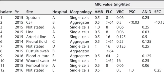
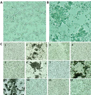
The characteristics of the 12 isolates of C. aurisemployed in the current study are detailed in Table 1, with antifungal MIC values determined at the MRL. Initial attempts to generate suspensions ofC. aurisisolates in phosphate-buffered saline (PBS) for larval inoculation revealed striking strain-specific differences in phenotypic behavior. While most isolates readily formed homogeneous suspensions upon thorough vortex mixing, the resulting suspensions seen with 4 independent isolates from 3 different referring hospitals were grossly particulate and contained individual yeast cells mixed with large aggregations (“aggregate” strains) (Table 1 and Fig. 1). For these 4 isolates, aggregates could not be physically disrupted by vigorous vortex mixing or by detergent treatments (data not shown). Since the aggregates were too large to permit larval inoculation and since cell numbers within the aggregates could not be accurately quantified, homo-geneous suspensions were instead achieved by allowing initial suspensions to settle for TABLE 1 Origin of theCandida aurisstrains employed in this studya
Isolate Yr Site Hospital Morphology
MIC value (mg/liter)
AMB FLC VRC PSC ANID 5FC
1 2015 Sputum A Single cells 0.5 8 0.06 0.25
2 2015 CSF B Aggregates 0.5 ⬎64 0.5 ⬍0.03 ⬍0.125 3 2015 Not stated A Single cells 1.0 8 0.06 0.25
4 2015 Line A Single cells 0.5 8 0.06 0.03
5 2015 Arterial line A Single cells 0.5 16 0.125 0.5 6 2014 Pleural fluid C Aggregates 0.5 ⬎64 0.25 0.125 7 2016 Not stated D Single cells 1 16 0.125 0.25 8 2015 Pustule swab B Aggregates ⬎64
9 2016 Blood culture E Aggregates 0.5 64 2 0.125 10 2016 Wound swab Fb Single cells 1 ⬎64 16 0.25 11 2015 Femoral line A Single cells 0.5 8 0.06 0.06 12 2016 Not stated E Single cells 0.5 0.5 1.0 0.25
aThe antifungal susceptibility results expressed as MICs (in milligrams per liter) are given for those antifungal
agents requested by referring centers; the susceptibility tests were performed at the MRL. MICs were obtained using CLSI broth microdilution methodologies (26). Abbreviations: AMB, amphotericin B; FLC, fluconazole; VRC, voriconazole; PSC, posaconazole; ANID, anidualfungin; 5FC, flucytosine; CSF, cerebrospinal fluid.
bThe patient was transferred from hospital A.
on September 8, 2020 by guest
http://msphere.asm.org/
10 min, followed by removal of the supernatant containing individual yeast cells that had remained in suspension and adjustment of these individual cells to the appropriate concentration for injection into larvae.
In agreement with previous reports (10, 24), the pathogenicity of the common Candidaspecies at 37°C inG. mellonellawas directly related to the ability of individual species to produce hyphal filaments or pseudohyphae (Fig. 2; see also Fig. S1 in the supplemental material), with very little strain-to-strain variation in virulence within each species (see Fig. S1). Thus,C. albicansandC. tropicalisexhibited greater virulence than C. lusitaniae,C. guilliermondii, and members of theC. parapsilosisspecies complex, and virtually no larval killing was induced by those organisms that form only rudimentary pseudohyphae or no pseudohyphae (C. glabrata, C. nivariensis, C. krusei, C. kefyr, C. bracarensis, and Saccharomyces cerevisiae) (Fig. 2; see Table 2 for full statistical analyses).
Strikingly, despite most reports suggesting thatC. aurisdoes not form significant pseudohyphaein vitro(14, 15, 21),C. aurisstrains exhibited virulence inG. mellonella that was significantly higher (in terms of the kinetics of larval death and the number of larvae killed) than that seen with most other common pathogenic yeast species, with overall pathogenicity approaching that observed with C. albicans and C. tropicalis isolates (Fig. 2 and Table 2). Dissection of representative larvae that had been inocu-lated with the various strains and incubated at 37°C for 18 h revealed significant hyphal FIG 1 Microscopic appearance of non-aggregate-forming isolates (A) and aggregate-forming isolates (B) of
C. aurisin PBS suspensions. Suspensions were subjected to vortex mixing for 1 min prior to examination at
ⴛ1,000 magnification. (C) The 12 isolates ofC. aurisemployed in the current study (ⴛ100 magnification).
on September 8, 2020 by guest
http://msphere.asm.org/
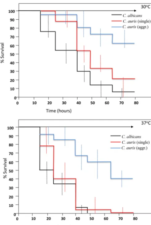
proliferation in hemolymph form larvae inoculated withC. albicans(Fig. 3A). However, no hyphal or pseudohyphal formation was observed in larvae infected with anyC. auris strains at 18 h or any time postinfection (Fig. 3B to D). Interestingly, in larvae that had received nonaggregating strains ofC. auris, larval dissection revealed large numbers of individual budding yeast cells, including in phagocytic cells (Fig. 3B and E). However, in larvae inoculated with individual yeast cells prepared from aggregate-forming strains of C. auris, hemolymph contained large aggregates ofC. auriscells, with few individual yeast cells, indicating that the ability to produce large aggregates had been maintained in vivo(Fig. 3C and E). In the light of this differential behavior of C. aurisisolates in G. mellonella, further experiments compared larval killing with aggregate-forming versus non-aggregate-forming strains, with larvae incubated at both 30°C and 37°C. Strikingly, nonaggregate strains exhibited significantly greater virulence than aggregate-forming strains at both temperatures (Fig. 4 and Table 2) (P⫽0.02), with nonaggregate isolates showing virulence that was indistinguishable from that of C. albicansstrains at 37°C (Fig. 4).
In the current report, we present for the first time a comparative study of the pathogenicities of isolates ofCandida aurisand those of other common pathogenic FIG 2 The virulence of Candidaspecies inGalleria mellonellalarvae at 37°C is species specific. Kaplan-Meier plots of G. mellonellasurvival after injection with 106 CFU/larva of the indicated
Candidaspecies, organized as those that produce true hyphae (top panel), pseudohyphae (middle panel), or no hyphae/pseudohyphae (bottom panel), are shown. Equivalent plots obtained with
C. aurisisolates are included in all three panels for comparison. Four strains were tested per species, with 15 larvae per strain (60 larvae per species), except forC. auris, where 12 strains were included, with 10 larvae per strain. Experiments were performed in duplicate; plots represent the combined (additive) data from all strains and all experiments. No larval killing was observed in control larvae injected with an equivalent volume of PBS.
on September 8, 2020 by guest
http://msphere.asm.org/
Candida species and the somewhat surprising finding that C. auris virulence is comparable to that seen withC. albicans in the invertebrateG. mellonella model, despite the fact thatC. aurisisolates do not undergo significant filamentation in this model organism. This finding is all the more striking since C. aurisyeast cells are TABLE 2 Statistical analyses of species-specific differences in pathogenicitya
Species
Pathogenicity differencePvalue for species:
1 2 3 4 5 6 7 8 9 10 11 12 13 14 15
1.C. albicans
2.C. tropicalis ns
3.C. auris(all) ns ns
4.C. auris(single) ns ns ns
5.C. auris(aggregative) 0.008 0.007 ns 0.02
6.C. parapsilosis 0.04 0.01 ns 0.04 ns
7.C. orthopsilosis 0.04 0.03 ns 0.05 ns ns
8.C. lusitaniae 0.01 0.01 ns 0.04 ns ns ns
9.C. guilliermondii 0.001 0.001 0.01 0.001 ns ns ns 0.02
10.C. glabrata 0.001 0.001 0.001 0.001 0.01 0.01 0.01 0.01 0.02
11.C. krusei 0.001 0.001 0.001 0.001 0.002 0.003 0.002 0.003 0.009 ns
12.C. nivariensis 0.001 0.001 0.001 0.001 0.001 0.001 0.003 0.004 0.009 ns ns
13.C. bracarensis 0.001 0.001 0.001 0.001 0.001 0.001 0.001 0.001 0.007 0.03 ns ns
14.C. kefyr 0.001 0.001 0.001 0.001 0.001 0.001 0.001 0.001 0.007 0.03 ns ns ns
15.S. cerevisiae 0.001 0.001 0.001 0.001 0.001 0.001 0.001 0.001 0.007 0.03 ns ns ns ns
aPvalues of⬍0.05 as determined using the Mann-Whitney (two-sample Wilcoxon) test are given for all species combinations where a given species (horizontal axis) was
more pathogenic than another (vertical axis). ns, not statistically significant (P⬎0.05). 1,C. albicans; 2,C. tropicalis; 3,C. auris(all); 4,C. auris(single); 5,C. auris(aggregative);
6,C. parapsilosis; 7,C. orthopsilosis; 8,C. lusitaniae; 9,C. guilliermondii; 10,C. glabrata; 11,C. krusei; 12,C. nivariensis; 13,C. bracarensis; 14,C. kefyr; 15,S. cerevisiae.
FIG 3 Microscopic appearance of hemolymph from infected larvae. (A to D) Hemolymph recovered after 18 h at 37°C in larvae inoculated withC. albicans(A), a nonaggregating strain ofC. auris(strain 1) (B), and single cells prepared from an aggregate-forming isolate ofC. auris(strain 2) (C and D). The hemolymph was stained with Calcofluor fluorescent enhancer after KOH treatment and examined under UV illumi-nation (A to C) or was examined directly by light microscopy (D and E). Panel E shows a single phagocytic cell containing many individual buddingC. auriscells. Magnification in all panels wasⴛ400. Scale barⴝ10m.
on September 8, 2020 by guest
http://msphere.asm.org/
more comparable in size and growth rate toC. glabratathan toC. albicans(Fig. 2 and data not shown). Moreover, we have demonstrated the novel finding that certain C. aurisisolates form large aggregates of cells bothin vitroand in vivoin inoculated larvae, even when larvae were inoculated with individual cells prepared from aggregating isolates. Microscopic examination of these aggregates suggests that they form due to reduced daughter cell liberation after budding (see, for example, Fig. 3C), rather than due to flocculation of individual budding cells. This contention would certainly be supported by our inability to disrupt the aggregates with intense vortex mixing and detergent treatments. InG. mellonella, aggregate-forming strains exhibit less virulence than those strains that exist as single budding cells. Further studies will be required to determine if aggregate-forming strains produce less dissemination during infections in humans or, conversely, whether the ability to form large aggregates protects those strains against phagocytic attack or the effects of antifungal agents or detergents used to clean hospital environments.
FIG 4 Virulence of aggregate-forming and nonaggregate strains ofCandida auris compared to
C. albicansinGalleria mellonellalarvae at 30°C (upper panel) and 37°C (lower panel). Kaplan-Meier plots ofG. mellonellasurvival after injection with 106 CFU/larva ofCandida albicans(black line), nonaggregatingC. aurisstrains (red line), and aggregate-formingC. aurisstrains (blue line) are shown. Four strains were tested forC. albicans, with 15 larvae per strain, and 8 and 4 strains were tested for nonaggregate and aggregate-formingC. auris, respectively (with 10 larvae per strain). Experiments were performed in duplicate; plots represent the combined (additive) data from all strains and all experiments. Error bars represent the maximum and minimum larval killing observed with different isolates of each species at each time point. No larval killing was observed in control larvae injected with an equivalent volume of PBS (arrowed lines).
on September 8, 2020 by guest
http://msphere.asm.org/
MATERIALS AND METHODS
Fungal strains.AllC. aurisisolates were identified by ribosomal DNA (rDNA) gene sequencing targeting the 28S rRNA or by internal transcribed spacer 1 (ITS1) regions and matrix-assisted laser desorption ionization–time of flight (MALDI-TOF) analysis or by a combination of the two methods exactly as described previously (25). For the otherCandidaspecies included for comparison, where possible, clinical isolates were from deep-seated infections. Identity to the species level was confirmed by sequencing or MALDI-TOF analysis in all cases.
Killing assays in G. mellonella.Killing assays were performed inGalleria mellonella exactly as described previously (10), using final (sixth) instar larvae (Livefood UK Ltd., Rooks bridge, Somerset, United Kingdom) weighing approximately 300 mg each that were free of gray markings and that had been maintained at room temperature in the dark and inoculated within 48 h of receipt. Suspensions of individualCandidaisolates that had been grown on Sabouraud’s agar for 24 h at 37°C were harvested by gentle scraping of colony surfaces with sterile plastic loops, washed twice in sterile PBS, counted in hemocytometers, and adjusted to 105cells/l in sterile PBS. Individual larvae were inoculated in the left
rear proleg with 1⫻106yeast cells–PBS (final inoculum volume, 10l) using a 10-l Hamilton syringe
fitted with a 26-gauge blunt needle. At least 10 larvae were inoculated per isolate per experiment (experiments employed 4 independent isolates of eachCandidatest species [12 isolates in the case of
C. auris]). Control groups of larvae received 10l of sterile PBS in exactly the same manner. Inoculated larvae were incubated at 30°C or 37°C and scored for viability at 8-h intervals as described previously (10). Differences in resulting Kaplan-Meier survival plots were evaluated using the Mann-Whitney (two-sample Wilcoxon) test. In some experiments, fungal cell filamentation postinfection was assessed by sacrificing representative larvae from each inoculum group at 24 h postinfection and aseptic collection of the fat body/solid internal structures and hemolymph followed by microscopic examination (10).
Antifungal susceptibility testing ofC. aurisisolates.Broth microdilution determination of yeast MICs was performed according to CLSI method M27-A3 (26) in round-bottomed 96-well plates with yeast blastospore suspensions prepared in saline solution and then diluted into RPMI 1640 and adjusted to a final concentration of 2.5⫻103CFU/ml. Inoculated plates were incubated for 24 to 48 h at 35°C. MICs
were read at 24 and 48 h as the concentration of drug that elicited 100% inhibition of growth (amphotericin B) or significant (approximately 50%) inhibition of growth compared with that of a drug-free control (fluconazole, voriconazole, posaconazole, anidulafungin, and flucytosine).
SUPPLEMENTAL MATERIAL
Supplemental material for this article may be found at http://dx.doi.org/10.1128/ mSphere.00189-16.
Figure S1, TIF file, 0.05 MB.
ACKNOWLEDGMENT
We are grateful to the other members of the United Kingdom MRL for their assistance with data collation and phenotypic and molecular analyses of isolates.
REFERENCES
1. Marr KA. 2004. InvasiveCandidainfections: the changing epidemiology. Oncology18:9 –14.
2. Nucci M, Marr KA. 2005. Emerging fungal diseases. Clin Infect Dis
41:521–526.http://dx.doi.org/10.1086/432060.
3. Pfaller MA, Jones RN, Doern GV, Fluit AC, Verhoef J, Sader HS, Messer SA, Houston A, Coffman S, Hollis RJ. 1999. International surveillance of bloodstream infections due toCandida species in the European SENTRY program: species distribution and antifungal suscep-tibility including the investigational triazole and echinocandin agents. SENTRY Participant Group (Europe). Diagn Microbiol Infect Dis35:19 –25.
http://dx.doi.org/10.1016/S0732-8893(99)00046-2.
4. Pfaller MA, Jones RN, Messer SA, Edmond MB, Wenzel RP. 1998. National surveillance of nosocomial bloodstream infection due to species ofCandidaother thanCandida albicans: frequency of occur-rence and antifungal susceptibility in the SCOPE Program. Diagn Microbiol Infect Dis 31:327–332. http://dx.doi.org/10.1016/S0732 -8893(97)00240-X.
5. Ruhnke M. 2006. Epidemiology ofCandida albicansinfections and role of non-Candida albicans yeasts. Curr Drug Targets7:495–504.http:// dx.doi.org/10.2174/138945006776359421.
6. Snydman DR. 2003. Shifting patterns in the epidemiology of nosoco-mial Candida infections. Chest 123:500S–503S. http://dx.doi.org/ 10.1378/chest.123.5_suppl.500S.
7. Wingard JR. 1994. Infections due to resistantCandida species in patients with cancer who are receiving chemotherapy. Clin Infect Dis 19(Suppl 1):S49 –S53. http://dx.doi.org/10.1093/clinids/ 19.Supplement_1.S49.
8. Wright WL, Wenzel RP. 1997. Nosocomial Candida. Epidemiology, transmission, and prevention. Infect Dis Clin North Am11:411– 425.
http://dx.doi.org/10.1016/S0891-5520(05)70363-9.
9. Moran GP, Coleman DC, Sullivan DJ. 2011. Comparative genomics and evolution of pathogenicity in human pathogenic fungi. Eukaryot Cell
10:34 – 42.http://dx.doi.org/10.1128/EC.00242-10.
10. Borman AM, Szekely A, Linton CJ, Palmer MD, Brown P, Johnson EM. 2013. Epidemiology, antifungal susceptibility, and pathogenicity of Can-dida africanaisolates from the United Kingdom. J Clin Microbiol51:
967–972.http://dx.doi.org/10.1128/JCM.02816-12.
11. Satoh K, Makimura K, Hasumi Y, Nishiyama Y, Uchida K, Yamaguchi H. 2009.Candida aurissp. nov., a novel ascomycetous yeast isolated from the external ear canal of an inpatient in a Japanese hospital. Microbiol Immunol 53:41– 44. http://dx.doi.org/10.1111/j.1348 -0421.2008.00083.x.
12. Kim MN, Shin JH, Sung H, Lee K, Kim EC, Ryoo N, Lee JS, Jung SI, Park KH, Kee SJ, Kim SH, Shin MG, Suh SP, Ryang DW. 2009.Candida haemuloniiand closely related species at 5 university hospitals in Korea: identification, antifungal susceptibility, and clinical features. Clin Infect Dis48:e57– e61.http://dx.doi.org/10.1086/597108.
13. Chowdhary A, Anil Kumar V, Sharma C, Prakash A, Agarwal K, Babu R, Dinesh KR, Karim S, Singh SK, Hagen F, Meis JF. 2014. Multidrug-resistant endemic clonal strain ofCandida auris in India. Eur J Clin Microbiol Infect Dis33:919 –926.http://dx.doi.org/10.1007/s10096-013 -2027-1.
14. Lee WG, Shin JH, Uh Y, Kang MG, Kim SH, Park KH, Jang HC. 2011. First three reported cases of nosocomial fungemia caused byCandida
on September 8, 2020 by guest
http://msphere.asm.org/
auris. J Clin Microbiol 49:3139 –3142. http://dx.doi.org/10.1128/ JCM.00319-11.
15. Chowdhary A, Sharma C, Duggal S, Agarwal K, Prakash A, Singh PK, Jain S, Kathuria S, Randhawa HS, Hagen F, Meis JF. 2013. New clonal strain ofCandida auris, Delhi, India. Emerg Infect Dis19:1670 –1673.
http://dx.doi.org/10.3201/eid1910.130393.
16. Sarma S, Kumar N, Sharma S, Govil D, Ali T, Mehta Y, Rattan A. 2013. Candidemia caused by amphotericin B and fluconazole resistant Can-dida auris. Indian J Med Microbiol31:90 –91.http://dx.doi.org/10.4103/ 0255-0857.108746.
17. Kumar D, Banerjee T, Pratap CB, Tilak R. 2015. Itraconazole-resistant
Candida auriswith phospholipase, proteinase and hemolysin activity from a case of vulvovaginitis. J Infect Dev Ctries9:435– 437.
18. Magobo RE, Corcoran C, Seetharam S, Govender NP. 2014.Candida auris-associated candidemia, South Africa. Emerg Infect Dis 20:
1250 –1251.http://dx.doi.org/10.3201/eid2007.131765.
19. Rodero L, Cuenca-Estrella M, Córdoba S, Cahn P, Davel G, Kaufman S, Guelfand L, Rodríguez-Tudela JL. 2002. Transient fungemia caused by an amphotericin B-resistant isolate ofCandida haemulonii. J Clin Microbiol 40:2266 –2269. http://dx.doi.org/10.1128/JCM.40.6.2266 -2269.2002.
20. Muro MD, Motta Fde A, Burger M, Melo AS, Dalla-Costa LM. 2012. Echinocandin resistance in twoCandida haemuloniiisolates from pedi-atric patients. J Clin Microbiol50:3783–3785.http://dx.doi.org/10.1128/ JCM.01136-12.
21. Kathuria S, Singh PK, Sharma C, Prakash A, Masih A, Kumar A, Meis JF, Chowdhary A. 2015. Multidrug-resistantCandida aurismisidentified asCandida haemulonii: characterization by matrix-assisted laser
desorp-tion ionizadesorp-tion–time of flight mass spectrometry and DNA sequencing and its antifungal susceptibility profile variability by Vitek 2, CLSI broth microdilution, and Etest method. J Clin Microbiol53:1823–1830.http:// dx.doi.org/10.1128/JCM.00367-15.
22. Girard V, Mailler S, Chetry M, Vidal C, Durand G, van Belkum A, Colombo AL, Hagen F, Meis JF, Chowdhary A. 2016. Identification and typing of the emerging pathogenCandida aurisby matrix-assisted laser desorption ionisation time of flight mass spectrometry. Mycoses59:
535–538.http://dx.doi.org/10.1111/myc.12519.
23. Oh BJ, Shin JH, Kim MN, Sung H, Lee K, Joo MY, Shin MG, Suh SP, Ryang DW. 2011. Biofilm formation and genotyping ofCandida haemu-lonii,Candida pseudohaemulonii, and a proposed new species (Candida auris) isolates from Korea. Med Mycol 49:98 –102. http://dx.doi.org/ 10.3109/13693786.2010.493563.
24. Cotter G, Doyle S, Kavanagh K. 2000. Development of an insect model for the in vivo pathogenicity testing of yeasts. FEMS Immunol Med M i c r o b i o l 2 7 :1 6 3 – 1 6 9 . h t t p : / / d x . d o i . o r g / 1 0 . 1 1 1 1 / j . 1 5 7 4 -695X.2000.tb01427.x.
25. Fraser M, Brown Z, Houldsworth M, Borman AM, Johnson EM. 2016. Rapid identification of 6328 isolates of pathogenic yeasts using MALDI-ToF MS and a simplified, rapid extraction procedure that is compatible with the Bruker Biotyper platform and database. Med Mycol54:80 – 88.
http://dx.doi.org/10.1093/mmy/myv085.
26. Clinical and Laboratory Standards Institute. 2008. Reference method for broth dilution susceptibility testing of yeasts: approved standard, 3rd ed. Document M27-A3. Clinical and Laboratory Standards Institute, Wayne, PA.