International Journal of Medical Science and Current Research (IJMSCR)
Available online at: www.ijmscr.com
Volume 2, Issue 5,Page No: 182-189
September-October 2019
Medicine ID-101739732
IJMSCR
Morphological Variation of the Liver - A Cadaveric Study
Dr Rachna Agrawal1, Dr Manish Kumar Singhal2*
1Senior Demonstrator, Dept of Anatomy, 2Assosiate Professor, Dept of Pathology,
Government Medical College, Bharatpur, Rajasthan, India
Corresponding Author Dr Manish Kumar Singhal
Assosiate Professor, Dept of Pathology, Government Medical College, Bharatpur, Rajasthan, India
Type of Publication: Original Research Paper Conflicts of Interest: Nil
ABSTRACT Introduction
Liver is the largest abdominal organ located in right hypochondrium, epigastrium and left hypochondrium in upper abdominal cavity. The major fissures are important landmarks for interpreting the lobar anatomy and locating the liver lesions.
Most common morphological variations of liver are irregularities in the form, shape, and presence of number of accessory lobes, accessory fissures or abnormal ligaments. The developmental anomalies of liver may cause confusion to clinician during procedures like biopsy, transplantation & other important surgical or radiological procedures. Knowledge and awareness of these anomalies is useful to the clinician to rule out diseases, to pathologist during procedures like biopsy, surgeons during segmental resection of liver and radiologist when interpreting liver radiologic findings.
Aims & Objective: Objective of the present study was to study morphology of liver and its variations.
Materials and Methods:The 95 liver specimens available in the department of Anatomy, SMS Medical College, Jaipur, Rajasthan and Government medical college, Bharatpur , Rajasthan, were studied. The liver were studied in detail for the shape, accessory fissures, and accessory lobes. Morphological variations like caudate process, accessory fissures, Pons Hepatis, lingual process, papillary process,accessory lobes and variations in shapes of caudate and quadrate lobes were observed and reported.
Result: In the present study out of the 95 specimens, 61(64.21%) were normal without any accessory fissures or lobes and with normal contour. Out of the remaining 34 specimens, 26(27.36%) specimens had accessory fissures on the left lobe, right lobe, caudate lobe, and quadrate lobe, which resulted in the formation of accessory lobes.
Conclusion:This study highlights some of the variations in the lobes and fissures of the liver. Knowledge of variations like atrophy, agenesis, and presence of accessory fissures or lobes, absence of normal fissure or lobe is important for anatomist as well as for
pathologist, radiologist and hepatobiliary surgeons.
Keywords: Liver, Morphology, Quadrate, Caudate, Variations, Accessory fissure (AF).
INTRODUCTION
Liver is the most massive viscera, largest gland of the body and second largest organ in the human after skin with 1.5kg weight in an average adult.
It occupy a substantial portion of abdominal cavity, that is, right hypochondrium and epigastrium, and extending into left hypochondrium as far as left lateral line [1].
It is a wedge shaped organ with its narrow end pointing towards left. It is convex in the front, to the right, above, and behind, and is somewhat concave
Anatomically liver divided into four lobes and eight segments. It is divided into anatomical right and left lobe by the attachment of falciform ligament, fissure for ligamentum venosum and fissure for ligamentum teres. The caudate and quadrate lobes are parts of the right lobe of liver separated from each other by the porta hepatis. The gall bladder fossa is situated on the inferior surface of right lobe and fundus of the gall bladder is situated beyond the inferior border of the liver [1].
Pag
e
183
Pag
e
183
Pag
e
183
Pag
e
183
Pag
e
183
Pag
e
183
Pag
e
183
Pag
e
183
Pag
e
183
Pag
e
183
Pag
e
183
Pag
e
183
Pag
e
183
Pag
e
183
Pag
e
183
Pag
e
183
Pag
e
183
Pag
e
183
Pag
e
183
Pag
e
183
Pag
e
183
the inferior border, on the left by fissure for ligamentum teres, behind by porta hepatis, and on the right by the fossa for the gall bladder (Fig. 1).
The caudate lobe is visible on the posterior surface, bounded on the left by fissure for ligamentum venosum, below by porta hepatis, and on the right by the groove for inferior vena cava. Above, it continues into the superior surface. Below and to the right, it has a narrow caudate process. Below and to left it has a small round papillary process (Fig. 1).
In contrast to anatomic variations of hepatic vasculature and biliary ducts, the presence of accessory liver lobes is rare. Accessory liver lobes are defined as morphologic variations of the liver and are related to excessive development [1,2].This may confuse the clinicians and pathologist as multinodular liver.
Accessory lobes of the liver have different size, shape, situation, connection with main organ. These abnormalities in the anatomy of human liver have the unspecified clinical significance.
The more common gross abnormalities are irregularities in form, number of lobules, and in the presence of cysts. A less common abnormality is occurrence of one or more accessory liver or lobes [3].
Riedel’s lobe is the most well-known of accessory liver lobes, corresponding to hypertrophy of segments V and VI. They can be pedunculated or sessile according to their attachment to the liver. They are most often located on the right liver, attached by a pedicle that contains vessels and bile ducts.
Knowledge of variations like atrophy, agenesis, and presence of accessory fissures or lobes, absence of normal fissure or lobe can cause diagnostic error in interpretation for anatomists, pathologists,radiologists and surgeons. It is more significance in the era of diagnostic imaging and minimally invasive surgical approaches. Awareness helps to avoid fatal or serious
complications and assist in planning appropriate surgical approaches.
Materials and Methods
Present study was conducted on 95 liver specimens available in the Department of Anatomy, SMS Medical College, Jaipur, Rajasthan and Government medical college, Bharatpur, Rajasthan. The liver specimens had been removed from embalmed adult human cadavers during routine dissection for medical undergraduate students teaching and then preserved in 10% of formalin.
Lobe of the liver as right lobe, left lobe, caudate lobe, and quadrate lobe was morphologically observed for the size, shape, accessory fissures, and accessory lobes and classified according to Netter’s classification.
Results
In the present study the livers with normal surfaces, fissures, and borders were considered normal. Out of the 95 specimens, 61(64.21%were normal without any accessory fissures or lobes and with normal contour (Fig. 1). Out of the remaining 34 specimens, 26(27.36%) specimens, even though they appear normal, they had accessory fissures on the left lobe, right lobe, caudate lobe, and quadrate lobe, which resulted in the formation of accessory lobes.
Classification of liver according to Netter’s
1. Netter Type 1 (Very small left lobe deep
costal impressions)
2. Netter Type 2 (Complete atrophy of left lobe)
3. Netter Type 3 (Transverse saddle like liver,
relatively large left lobe)
4. Netter Type 4 (Tongue like process of right
lobe)
5. Netter Type 5 (Very deep renal impression
and corset constriction)
6. Netter Type 6 (Diaphragmatic grooves.
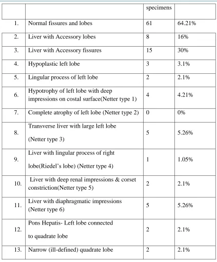
Table 1: Showing the incidence of normal and variant livers (Netter’s classification).
S. No. Morphological features Number
of
184
184
184
184
184
18
4
184
184
184
184
184
184
184
184
184
184
184
184
184
184
184
specimens
1. Normal fissures and lobes 61 64.21%
2. Liver with Accessory lobes 8 16%
3. Liver with Accessory fissures 15 30%
4. Hypoplastic left lobe 3 3.1%
5. Lingular process of left lobe 2 2.1%
6. Hypotrophy of left lobe with deep
impressions on costal surface(Netter type 1) 4 4.21%
7. Complete atrophy of left lobe (Netter type 2) 0 0%
8.
Transverse liver with large left lobe
(Netter type 3)
5 5.26%
9.
Liver with lingular process of right
lobe(Riedel’s lobe) (Netter type 4)
1 1.05%
10. Liver with deep renal impressions & corset
constriction(Netter type 5) 2 2.1%
11. Liver with diaphragmatic impressions
(Netter type 6) 5 5.26%
12.
Pons Hepatis- Left lobe connected
to quadrate lobe
2 2.1%
13. Narrow (ill-defined) quadrate lobe 2 2.1%
Table 2: Showing incidence of accessory fissures and accessory lobes in various lobes of liver.
S. No. Lobe Accessory
fissures
Accessory lobes
1 Right lobe 9 2
2 Left lobe 6 1
3 Caudate lobe 5 4
Pag
e
185
Pag
e
185
Pag
e
185
Pag
e
185
Pag
e
185
Pag
e
185
Pag
e
185
Pag
e
185
Pag
e
185
Pag
e
185
Pag
e
185
Pag
e
185
Pag
e
185
Pag
e
185
Pag
e
185
Pag
e
185
Pag
e
185
Pag
e
185
Pag
e
185
Pag
e
185
Pag
e
185
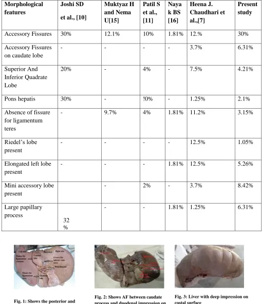
Table- 3: Comparison between present study and other studies showing variations in Morphological features of liver.
Morphological features
Joshi SD
et al., [10]
Muktyaz H and Nema U[15]
Patil S et al., [11]
Naya k BS [16]
Heena J. Chaudhari et al.,[7]
Present study
Accessory Fissures 30% 12.1% 10% 1.81% 12.% 30%
Accessory Fissures on caudate lobe
- - - - 3.7% 6.31%
Superior And Inferior Quadrate Lobe
20% - 4% - 7.5% 4.21%
Pons hepatis 30% - !0% - 1.25% 2.1%
Absence of fissure for ligamentum teres
- 9.7% 4% 1.81% 11.2% 3.15%
Riedel’s lobe present
- - - - 12.5% 1.05%
Elongated left lobe present
- - - 1.81% 12.5% 5.26%
Mini accessory lobe present
- 2% - 3.7% 8.42%
Large papillary process
32 %
- - 1.81% 1.25% 6.31%
Fig. 1: Shows the posterior and inferior surfaces of normal liver
Fig. 2: Shows AF between caudate process and duodenal impression on the inferior surface of right lobe
186
186
186
186
186
186
186
186
186
186
186
186
186
186
186
186
186
186
186
186
186
Fig.4: Accessory lobes are present in both caudate and quadrate lobes in one Specimen
Fig.5: Shows accessory fissures and accessory lobes in caudate lobe of liver
Fig.6: Shows accessory fissure between caudate process and papillary process of caudate lobe
Fig. 7: Shows diaphragmatic impressions on superior surface of right lobe of liver
Fig.8: Shows AF over various areas of right lobe of liver and elongated left lobe (Netter type 3)
Fig. 9: Shows AF over various areas of the left lobe of liver
Fig. 10: Shows hypoplastic left lobe of liver (Netter type 1)
Fig. 11: Shows deep notch on diaphragmatic and superior surface of liver.
Fig. 12: Liver with costal grooves over superior surface
Fig. 13: Shows lingular process of left lobe( Netter type 5 )
Fig. 14: Shows AFs and accessory lobes in caudate Lobe and quadrate lobe of liver
Pag
e
187
Pag
e
187
Pag
e
187
Pag
e
187
Pag
e
187
Pag
e
187
Pag
e
187
Pag
e
187
Pag
e
187
Pag
e
187
Pag
e
187
Pag
e
187
Pag
e
187
Pag
e
187
Pag
e
187
Pag
e
187
Pag
e
187
Pag
e
187
Pag
e
187
Pag
e
187
Pag
e
187
Fig. 16: Shows complete transverse fissure dividing quadrate lobe into superior and inferior lobes.
Fig. 17: Shows triangular quadrate lobe of liver.
lobe of liver
Fig. 19: Shows very narrow quadrate lobe of liver.
Fig. 20: Shows quadrate lobe continuous with left lobe( Pons Hepatis)
Fig. 21: Shows AFs over right, left, caudate, and quadrate lobes of liver
Total 95 embalmed human livers were studied among which 61 livers (64.21%) were normal in their external appearance,). Out of the remaining 34 specimens, 26 (27.36%) specimens had accessory fissures on the left lobe, right lobe, caudate lobe, and quadrate lobe, which resulted in the formation of accessory lobes shown in table -1. 9 (9.47%) specimens had accessory fissures on the different lobes, which resulted in the formation of accessory lobes [Table-2/ Fig-2,3,4,5,6,8,9,13,14]. A complete transverse fissure dividing quadrate lobe into a superior and an inferior lobe were seen in 4(4.21%) specimens [Table-3/Fig-16].
Variable size and shape of pons hepatis joining the left lobe with quadrate lobe was seen in 2(2.1%) specimen [Table -3/Fig-20].Absence of fissure for ligamentum teres was seen in 3(3.1%) specimens [Table -3/Fig-20] .The Riedel’s lobe was present in 1(1.05%) specimen [Table -3/Fig-15].
Elongated left lobe present in 5(5.26%) specimen [Table -1/Fig-8] while a mini accessory lobe above quadrate lobe was seen in 3(3.7%) specimens [Table -3/Fig-21]. Large papillary process was present in 6(6.31%) specimen [Table -3/Fig-6, 14].
Type 1 (Very small left lobe, deep costal impressions) was present in 4 (4.21 %) specimens [Table -1/Fig-3, 10].Type 2 (Complete atrophy of left lobe) were not found in any of the livers.
Type 3 (Transverse saddle like liver, relatively large left lobe) was present in 5 (5.26 %) specimens [Table -1/Fig-8]. Type 4 (Tongue like process of right lobe) was present in 1 (1.05 %) specimen [Table -1/Fig-15].
Type 5 (Very deep renal impression and corset constriction) was present in 2 (2.1%) specimen [Table -1/Fig-13]. Type 6 (Diaphragmatic grooves) was present in 5 (5.26 %) specimens [Table -1/Fig-11, 12].
In 9 specimens, accessory fissures were seen in different areas of the right lobe (Table 2). Out of these 2 specimens showed accessory fissures between caudate process and duodenal impression (Fig. 2).
In 6 specimens, accessory fissures were noted over various areas of left lobe of liver (Fig.5, 6) (Table 2).
Out of 34, 5 specimens showed accessory fissures and accessory lobes in the caudate lobe (Table 2) (Fig. 4). In 1 specimen the fissure was found to be between the caudate process and papillary process (Fig. 5).
Out of 6 specimens with accessory fissures in quadrate lobe, 1 specimen shows a complete transverse fissure dividing into a superior and an inferior lobe (Fig. 16). Quadrate lobe varies in shape from triangular (Fig. 17) to irregular (Fig. 18) and it also varies in size from very narrow (Fig. 19) to ill-defined and also continuous with left lobe due to the presence of an incomplete fissure for ligamentum teres (Fig. 20).In 1 specimen accessory fissures are present over the right, left, caudate, and the quadrate lobes (Fig. 21). Accessory lobes are present in both the caudate and quadrate lobes in one specimen (Fig. 14).
Discussion
188
188
188
188
188
188
188
188
188
188
188
188
188
188
188
188
188
188
188
188
188
lobe is very important surgically and radiologically as it might be mistaken as lymph node, and be accidentally removed during the surgery or while dissection around the porta hepatis[4].
Of all the digestive organs, the liver is the one which starts its organogenesis early during 3rd week of intrauterine life and develops most rapidly [5]. Gross abnormalities of the liver are rare in spite of its complex development.The more common gross abnormalities are irregularities in form and less common abnormality is the occurrence of one or more accessory livers or lobes [3].
We compare our study to various authors which is shown in table -3. Bradley [6] has done much to elucidate the development of liver.
Heena J. Chaudhari et al. [7]study 80 liver in which 14 livers (17.5%) were normal in their external appearance ,66(82.5%) specimens showed anomalies in fissures, lobes, shape and size of lobe, six types of variations according to Netter’s classification shown in table -3. According to Auh YH et al.[8], the accessory hepatic fissures are potential sources of diagnostic errors during imaging.
According to Torzilli G et al.[9] used intraoperative ultrasonography during liver surgery and detected location and extent of the lesions to reduce more
tissue dissections and iatrogenic trauma.So
knowledge of variations in liver is important during radiological investigation and surgery [7].
Joshi SD et al [10]., studied 90 specimens of liver and he observed that 30% of liver had variable diameter of pons hepatis, joining quadrate lobe and left lobe, which bridges the fissure for ligamentum teres, so normal appearance of the fissure would not be possible as well as diameter of both right and left lobe of liver may be mistaken [13]. Patil S et al., also observed similar findings in 10% specimen out of 50 liver specimens [11].
Prominent vertical groove on anterosuperior surface were found in 6% of the liver by Joshi SD et al., [10].A higher incidence of these grooves was observed by Macchi et al., [12].
Joshi et a.[10]lreported13 notching along inferior
border of caudate lobe in 18% of livers, vertical fissure in 30% and prominent papillary process in
32% of livers in their extensive study on lobes and fissures.
Absence of fissure for ligamentum teres was present in 11.2% specimens and in 9.7% cases out of 41 livers by Muktyaz H and Nema U [15].Nayak BS found prominent papillary process and long caudate process in 1.25% specimen which is almost similar to our study. [16]
The variations in the anatomy of human liver have been classified as congenital or acquired [7].The congenital anomalies of liver can be divided into anomalies due to defective [9]liver can lead to gastric volvulus, whereas defective development of right lobe may remain latent or progress to portal hypertension . The excessive development of liver results in the formation of accessory lobes of liver which may carry the risk of torsion [13].
Sonographically multiple accessory fissures also may be confused at first look with a macronodular liver, so a supplementary CT scan is necessary. Multiple hepatic fissures and lobes were more common on the under surface of liver, opposite to the quadrate lobe, and in the left lobe [14]. In the present study, 30% of livers are with accessory fissures over their various parts especially on the undersurface in accordance with Cullen [14].
In the present study 6 specimens have accessory fissures in quadrate lobe alone, whereas in 1 specimen, accessory fissures are present both in caudate and quadrate lobes (Fig. 16) and in another liver all the lobes, that is, right, left, caudate, and quadrate lobes have accessory lobe abnormalities of the liver can cause diagnostic difficulties and confusions during procedures like biopsy.
The embryological basis of anomalies of liver morphology occurs in the course of organogenesis. The Defective development of left lobe of liver can
lead to gastric volvulus whereas defective
development of right lobe may progress to portal hypertension.
Conclusion
Pag
e
189
Pag
e
189
Pag
e
189
Pag
e
189
Pag
e
189
Pag
e
189
Pag
e
189
Pag
e
189
Pag
e
189
Pag
e
189
Pag
e
189
Pag
e
189
Pag
e
189
Pag
e
189
Pag
e
189
Pag
e
189
Pag
e
189
Pag
e
189
Pag
e
189
Pag
e
189
Pag
e
189
for management of hepatic diseases and correct
interpretation of radiographs which prevents
misdiagnosis of cystic lesions or any macroscopic pathological lesions of the liver.
Awareness helps to avoid fatal or serious complications and assist in planning appropriate surgical approaches.
Our study would certainly throw light on the importance of such variant appearance and enlighten the morphologists, clinicians, pathologist,surgeons and embryologists to update the knowledge of
morphological variations.
References
1. C. Rouiller, Ed., the Liver: Morphology,
Bio-Chemistry, Physiology, vol. 2, Academic Press, New York, NY, USA, 1964.
2. Standring S. Gray’s Anatomy: The
anatomical basis of clinical practice 40th ed.
New York: Churchill Livingstone 2008; 3441-46.
3. F.H. Netter, Atlas de Anatomia Humana,
Artmed, Porto Alegre, Brazil, 2000.
4. Daver GB, Bakhshi GD, Patil A, Ellur S, Jain
M, Daver NG. Bifid liver in a patient with diaphragmatic hernia. Indian J Gastroenterol. 2005; 24(1):27-28.
5. Meirelles GSP, G. D’Ippolito. Liver pseudo
lesions in helical CT: pictorial essay. [5] Radiol Bras. 2003; 36:229-35.
6. O. C. Bradley, “A contribution to the
morphology and development f the
mammalian liver,” Journal of Anatomy and Physiology, vol. 43, pp. 1–42, 1908.
7. Heena J. Chaudhari et al., Morphological
Study of Human Liver and Its Surgical
Importance, Journal of Clinical and
Diagnostic Research. 2017 Jun, Vol-11(6): AC09-AC12
8. Y. H. Auh, W. A. Rubenstein, and K.
Zirinsky, “Accessory fissures of the liver: CT and sonographic appearance,” American Journal of Roentgenology, vol. 143, no. 3, pp. 565–572, 1984.
9. Torzilli G, Procopio F, Palmisano A,
Donadon M, Del Fabbro D, Marconi M, et al. Total or partial anatomical resection of segment 8 using the ultrasound guided -finger compression technique. HPB (oxford).2011; 13(8):586-91.
10.Joshi SD, Joshi SS, Athavale SA. Some
interesting observations on the surface features of the liver and their clinical
implications, Singapore Med J. 2009;
50(7):715-19.
11.Patil S, Sethi M, Kakar S. Morphological
study of human liver and its surgical [9] importance. Int J Anat Res. 2014; 2(2):310-14.
12.Macchi V, Feltrin G, Parenti A, De Caro R.
Diaphragmatic sulci and portal fissures. [10] J Anat. 2003; 202(3):303-08.
13.G. B. Daver, G. D. Bakhshi, A. Patil, S. Ellur,
M. Jain, and N. G. Daver, “Bifid liver in a patient with diaphragmatic hernia,” Indian Journal ofGastroenterology, vol. 24,no. 1, pp. 27–28, 2005.
14.T. S. Cullen, “Accessory lobes of the liver,”
Archives of Surgery, vol. 11, pp. 718–764, 1925.
15.Muktyaz H, Nema U.Morphological
variations of liver lobes and its clinical significance in north Indian population. GJMMS.2013;1(1):1-5.
16.Nayak BS. A study on the anomalies of liver