International Journal of Medical Science and Current Research (IJMSCR)
Available online at: www.ijmscr.com
Volume2, Issue 2,Page No: 49-56
March-April 2019
49
Medicine ID-101739732
Histopathological Spectrum of Endometrial Lesions in Patients Presenting With
Abnormal Uterine Bleeding
1Dr.Majid Ahmad Khan, 2Dr.Subuh Parvez Khan, 3Dr.Mudasir Ahmad Khan, 4Dr.Mukhtar Ahmad Khan, 5Prof. Abdul Rasheed
1Department of Pathology, Government Medical College,Anantnag, 2
Department of Haematopathology, Sher e Kashmir Institute of Medical Sciences,Srinagar
3Department of Anatomy, Government Medical College, Srinagar 4Department of ENT, Government Medical College, Rajouri ,
5Department of Pathology, SKIMS Medical College,
J&K, India
*Corresponding Author:
Dr Subuh Parvez Khan
Senior Resident,Department of Haematopathology, Sher e Kashmir Institute of Medical Sciences,Srinagar,J&K,India
Type of Publication: Original Research Paper Conflicts of Interest: Nil
ABSTRACT
INTRODUCTION: Abnormal uterine bleeding (AUB) is a very common gynaecological condition affecting all age group.AUB is defined as changes in frequency of menses, duration of flow or amount of blood flow. The causes of abnormal uterine bleeding include a wide spectrum of diseases of the reproductive system and non-gynecological causes as well. Dysfunctional Uterine Bleeding is a diagnosis of exclusion when there is no underlying medical pathology. Dilation and curettage is a diagnostic as well as therapeutic procedurefor the AUB workup.
AIMS AND OBJECTIVES:
1. To Study the histopathological pattern of Endometrium in patients presenting with abnormal uterine bleeding.
2. To compare the histopathological pattern of Endometrium in patients presenting with abnormal uterine bleeding in various age groups and to ascertain the underlying pathology responsible for such abnormal bleeding.
MATERIAL AND METHOD:The study was conducted in the Department of Pathology at a tertiary care centre of Kashmir valley .It was carried over a period of 1 and ½ years from June 2015 to November 2016.The Study Material included specimens consisting of Endometrial Samples (Endometrial curettage and biopsy) and hysterectomy Specimens. Patients with isolated endometrial causes of abnormal uterine bleeding like Endometrial Hyperplasia, Endometrial Polyp, Chronic Endometritis and Endometrial Carcinoma were included in this study. Patients with Leiomyomas, Cervical Pathology, Vaginal Pathology, Hemostatic Disorders, were excluded. RESULTS: A total of 150 cases were studied during a period of 1 and ½ year. Maximum incidence of AUB was observed in reproductive age group, followed by perimenopausal and post- menopausal age group. The most common symptom was menorrhagia (55.64%). Out of 150 cases, 144 cases (96%) were benign whereas 6 cases (4%) were malignant .Proliferative Endometrium (40/150; 27%) was more common in reproductive age where as hyperplasia (57/150; 38%) was more common in perimenopausal and post-menopausal women. In endometrial hyperplasia, maximum number of cases (46 cases,31%) showed non atypical endometrial hyperplasia and 11 cases (7%) showed atypical Endometrial hyperplasia. All cases of endometrial carcinoma were noticed in age group of more than 50 years.
CONCLUSION:The histopathological study of endometrium in patients presenting with Abnormal Uterine Bleeding plays an important role in diagnosing various histopathological patterns, aetiopathological factors and management of abnormal uterine bleeding. In our Study, functional causes of Abnormal Uterine Bleeding (like proliferative and secretory endometrium) were much more common in reproductive age group whereas in perimenopausal and post-menopausal age group, organic lesions (like endometrial hyperplasia, endometrial carcinoma) were responsible for Abnormal Uterine Bleeding. Thus endometrial curettage is recommended in women of perimenopausal and post-menopausal age group presenting with Abnormal Uterine Bleeding to rule out pre-neoplastic conditions (like atypical endometrial hyperplasia) and malignancy which has excellent prognosis if detected early.
Pag
e
50
Pag
e
50
Pag
e
50
Pag
e
50
Pag
e
50
Pag
e
50
Pag
e
50
Pag
e
50
Pag
e
50
Pag
e
50
Pag
e
50
Pag
e
50
Pag
e
50
Pag
e
50
Pag
e
50
Pag
e
50
Pag
e
50
Pag
e
50
Pag
e
50
Pag
e
50
Pag
e
50
INTRODUCTION
Abnormal uterine bleeding (AUB) is a very common gynaecological condition affecting all age groups [1]. AUB is defined as changes in frequency of menses, duration of flow or amount of blood flow . Dysfunctional Uterine Bleeding is diagnosis of exclusion when there is no underlying medical pathology [2]. The causes of abnormal uterine bleeding include a wide spectrum of diseases of the reproductive system and non-gynecologic causes as well. Organic cause of abnormal uterine bleeding may be subdivided into reproductive tract disease, iatrogenic causes and systemic disease. In about 25% of the patients, the abnormal uterine bleeding is the result of a well defined organic abnormality [3]
. Dilation and curettage is a diagnostic as well as therapeutic procedure [4] for the AUB workup. The sensitivity of endometrial biopsy for the detection of endometrial abnormalities has been reported to be as high as 96% [5, 6].
AIMS AND OBJECTIVES:
3. To Study the histopathological pattern of Endometrium in patients presenting with abnormal uterine bleeding.
4. To compare the histopathological pattern of Endometrium in patients presenting with abnormal uterine bleeding in various age groups and to ascertain the underlying pathology responsible for such abnormal bleeding.
MATERIAL AND METHOD:
The study was conducted in the Department of Pathology at a tertiary care centre of Kashmir valley .It was carried over a period of 1 and ½ years from June 2015 to November 2016.This study was approved by Ethical Committee of the institution.The Study Material included specimens consisting of Endometrial Samples (Endometrial curettage and biopsy) and hysterectomy Specimens. Inclusion criteria included Patients with isolated endometrial causes of abnormal uterine bleeding like Endometrial Hyperplasia, Endometrial Polyp, Chronic Endometritis and Endometrial Carcinoma.Patients with Leiomyomas, Cervical Pathology, Vaginal Pathology, Hemostatic Disorders, were excluded.
The gross morphology was recorded with total submission of endometrial samples and representative tissue was taken from the hysterectomy specimens. The tissue bits were processed and paraffin blocks were prepared. Tissue sections were cut and stained with hematoxylin and eosin stain (H&E).Statistical analysis was done using SPSS version 16.0 software.
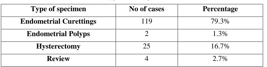
RESULTS: A total of 150 cases of Endometrial
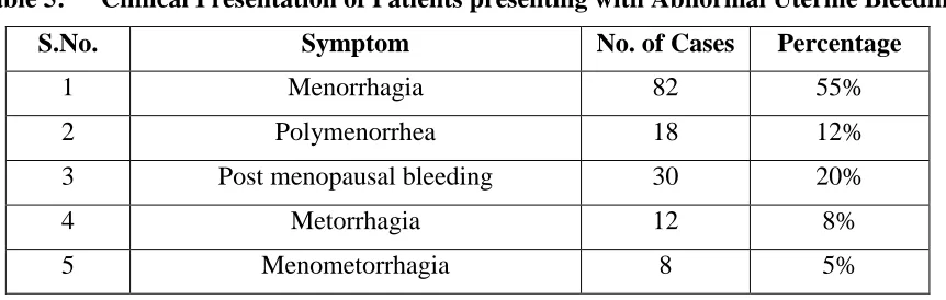
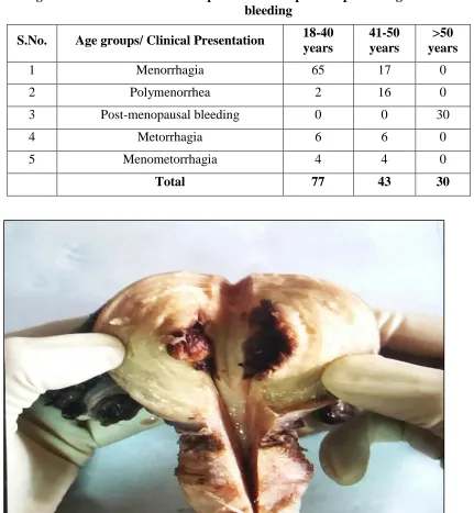
Curettings/specimens were included in the study. Types of specimens /biopsies received are shown in Table 1.Majority of the specimen were benign(96%)(Table 2).The age wise distribution of cases of AUB with histopathological pattern of endometrium is shown in Table 3. The distribution of endometrial lesions according to the histopathology is shown in Table 4. Menorrhagia was the most common clinical presentation and was seen in 82 patients presenting with abnormal uterine bleeding(Table 5) Distribution of Endometrial causes of AUB according to Age Group of Patients is shown in Table 6. Menorrhagia was the most common clinical presentation in 18 – 40 years age group, Polymenorrhea was the most common clinical presentation in 41-50 years age group and Post-menopausal bleeding was the most common clinical presentation in >50 years age group.(Table 7)
DISCUSSION:
Pag
e
51
Pag
e
51
Pag
e
51
Pag
e
51
Pag
e
51
Pag
e
51
Pag
e
51
Pag
e
51
Pag
e
51
Pag
e
51
Pag
e
51
Pag
e
51
Pag
e
51
Pag
e
51
Pag
e
51
Pag
e
51
Pag
e
51
Pag
e
51
Pag
e
51
Pag
e
51
Pag
e
51
their study found 10 (2.48%) cases of endometrial carcinoma mostly seen in postmenopausal women like in our study and 393 cases (98%) were non neoplastic lesions.[9] In our study, in reproductive age group Proliferative endometrium (functional cause) was the most common finding accounting for 39 cases (48%) followed by Secretory endometrium (functional cause) 18 cases (22%) and Endometrial hyperplasia (organic lesion) 17 cases (21%). In perimenopausal age group endometrial hyperplasia was the most common finding accounting for 24 cases (53%) followed by proliferative endometrium 11 cases (24%) and in postmenopausal age group endometrial hyperplasia was the most common finding accounting for 16 cases (70%). Diagnosis of endometrial hyperplasia is important as they are precursors of endometrial carcinoma. The calculated risk of progression of hyperplasia to cancer is 5-10%.
[10]
According to the new WHO classification [11], endometrial hyperplasia is classified into non atypical endometrial hyperplasia and atypical endometrial hyperplasia.Chronic Endometritis was diagnosed in 6% cases. All cases were observed in 35 - 50 years age i.e. peri-menopausal age group. The endometritis was diagnosed on the basis of presence of plasma cells. Chronic endometritis is often a result of intra uterine contraceptive devices (IUCD), pregnancy and incomplete abortions.[12] This pathology needs to be diagnosed and kept in mind while dealing with a case of AUB because with specific treatment, endometrium can be reverted back to normal state. Endometrial polyps (5/150; 3.33%) were seen in reproductive age group and peri-menopausal age group in equal amount. Polyp in reproductive age group was benign functional polyp and in perimenopausal age group was benign hyperplastic polyp. Functional polyp is peculiar to the cyclic nature of endometrium in reproductive age group. There is compelling difference between endometrial polyp and normal endometrium in receptor expression, cell proliferation and apoptosis regulation suggesting that polyp may provide a suitable background for the advancement of malignancy.[13]Khare A et al (2012) in their study, found the following observation. In reproductive age group, proliferative endometrium was the most common finding (26.8%) followed by irregular maturation (25%). Complex hyperplasia was seen in 6 cases, out of which 1 case showed atypia. Nineteen
cases (16.4%) showed associated endometritis. No case of malignancy was observed in this group. In perimenopausal age group, simple hyperplasia was the most frequent finding (29.8%).Complex hyperplasia was seen in 3 cases, out of which 1 revealed atypia. Three cases of malignancy (6.4%) were reported.In postmenopausal age group, most frequent finding was complex hyperplasia seen in 8 cases (33.3%), out of which 2 cases showed atypia. Six cases (25%) of simple hyperplasia and 4 cases (16.7%) of malignancy were reported.[14] Dadhania B
et al (2013) in their study found that the most
common pattern was proliferative endometrium (32/150) followed by secretory endometrium (23/150) and hyperplasia in (40/150). [7] Bolde SA et
al (2014) in their study reported that the most
Pag
e
52
Pag
e
52
Pag
e
52
Pag
e
52
Pag
e
52
Pag
e
52
Pag
e
52
Pag
e
52
Pag
e
52
Pag
e
52
Pag
e
52
Pag
e
52
Pag
e
52
Pag
e
52
Pag
e
52
Pag
e
52
Pag
e
52
Pag
e
52
Pag
e
52
Pag
e
52
Pag
e
52
years. Similar distribution of cases was observed by Abid M et al.[16]Increased prevalence of lesions in higher age group could be due to the fact that endometrium is exposed to estrogen for a longer period of time as compared to patients with younger age group. Khare A et al (2012) in their study found 62% of cases in reproductive age group followed by 25.1% cases in perimenopausal age group and 12.8% of cases in postmenopausal age group.[14] Forae GD
et al (2013) in their study found 89.6% of cases in
reproductive and perimenopausal age groups and 10.4% of cases in postmenopausal age group.[12]Goel
A et al (2016) in their study found 37.5% of cases in
reproductive age group and 36% in perimenopausal age groups.[15]In our study, menorrhagia was the most common clinical presentation and was seen in 82 patients presenting with abnormal uterine bleeding, followed by postmenopausal bleeding in 30 patients.
Goel A et al (2016) in their study of 264 cases found
most common pattern of bleeding was menorrhagia (62.5%) followed by metorrhagia (14%), post-menopausal bleeding (12%), menometorrhagia (6.1%) and polymenorrhea (5.3%).[15] Jetley S et al
(2013) in their study found most common clinical
presentation was represented by menorrhagia (46.4%) followed by metrorrhagia (20%), menometorrhagia and polymenorrhagia.[18] Bolde SA
et al (2014) in their study found most common
clinical presentation was represented by menorrhagia (46.86%).[8] Masood A et al (2014) in their study found most common clinical presentation was represented by menorrhagia (70%) followed by metrorrhagia (15%) and polymenorrhagia (15%). [19]
CONCLUSION:
To conclude, the histopathological study of endometrium in patients presenting with Abnormal Uterine Bleeding plays an important role in diagnosing various histopathological patterns, aetiopathological factors and management of abnormal uterine bleeding. In our Study, functional causes of Abnormal Uterine Bleeding (like proliferative and secretory endometrium) were much more common in reproductive age group whereas in perimenopausal and post-menopausal age group, organic lesions (like endometrial hyperplasia, endometrial carcinoma) were responsible for Abnormal Uterine Bleeding. Thus endometrial curettage is recommended in women of perimenopausal and post-menopausal age group
presenting with Abnormal Uterine Bleeding to rule out pre-neoplastic conditions (like atypical endometrial hyperplasia) and malignancy which has excellent prognosis if detected early.
BIBLIOGRAPHY:
1. Awwad JT, Toth TL, Schiff I. Abnormal uterine bleeding in the perimenopause. International Fertility & Menopausal Studies. 1993; 38(5): 261-269.
2. Speroff L, Fritz MA. Menopause and the perimenopausal transition. In Clinical gynaecological endocrinology and infertility. 7th edition, Jaypee Brothers Med Publishers (P) Ltd. 2005; 621-688.
3. Brenner PF. Differential diagnosis of AUB. Am J ObstetGynecol; 1996; 175:766–769. 4. Rosai J. Rosai and Ackerman’s surgical
pathology. 9th edition, Volume 2, 2004; 1569-1635.
5. Albers JR, Hull SK, Wesley RM. Abnormal uterine bleeding. AmFam Phys. 2004; 69:1915–1926.
6. Litta P, Merlin F, Saccardi C. Role of hysteroscopy with endometrial biopsy to rule out endometrial cancer in post-menopausal women with abnormal uterine bleeding. Maturitas.2005; 50:117–123.
7. Dadhania B, Dhruva G, Agravat A, Pujara K. Histopathological Study of Endometrium in Dysfunctional Uterine Bleeding. Int. J Res Med. 2013; 2(1): 20-24.
8. Bolde SA, Smita S. Pudale, Gopal A. Pandit, Pushkar P. Matkari. Histopathological study of endometrium in cases of abnormal uterine bleeding. Int J Res Med Sci., 2014 2(4):1378-1381.
9. Vaidya S, M Lakhey, S Amatya Vaidya, PK Sharma, S Hirachand, S Lama et al. Histopathological pattern of abnormal uterine bleeding in endometrial biopsies.Nepal Med Coll J. 2013; 15(1): 74-77.
10. Baak JP, Mutter GL. EIN and WHO 94. J Clin Pathol 2005; 58:1-6.
11. Emons G, Beckmann MW, Schmidt D, Mallmann P. New WHO Classification of Endometrial Hyperplasias. Geburtshilfe und Frauenheilkunde. 2015; 75(2):135-136. 12. Jairajpuri ZS, Rana S, Jetley S. Atypical
Pag
e
53
Pag
e
53
Pag
e
53
Pag
e
53
Pag
e
53
Pag
e
53
Pag
e
53
Pag
e
53
Pag
e
53
Pag
e
53
Pag
e
53
Pag
e
53
Pag
e
53
Pag
e
53
Pag
e
53
Pag
e
53
Pag
e
53
Pag
e
53
Pag
e
53
Pag
e
53
Pag
e
53
endometrium - A study of 638 cases. Al Ameen J Med Sci 2013; 6: 21-28.
13. Doraiswami S, Johnson T, Rao S, Rajkumar A, Vijayaraghavan J, Panicker VK. Study of endometrial pathology in abnormal uterine bleeding. Journal of obstetrics and gynaecology of India 2011; 61(4):426–430. 14. Khare A, R. Bansal, S. Sharma, P. Elhence,
N. Makkar, Y. Tyagi. Morphological spectrum of endometrium in patients presenting with Dysfunctional uterine bleeding. People’s Journal of Scientific Researc; 2012; 5: 13-16.
15. Goel A, Shanthi V, Rao NM, Jain P, Byna SS, Conjeevaram J. Spectrum of Endometrial Lesions in Patients with Abnormal Uterine
Bleeding.Annals of Pathology and Laboratory Medicine, 2016; 03(05).
16. Abid M, Hashmi AA, Malik B, Haroon S, Faridi N, Edhi MM et al. Clinical pattern and spectrum of endometrial pathologies in patients with abnormal uterine bleeding. BMC women’s health 2014; 14:132.
17. Forae GD, Aligbe JU. Journal of Basic and Clinical Reproductive Sciences.2013, 2(2). 18. Jetley S, Safia Rana, Zeeba Shamim
Jairajpuri. Journal of Mid-Life Health Year. 2013; 4 (4): 216-220.
19. Masood A , Waris E. Histopathological Patterns of Endometrium in Women with Abnormal Uterine Bleeding in Akhtar Saeed Medical College. Proceeding S.Z.P.G.M.I. 2014; 28(2): 75-79.
Table 1: Types of Samples Received
Type of specimen No of cases Percentage
Endometrial Curettings 119 79.3%
Endometrial Polyps 2 1.3%
Hysterectomy 25 16.7%
Review 4 2.7%
Table 2: Distribution of Endometrial Curettings / Hysterectomy Specimens into Benign & Malignant Lesions
MALIGNANT LESIONS BENIGN LESIONS TOTAL
Pag
e
54
Pag
e
54
Pag
e
54
Pag
e
54
Pag
e
54
Pag
e
54
Pag
e
54
Pag
e
54
Pag
e
54
Pag
e
54
Pag
e
54
Pag
e
54
Pag
e
54
Pag
e
54
Pag
e
54
Pag
e
54
Pag
e
54
Pag
e
54
Pag
e
54
Pag
e
54
Pag
e
54
Table 3: Age Wise Distribution of cases of AUB with histopathological pattern of endometrium
Age groups/ Histopathology of endometrium
‹ 20 years
21-30 years
31-40 years
41-50 years
51-60 years
> 60 years
Proliferative Endometrium 1 9 29 11 0 0
Secretory Endometrium 1 9 8 2 0 0
Hyperplasia 0 3 14 24 6 10
Endometrial Polyp 0 0 2 2 1 0
Chronic Endometritis 0 0 4 5 0 0
Endometrial Carcinoma 0 0 0 0 5 1
Non Diagnostic 0 0 2 1 0 0
Total 2 21 59 45 12 11
Table 4: Distribution of Endometrial causes of Abnormal Uterine Bleeding according To Histopathology
S. No. Histopathological Type No. of
cases Percentage
1 Non atypical endometrial hyperplasia 46 31%
2 Proliferative Endometrium 40 27%
3 Secretory Endometrium 30 20%
4 Atypical Endometrial hyperplasia 11 7%
5 Chronic Endometritis 9 6%
6 Endometrial Polyp 5 3%
7 Endometrial Carcinoma 6 4%
8 Non Diagnostic 3 2%
Table 5: Clinical Presentation of Patients presenting with Abnormal Uterine Bleeding
S.No. Symptom No. of Cases Percentage
1 Menorrhagia 82 55%
2 Polymenorrhea 18 12%
3 Post menopausal bleeding 30 20%
4 Metorrhagia 12 8%
Pag
e
55
Pag
e
55
Pag
e
55
Pag
e
55
Pag
e
55
Pag
e
55
Pag
e
55
Pag
e
55
Pag
e
55
Pag
e
55
Pag
e
55
Pag
e
55
Pag
e
55
Pag
e
55
Pag
e
55
Pag
e
55
Pag
e
55
Pag
e
55
Pag
e
55
Pag
e
55
Pag
e
55
Table 6: Distribution of Endometrial causes of AUB according to Age Group of Patients
S. No. Age group (years) No. of cases Percentage
1 18 – 40 years (reproductive) 60 40%
2 41 – 50 years (perimenopausal) 59 39%
3 >50 years (postmenopausal) 31 21%
Total 150
Table 7: The age wise distribution of clinical presentation of patients presenting with abnormal uterine bleeding
S.No. Age groups/ Clinical Presentation 18-40
years
41-50 years
>50 years
1 Menorrhagia 65 17 0
2 Polymenorrhea 2 16 0
3 Post-menopausal bleeding 0 0 30
4 Metorrhagia 6 6 0
5 Menometorrhagia 4 4 0
Total 77 43 30
Fig. 1: Gross photograph of hysterectomy specimen showing endometrial polyp and increased endometrial
Pag
e
56
Pag
e
56
Pag
e
56
Pag
e
56
Pag
e
56
Pag
e
56
Pag
e
56
Pag
e
56
Pag
e
56
Pag
e
56
Pag
e
56
Pag
e
56
Pag
e
56
Pag
e
56
Pag
e
56
Pag
e
56
Pag
e
56
Pag
e
56
Pag
e
56
Pag
e
56
Pag
e
56
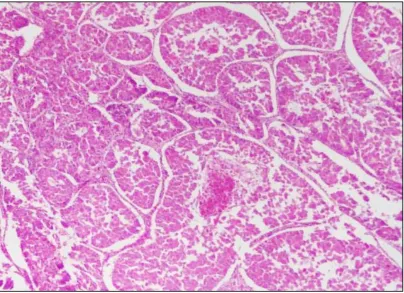
Fig. 2: Photomicrograph showing secretory endometrium. (40X, H&E)