* To whom all correspondence should be addressed.
Feature Based Information Fusion Through Gabor Wavelet
Transformation and Independent Component Analysis
K. Kumar
Department of Electronics and Instrumentation, Sathyabama University, Chennai, India. DOI: http://dx.doi.org/10.13005/bbra/1713
(Received: 01 January 2015; accepted: 09 February 2015)
In this paper a method is proposed for multichannel image registration which combines the information from different methods to form a joint registration. This method uses feature based information fusion through Gabor wavelet transformation and Independent component analysis technique. This technique is more robust and accurate. The medical images are analyzed and registered to find the various diseases like brain tumour and hemorrhage.
Key words: Gabor filter, Independent component analysis, multichannel image registration.
In medical applications, one is concerned with processing of chest X-rays, projection images of tomography and other medical images that occur in radiology and nuclear magnetic resonance (NMR) These images can be used for patient testing and monitoring or for detection of tumors’ or other disease in patients.In MRI, different protocols (T1,T2,FLAIR,mPrage and diffusion tensor imaging (DTI), etc can also be viewed as different methods.Every one of these methods uses some distinctive and often complementary characterization of the underlying anatomy and tissue microstructure. The main goal of our project is nonrigid inter-subject multichannel image registration method which combines information from different modalities/channels to produce a
unified joint registration. Multichannel images are produced using co-registered multimodality images of the same subject to utilize information across modalities exensively. Comparing to the current methods which combine the information at the image/intensity level.
The suggested method uses feature-level information fusion method to spatio-adaptively combine the complementary information from different modalities that characterize different tissue types, through Gabor wavelets transformation and Independent Component Analysis (ICA), to produce a robust inter-subject registration.
the areas where the driving modality does not characterize the underlying tissue type well. The proposed multichannel image registration method can integrate and enhance complementary information while eliminating the less reliable/ redundant information from different channels and leads to a more accurate and robust inter-subject registration at low computational cost. Image registration is the process of overlaying two or more images of the same scene taken at various points and times and by various sensors. Image registration is a important step in all image analysis tasks in which the final information is gained from the combination of various data sources. Image registration or image alignment algorithms can be classified into intensity-based and feature-based. One of the images is referred to as the reference or source and the second image is referred to as the target or sensed. Image registration involves the target image to align with the refererred image.
Methods
Module descriptions Module 1
For various method, we use Gabor wavelets, as Gabor wavelet transformation has been shown to be optimal in the sense of minimizing the joint uncertainty in space and frequency, and has been widely used for feature extraction, and hence, more appropriate for the purpose of matching/registration.
Module 2
To incorporate all the relevant information regarding each particular tissue type from different modalities, and thus facilitate the subsequent “Choose-Max” information fusion scheme, we apply the independent component analysis (ICA) on the Gabor features extracted. ICA has been successfully applied in MRI enhancement, functional MRI (fMRI) analysis and blind source separation.
Module 3
After the ICA step, each IC of Gabor features is “specialized” in depicting one particular tissue type. Therefore, by using the Choose-Max scheme on every voxel, we can select the optimal Gabor features from the corresponding IC to characterize the underlying structure.
Module 4
For every voxel, depending on the optimal IC obtained through ICA and
“Choose-Max” scheme, divergence metric is used to find the correspondence between two multichannel images.
Module 5
Based on the divergence metric, the image similiarity problem is defined as a cost function. By optimizing it, the deformation field of the registration is obtained.
Module 6
§ Image Acquisition and Preprocessing § Registration of Simulated Images § Registration of Real Images § Computational Efficiency
Feature Extraction
Image modalities have become available for clinical/research studies, for instance, X-ray computed tomography (CT), positron emission tomography (PET) and magnetic resonance imaging (MRI). In MRI, different protocols (T1, T2, FLAIR, mPRAGE and diffusion tensor imaging DTI), etc.) can also be viewed as different methods. Every one of these methods provides some distinctive and often complementary characterization of the underlying anatomy and tissue microstructure
Multichannel inter-subject registration is rendered challenging because there could be competing information from different methods.In image processing, a Gabor filter, is a linear filter used for edge detection. In the spatial domain, a 2D Gabor filter is a Gaussian kernel function modulated by a sinusoidal plane wave. The Gabor filters are self-similar: all filters can be generated from one mother wavelet by dilation and rotation. Gabor filters aredirectly related to Gabor wavelets, since they can be designed for a number of dilations and rotations. The filters are convolved with the signal, resulting in a so-called Gabor space. Among various wavelet bases, Gabor functions provide the optimal resolution in both the time (spatial) and frequency domains. Although the registration is for 3D images, to improve the computational cost, we use 3 perpendicular (axial, coronal, and sagittal) 2D Gabor filter banks to extract the features. A 2D Gabor filter can be viewed as a sinusoidal plane of particular frequency and orientation, modulated by a Gaussian envelope: G(x,y) = s(x,y) g(x,y)
s(x,y) = exp .
g(x,y) = exp
and characterize the spatial extent and bandwidth of along the respective axes, and are the shifting frequency parameters in the frequency domain. Using as the mother wavelet, a class of self-similar functions can be obtained by appropriate dilations and rotations of through:= where (xsin , a>1, indicates the number of orientations, S the number of scales in the multi resolution decomposition and a is the scaling factor . These parameters can be set according to reduce the redundant information (caused by the Non orthogonality of the Gabor wavelets) in the filtered images. Given an image I, the Gabor transform with orientation n and m scale can be computed as
Where indicates the complex conjugate. In our work, we set the Gabor filter to have S=4 scale levels and O=6 orientations. Gives the examples of the extracted Gabor features using the designed filter bank on T1, PD, T2 and FA images, respectively. As we can see, on different locations, scales and orientations, we need Gabor features from different modalities to best delineate the underlying structure.
=
Gabor features are obtained by convolve the input image and mother wavelet using below equation
Independent Components of the Extracted Gabor Features
To incorporate all the relevant information regarding each particular tissue type from different modalities, and thus facilitate the subsequent “Choose-Max” information fusion scheme, we apply the independent component analysis (ICA) on the Gabor features extracted. ICA has been successfully applied in MRI enhancement functional MRI (fMRI) analysis and blind source separation. The basic theory of applying ICA in multichannel image analysis can be briefly described as follows.Let X=denote the multichannel image set generated by using different imaging modalities or scanning parameters.
n is the number of modalities/channels Let X is the signal we observed and it can be considered as the linear mixture of many independent sources, for instance water, blood, fat, GM, WM, CSF, and muscle, etc. For applying ICA for the analysis of multichannel images, there are two steps: training and decomposition. In the first step, a number of voxels are randomly selected from a few sample multichannel images. After the above optimization, when the algorithm converges and the error is below a specified threshold, the obtained decomposition matrix W can be applied
Block Diagram:
Input images
Tissue-Pertinent
Information Fusion Feature
Extraction
Dis-Similarity Metric
Cost Function and Optimization
Registration
Fig. 1. Input Images a) T1 image b) T2 image c) Flair image d) PD image
(a) (b) (c) (d)
decomposition, each independent component image is “specialized” for capturing one tissue type and contains all the relevant information (taken from all the modalities) pertaining to that tissue type. Because of this “purity,” the decomposed independent component image provides higher contrast and better characterization than the original image. It is worthwhile to clarify that although each tissue type will be highlighted in one IC.
In this work, instead of applying ICA to the multichannel image, ICA is applied to the
multivariate Gabor features, since our goal is to separate the information in Gabor feature space according to different tissue types. ICA is performed on all the Gabor features obtained from different methods. We have proposed a general framework for multichannel image registration. Here we demonstrate its applicability on the combination of a T1 and DTI image as an example, as these images are routinely acquired in a clinical study. Specifically, we use a multichannel image created by combining T1 and five DTI-derived scalar images to illustrate and test the proposed algorithm.
Choose Max fusion using feature based method
The aim of the study was to address registration of images acquired from the same sensor under different conditions Image similarity-based methods are broadly used in medical imaging. A basic image similarity-based method consists of a transformation model which is applied to reference image coordinates to locate their corresponding coordinates in the target image space, an image similarity metric, which quantifies
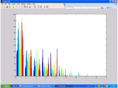
Fig. 5. histogram of the registered image
Table 1. Summarization Of The Experimental Results On The Real Images(t1,T2,Pd,Fa) After Ica
Images Max Mean Standarddeviation
T1 0.9373 0.1920 0.2094
After Ica 7.9799 2.3716 1.4864
Pd 0.5686 0.1557 0.1692
After Ica 4.4333 1.8463 1.1418
T2 0.7412 0.1408 0.1447
After Ica 7.1313 1.5746 1.0004
Fa 0.9961 0.1960 0.2210
After Ica 10.4132 2.2409 1.8511
Table 2. Summarization Of The Final Experimental Results On The Real Images(t1,T2,Pd,Fa)
Images Max Mean Std Deviation
T1 .9373 0.1920 0.2094
Pd .5686 0.1557 0.1692
T2 0.7412 0.1408 0.1447
Fa .9961 0.1960 0.2210
Fused image .9204 1.8866 1.3755 Registered 8.4030 1.4783 0.3451 Image
Fig. 3. Fused Image
the degree of correspondence between features in both image spaces achieved by a given method to maximize image similarity by changing the transformation parameters. The choice of an image similarity measure depends on the nature of the images to be stored. Some of the measures include Cross Correlation, Mutual Information, Mean-square difference and Ratio Image Uniformity. Mutual Information and its variant, Normalized Mutual Information, are the most popular image similarity measures for registration of multimodality images. Cross-correlation, Mean-square difference and Ratio Image Uniformity are commonly used for registration of images of the same modality.
Mutual information is an information theory measure of the statistical dependence between two random variables or the amount of information that one variable contains about the other. It can be qualitatively considered as a measure of how well one image explains the other. After the ICA step, each IC of Gabor features is “specialized” in depicting one particular tissue type. Therefore, by using the Choose-Max scheme on every voxel, we can select the optimal Gabor features from the corresponding IC to characterize the underlying structure. As the tissue type could be different for different voxels, different independent components (IC) are needed to acquire the best characterization. Therefore, “Choose-Max” scheme is adopted to select the optimal IC according to the underlying tissue type. In this method, the information is kept and enhanced while less reliable/redundancy is reduced, via information fusion. Image similarity-based methods are broadly used in medical imaging. A basic image similarity-based method consists of a transformation model which is applied to reference image coordinates to locate their corresponding coordinates in the target image space, an image similarity metric, which quantifies the degree of correspondence between features in both image spaces achieved by a given transformation, and an optimization algorithm.
RESULTS
The choice of an image similarity measure depends on the nature of the images to be saved. General examples of image comparison methods include Cross Correlation, Mutual Information,
Mean-square difference and Ratio Image Uniformity. Mutual Information and its variant, Normalized Mutual Information, are the procedures for registration of multimodality images. Cross-correlation, Mean-square difference and Ratio Image Uniformity are commonly used for registration of images of the same modality.For each voxel, based on the optimal IC obtained through ICA and “Choose-Max” scheme, dissimilarity metric is defined to find the correspondence between two multichannel images.
Image Registration Methodology
Image registration, is commonly used in remote sensing, biomedical screening etc. In common its uses can be divided into four main groups according to the manner of the image acquisition: Different viewpoints (multi view analysis). Images of the same scene are obtained from various viewpoints. The objective is to gain larger a 2D view or a 3D representation of the scanned image.
CONCLUSION
A multichannel inter-subject image registration framework that combines information from different modalities based on feature-level information fusion. The registration produces spatially normalized images of all the modalities acquired in the study. Thus the statistical analysis done on these jointly spatially normalized images is more comparable, as a unified registration scheme has been used to register them.
we also plan to apply this method on clinical studies for joint comparative statistics on T1 and DTI.
REFERENCES
1. M. Jenkinson, P. Bannister, J. Brady, and S. Smith, “Improved optimization for the robust and accurate linear registration and motion correction of brain images,” NeuroImage, 2002; 17(2): 825–841.
2. H.-J. Park, M. Kubicki, and M. Shenton, “Spatial normalization of diffusion tensor MRI using multiple channels,” Neuroimage, 2003; 20, pp. 1995–2009.
3. J. P. Thirion, “Non-rigid matching using demons,” in Proc. IEEE Conf. Comput. Vis. Pattern Recognit. (CVPR), 1996, pp. 245–251. 4. B. Avants, J. Duda, H. Zhang, and J. Gee, “Multivariate normalization with symmetric diffeomorphisms for multivariate studies,” in Med. Image Computing Computer-Assisted Intervent. (MICCAI), 2007, pp . 359–366. 5. J. Pluim, J. Maintz, and M. Viergever, “Mutual
information based registration of medical images: A survey,” IEEE Trans. Med. Imag., 2003; 22(8), 986–1004.
6. G. Tourassi, E. Frederick, M. Markey, and C. Floyd, “Application of the mutual information criterion for feature selection in computer-aided diagnosis,” Med. Phys., 2001; 28(12).
7. A. Andronache, M. von Siebenthal, G. Székely, and P. Cattin, “Nonrigid registration of multi-modal images using both mutual information and cross-correlation,” Med. Image Anal., 2008; 12(1), pp. 3–15.
8. C. Studholme, “Incorporating DTI data as a constraint in deformation tensor morphometry between T1 MR images,” in Inf. Process. Med. Imag. (IPMI), 2008, pp. 223–232.
9. P. Lorenzen, M. Prastawa, B. Davis, G. Gerig, E. Bullitt, and S. Joshi, “Multi-modal image set registration and atlas formation,” Med. Image Anal., 2006; 10, pp. 440–451.
10. B. Alfano, M. Ciampi, and G. De Pietro, “A wavelet-based algorithm for multimodal medical image fusion,” in Semantic Multimedia, 2007, pp. 117–120.
11. Z. Zhang and R. Blum, “A categorization of multiscale-decomposition-based image fusionschemes with a performance study for a digital camera application,” Proc. IEEE, 1999; 87(8), pp. 1315–1326.
12. P. J. Bassera and C. Pierpaoli, “Microstructural and physiological features of tissues elucidated
by quantitative-diffusion-tensor MRI,” J. Magn. Reson., 1996; 111(3); 209–219.
13. J. Daugman, “Complete discrete 2D Gabor transforms by neural networks for image analysis and compression,” IEEE Trans. Acoustics, 1988; 36(7); 1169–1179.
14. W. Ma and B. Manjunathi, “Texture features for browsing and retrieval of image data,” IEEE Trans. Pattern Anal. Mach. Intell., 1996; 18(8); 837–842.
15. A. Hyvarinen, J. Karhunen, and E. Oja, Independent Component Analysis. 2001; New York: Wiley.
16. S. Muraki, T. Nakai, Y. Kita, and K. Tsuda, “An attempt for coloring multichannel MR imaging data,” IEEE Trans. Vis. Comput. Graph., vol. 7, no. 3, pp. 265–273, 2001.
17. T. Nakai, S. Muraki, and E. Bagarinao, “Application of independent component analysis to magnetic resonance imaging for enhancing the contrast of gray and white matter,” NeuroImage, vol. 21, no. 1, pp. 251–260, 2004. 18. M. McKeown et al., “Analysis of fMRI data by blind separation into independent spatial components,” Human Brain Mapp., vol. 6, no. 3, pp. 160–188, 1998.
19. S. Makino, T.-W. Lee, and H. Sawada, Blind Speech Separation. New York: Springer, 2007. 20. J. Friedman and J. Tukey, “A projection pursuit
algorithm for exploratory data analysis,” IEEE Trans. Computers, vol. 23, no. 9, pp. 881–890, Sep. 1974.21. A. Hyvarinen, “Fast and robust fixed point algorithms for independent component analysis,” IEEE Trans. Neural Networks, vol. 10, no. 3, pp. 626–634, 1999. 22. C.-F. Westin, S. E. Maier, H. Mamata, A.
Nabavi, F. A. Jolesz, and R. Kikinis, “Processing and visualization of diffusion tensor MRI,” Med. Image Anal., vol. 6, no. 2, pp. 93–108, 2002. 23. D. Shen, “Image registration by local histogram
matching,” Pattern Recognit., vol. 40, pp. 1161– 1171, 2007.
24. A. Joshi, D. Shattuck, P. Thompson, and R. Leahy, “Surface-constrained volumetric brain registration using harmonic mappings,” IEEE Trans. Med. Imag., vol. 26, no. 12, pp. 1657– 1669, Dec. 2007.
25. G. Postelnicu, L. Zollei, and B. Fischl, “Combined volumetric and surface registration,” IEEE Trans. Med. Imag., vol. 28, no. 4, pp. 508–522, Apr. 2009.
27. D. Xu, S. Mori, D. Shen, P. C. M. V. Zijl, and C. Davatzikos, “Spatial normalization of diffusion tensor fields,” Magn. Reson. Med., vol. 50, no. 1, pp. 75–182, 2003.
28. Z. Xue, D. Shen, B. Karacali, J. Stern, D. Rottenberg, and C. Davatzikos, “Simulating deformations of MR brain images for validation of atlas-based segmentation and registration algorithms,” NeuroImage, vol. 33, no. 3, pp. 855–
866, 2006.
29. V. Arsigny, P. Fillard, X. Pennec, and N. Ayache, “Log-euclidean metrics for fast and simple calculus on diffusion tensors,” Magn. Reson. Med., vol. 56, pp. 411–421, 2006.