R E S E A R C H A R T I C L E
Open Access
The effect of a manual instrumentation technique
on five types of premolar root canal geometry
assessed by microcomputed tomography and
three-dimensional reconstruction
Ke-Zeng Li
1, Yuan Gao
1, Ru Zhang
1, Tao Hu
1*and Bin Guo
2*Abstract
Background:Together with diagnosis and treatment planning, a good knowledge of the root canal system and its frequent variations is a necessity for successful root canal therapy. The selection of instrumentation techniques for variants in internal anatomy of teeth has significant effects on the shaping ability and cleaning effectiveness. The aim of this study was to reveal the differences made by including variations in the internal anatomy of premolars into the study protocol for investigation of a single instrumentation technique (hand ProTaper instruments) assessed by microcomputed tomography and three-dimensional reconstruction.
Methods:Five single-root premolars, whose root canal systems were classified into one of five types, were scanned with micro-CT before and after preparation with a hand ProTaper instrument. Instrumentation
characteristics were measured quantitatively in 3-D using a customized application framework based on MeVisLab. Numeric values were obtained for canal surface area, volume, volume changes, percentage of untouched surface, dentin wall thickness, and the thickness of dentin removed. Preparation errors were also evaluated using a color-coded reconstruction.
Results:Canal volumes and surface areas were increased after instrumentation. Prepared canals of all five types were straightened, with transportation toward the inner aspects of S-shaped or multiple curves. However, a ledge was formed at the apical third curve of the type II canal system and a wide range in the percentage of unchanged canal surfaces (27.4-83.0%) was recorded. The dentin walls were more than 0.3 mm thick except in a 1 mm zone from the apical surface and the hazardous area of the type II canal system after preparation with an F3 instrument. Conclusions:The 3-D color-coded images showed different morphological changes in the five types of root canal systems shaped with the same hand instrumentation technique. Premolars are among the most complex teeth for root canal treatment and instrumentation techniques for the root canal systems of premolars should be selected individually depending on the 3-D canal configuration of each tooth. Further study is needed to demonstrate the differences made by including variations in the internal anatomy of teeth into the study protocol of clinical RCT for identifying the best preparation technique.
Keywords:Manual instruments, Microcomputed tomography, Root canal preparation, Root canal system, Three-dimensional imaging
* Correspondence: [email protected]; [email protected]
1State Key Laboratory of Oral Diseases, West China College of Stomatology,
Sichuan University, Chengdu, P.R. China
2Institute of Stomatology, Chinese PLA General Hospital, Beijing, P.R. China
Full list of author information is available at the end of the article
Background
The study of dental and root canal morphology is a critical theme in endodontic education, training, and treatment [1-5]. Together with diagnosis and treatment planning, a good knowledge of the root canal system and its frequent variations is an absolute necessity for successful root canal therapy [6]. Hess (1921) reported on the wide variation and complexity of root canal systems, establishing that a root with a tapering canal and a single foramen was the exception rather than the rule [7]. Weine (2004) categor-ized the canal systems in any one root into types I-IV [8] and Yoshioka et al. [9] (2004) added type V to Weine’s classification. Vertucci (2005) described a much more complex canal system and identified eight different pulp space configurations [4].
Among these classifications, Weine’s classification does not consider the possible positions for large auxili-ary canals or the position at which the apical foramina exit the root. Any combination of these factors could be present in any dimension of the canals, regardless of any specific configuration. Thus, Weine’s classification uses a simple, direct, and clinically oriented approach [8].
In addition, a number of conventional techniques have been used for evaluating root canal morphology and the shaping ability and cleaning effectiveness of various instruments [5,10-14]. Using the modified muffle sys-tem, the exposure of radiographs under reproducible conditions in two directions (buccolingual and mesiodis-tal) was guaranteed to take radiographs before, during and after root canal preparation [14]. Tooth decalcifica-tion allowed effective histological evaluadecalcifica-tion of the pre-paration [13], but the destruction of the specimens by the muffle system and decalcification may impede the simultaneous investigation of different parameters of root canal preparation. In addition, periapical radio-graphs provide only two-dimensional information about root canal morphology [12].
Recently, microcomputed tomography (micro-CT) has emerged as a powerful tool for the evaluation of root canal morphology [15]. Micro-CT technology allows noninvasive evaluation of both the external and internal morphology of a tooth in a detailed and accurate man-ner [2,3]. Though micro-CT is expensive and time-con-suming and not suitable for clinical use, it would be an effective way to examine the shape of the root canal after preparation [16,17] and obturation [18]. In com-parison, the cone-beam computed tomography (CBCT) designed for dental use can provide the clinician with an imaging modality that is capable of providing a 3D representation of the maxillofacial region with minimal distortion, and it can also enhance detection and map-ping the root canal system with the potential to improve the quality of root canal therapy [5,19,20].
Preparation of root canal systems includes both enlargement and shaping of the complex endodontic space together with its disinfection. A variety of instru-ments and techniques have been developed and described for this critical stage of root canal treatment [10]. Studies of the efficacy of various instruments for root canal preparation have typically been performed in simulated canals with simple anatomy [21,22] or in extracted human teeth with different curvatures [5,10,12-14,23,24].
Nickel-titanium (NiTi) rotary instruments were introduced to improve root canal preparation. Hand NiTi instruments can also be selected instead of rotary instruments in teeth with difficult canal anatomy and/or problematic handpiece access [25]. The ProTaper for hand use (HPT) appeared as an alternative NiTi instru-ment to the rotary ProTaper, embodying the same philosophy, indications, and sequence, but at a lower cost. The instrumentation is entirely manual, dispensing with the use of an electric motor [26]. The HPT instruments are recommended for use in reaming or “modified balanced forces” motion, differing from the motor-driven NiTi instruments [27]. Some limited stu-dies of HPT instruments have been carried out [26-28]. An evaluation of preparation efficacy that integrates the canal system classification with various instruments has not been performed.
Therefore, the object of this study was to demonstrate the difference made by including variations in internal anatomy of premolars into the study protocol for inves-tigation of a single instrumentation technique (HPT) using micro-CT and 3-D reconstruction. This is an observational report with limited numbers of teeth and techniques. And further study with large sample size is needed to obtain the clinically relevant conclusions.
Methods
Specimen selection and preparation
Preoperative radiographs of each selected tooth were first taken from the buccolingual and mesiodistal directions, then their canal system classifications were evaluated by an endodontist. After the radiographic eva-luation and without probing the canals for patency so as to avoid modifying the canals’apical anatomy, premolars whose canal systems were classified into one of four categories (types I, II, III, IV) according to Weine’s clas-sification [8] were scanned using a micro-CT system (μCT-80; Scanco Medical, Bassersdorf, Switzerland) with an isotropic voxel size of 36μm. Images were acquired from 622 slices of each tooth. From these images, a 3-D model of the tooth and canal system was constructed with a framework system (MeVisLab 2.0, MeVis Research, Bremen, Germany) on a personal computer (Athlon II X2, 2.8 GHz CPU, 2 Gbyte RAM, Windows XP) according to a customized application framework using MeVisLab [29]. The reconstructed 3-D model of the root canal system in each premolar was carefully examined. Unexpectedly, in addition to the four categories of Weine’s classification [8], we found one mandibular first premolar whose canal system was classified as type V based on the system of Yoshioka et al. [9], which was not recognized by the professional endodontist from the preoperative periapical radio-graphs. Finally, five representative single-root premolars (teeth A, B, C, D, and E), each of whose canal system was classified into one of five categories (types I, II, III, IV, and V), were included in this study. In addition to the reconstruction of the tooth and canal, we also calcu-lated the root canal diameter and showed its morphol-ogy with a 3-D color-coded image of the prepreparation canal systems.
After preparing a standard access cavity with #2 and #4 high-speed round carbide burs, using ample water cooling, each root canal was passively negotiated with a #10 K-file to the apical foramen. The working length of the canal was determined by observing the tip of the file protruding through the apical foramen and subtracting 1 mm from the recorded length.
In tooth E, however, the entrance of the lingual canal formed a perpendicular angle with the buccal canal, so we could not directly explore this canal with a #10 K-file. To find the lingual canal orifice, we adjusted the procedure: based on the length and location of the canal identified through the 3-D tooth model, a #40 K-file was used to remove the overhanging dentin above the orifice of the lingual canal, filing toward the occlusal surface. Chelating agents were also used to soften the overhanging dentin during this procedure. After the overhanging dentin was cleared, we explored the lingual canal with #8 and #10 K-files, then the working length was determined as per the aforemen-tioned method.
According to the manufacturer’s instructions, all root canals were explored with a #15 K-file after exploring with a #10 K-file, then the canals were prepared using a set of new HPT instruments (Dentsply Maillefer). Instrumental sequences followed the manufacturer’s instructions: first, canals were flared coronally with S1 (followed by SX if necessary), then their working lengths were measured and confirmed with a #15 K-file, after which they were prepared with S1, S2, F1, F2, and F3 at the working length, using a “modified balanced forces” motion.
During preparation, RC-Prep (Premier Products, Plymouth Meeting, PA) was used as the lubricant. Irrigation was performed with 2 ml of 1% sodium hypo-chlorite (NaOCl) solution after each instrument and canal patency was ascertained with a #10 K-file for each canal. All root canals were prepared by a single operator.
Micro-CT measurement and 3-D evaluation
After preparation with each HPT instrument at the working length, canals were dried with sterile paper points, then the teeth were scanned using the same micro-CT system. A series of micro-CT images were again obtained with the same isotropic voxel size of 36μm.
To evaluate the efficacy of root canal preparation, volumes of interest were selected extending from the cemento-enamel junction to the apex of the roots. Using the customized application framework MeVisLab [29], the canal models (pre- and postpreparation) were reconstructed and superimposed and the instrumenta-tion characteristics were quantitatively measured in 3-D. Numeric values were obtained for canal surface area, volume, volume changes, percentage of untouched surface, dentin wall thickness, and the thickness of dentin removed. The location of dentin removed or a lack of change during preparation were also demon-strated using the 3-D color images. Preparation errors such as canal straightening, ledging, elbow formation, or zipping were also evaluated using the color-coded reconstruction.
Results
Noninstrumented specimens
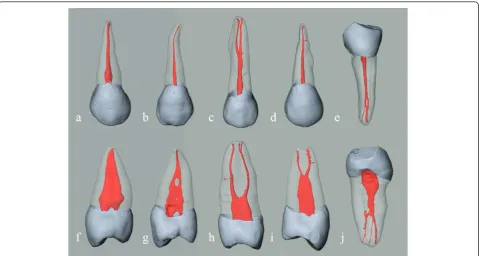
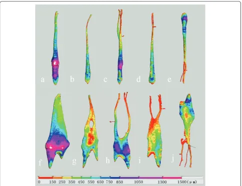
For the five single root premolars whose root canal sys-tems were classified into one of five types, volume ren-dering revealed detailed 3-D images of the root canal system, dentin, and enamel (Figure 1) and the various curves of the root canal were also shown in the 3-D model of the prepreparation canal (Figure 1, 2).
μm, except that the entrance of the additional canal in Tooth E is less than 60μm.
Effect after instrumentation
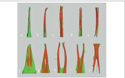
The gross canal anatomy of all teeth was substantially changed after root canal preparation with F3 (Figure 3). A gradual increase in the diameter along the length of the canal was noted. The canal volumes and surface areas were increased after instrumentation in all teeth. Superposition images of noninstrumented and instru-mented canals reveal a wide range (27.4-83.0%) in the proportion of surfaces unchanged during preparation. Table 1 shows the increases in canal volume (ΔV in mm3) and surface area (ΔAin mm2) and the percen-tages of unchanged surface (ΔP) for the five types canal systems. The type I canal system of Tooth A showed the least increases of canal volume and surface area (less than 5%) and largest unchanged surface (83%). The type II canal system of Tooth B and the type V canal system of Tooth E revealed the highest increases of canal volume and surface area (more than 146%), and least unchanged surface (less than 29%), and the additional canal of Tooth E remained untouched. In addition, the type III canal system of Tooth C and the type IV canal system of Tooth D had the middle increases of canal volume, surface area, and unchanged surface.
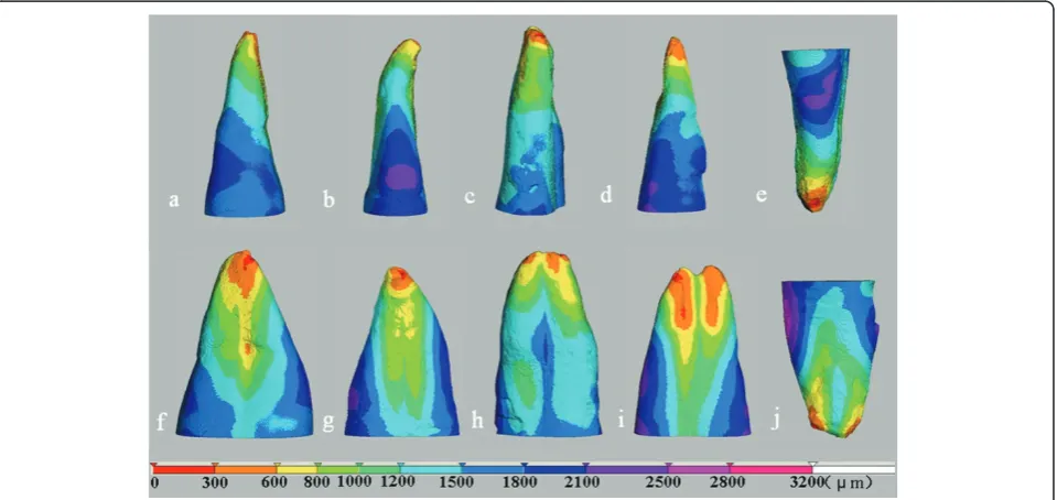
In the mesiodistal direction, all main canals were straightened after preparation with an F3 instrument and the straightening tended to occur toward the inner aspects of the curved parts of the root canal of teeth B, C, D and E (Figure 3). However, when viewed from the buccolingual direction, there was a transportation toward the outer aspects of the root canal and ledge for-mation at the apical third curve of Tooth B. Corre-sponding with the trend in canal transportation, more dentin was removed at the inner aspects of the curved parts and the outer aspect at the apical third curve of Tooth B (Figure 4).
The dentin wall thicknesses of canals after they were prepared with an F3 instrument are presented in Figure 5. Except for the mesial side of the lingual canal of Tooth D, all the dentin wall thicknesses were more than 0.3 mm. However, in a 1 mm zone near the apical foramen most of the dentin wall thicknesses were less than 0.3 mm. A hazardous zone was noted that was caused by the ledge formation in tooth B. No other pre-paration error or HPT instrument fractures occurred during the preparation of any canal.
Discussion
instrumentation technique (HPT) using micro-CT and 3-D reconstruction technology. Because of the diversity of clinical cases in endodontic therapy, we wanted to develop a study protocol that integrates the canal system classification with various instruments to evaluate the preparation efficacy using 3-D reconstruction techni-ques. As a pilot study, only five teeth of different cate-gories of root canal configuration (one each of types I, II, III, IV and V) were selected. The results showed the difference of morphological changes in the five types of root canal systems shaped with the same hand instru-mentation technique. However, because of the limited numbers of teeth and techniques, no statistical evalua-tion could be made concerning the instrumentaevalua-tion technique.
In the current study, color-coded images obtained by 3-D reconstruction gave insight into the morphological
changes in different types of root canal systems shaped with the same instrumentation technique.
In tooth A, there is a single flattened and straight root canal that was classified as type I canal system. After F3 instrumentation, 83% of the canal surface still remained unchanged. A similar result was also found for the widened canal after it was prepared by rotary ProTaper instruments [17].
complicated root canal anatomy with curves in multiple positions and planes could also have contributed [15]. When the morphology of the root canals after prepara-tion with S1, S2, F1, and F2 were also evaluated from 3-D canal models (data not shown), we found that signifi-cant transportation was created after the use of F1 and a ledge was formed after the use of F2. These results may suggest that the working length needs to be
reconfirmed after instrumentation of a type II canal system as in Tooth B using F1 [31].
Although there were multiple or S-shaped curves in the type III canal system of Tooth C and the type IV canal system of Tooth D, we achieved a good instrumentation effect in the coronal and middle third after the use of F3. However, there was an additional untouched canal surface caused by apical transportation in the apical third and we found it impossible to prepare the accessory canal in the apical third using HPT instruments, except by exploring it with small, pre-curved K-files. In clinical cases where the canal systems are like those in Tooth C and Tooth D, these unchanged surfaces may harbor microorganisms and allow for the presence of residual infection post treat-ment [32].
In the type V canal system of Tooth E, it is regrettable that we could not accomplish the instrumentation of the additional canal after preparing the other two canals with F3. The volume increased up to 146.3%, mainly caused by the removal of the dentin protuberance. In clinical cases that have type V canal systems like Tooth E, CT data and 3-D canal models could be useful to Figure 33-D color compound images showing the pre- and postpreparation effects. Prepreparation canal systems (green); the change in canal shapes post preparation (red). Mixed colors indicate superposition; green color alone shows the surfaces untouched during shaping. The images in the top and bottom row are viewed from buccolingual and mesiodistal directions, respectively. Letters indicate the same teeth as in Figure 1.
Table 1 Changes in canal volumes, surface areas, and the percentages of surface unchanged after preparation
Tooth (canal system classification) A (type
I)
B (type II)
C (type III)
D (type IV)
E (type V)
ΔVolume
(mm3) * 0.84 5.67 7.30 4.05 4.16
+4.8% +150.2% +96.7% +85.6% +146.3%
ΔArea (mm2) * 2.95 19.99 28.26 15.59 17.57
+3.4% +42.9% +42.4% +32.9% +49.0%
ΔPercentage (%)
83.0% 28.9% 41.1% 40.7% 27.4%
guide instrumentation and the use of a dental operating microscope and ultrasonic technique would be suitable for more efficiently exploring and finding the unusual canal access [1,33].
In the mesiodistal direction, all main canals were straightened after preparation with an F3 instrument and the straightening tended to occur toward the inner
aspects of the curved parts of the root canal of teeth B, C, D and E (Figure 3). Our results are consistent with those of Yang [34]. In previous studies [35,36], the ProTaper Universal instruments with noncutting tips showed better performance than the conventional ProTaper instruments for root canal transportation. However, Özer reported that all three rotary systems Figure 4Color-coded distance images showing the changes in canal shape during instrumentation. The distance also indicates the amount of dentin removed. The images in the top and bottom row are viewed from buccolingual and mesiodistal directions, respectively. Letters indicate the same teeth as in Figure 1.
(ProTaper Universal, Hero 642 Apical, FlexMaster) showed similar results during preparation of curved root canals and for transportation despite their noncutting tips [37].
For the five types of canal systems in this study, there was a wide range (27.4-83.0%) in the percentage of sur-face unchanged during preparation. The untouched area was distributed in the recesses of the flattened canal, the isthmus of the type II canal system, the outer aspects of multiple or S-shaped curves, lateral and accessory canals, and the additional canal of Tooth E. It has been shown that frequent and copious irrigation with sodium hypochlorite not only flushes out debris from the canal lumen, but also dissolves organic tissue in the nonin-strumented areas and the predentin layer [38]. More dentin debris can be removed from the isthmus, oval extensions in the root canal, and irregularities of the root canal wall by the use of ultrasonic irrigation with NaOCl as the irrigant [39,40]. Furthermore, in the mid-1990s, a method and device was presented that allowed cleansing of root canals without the need for manual instrumentation, and this noninstrumental hydrody-namic technique (NIT) showed an equal or even better cleanliness in all root sections than hand instrumenta-tion [41,42]. Thus, the untouched area within each type of canal system configuration, although not amenable to mechanical debridement, might be cleaned by these means.
In daily clinical practice, there are some cases in which conventional intraoral radiography and/or panoramic radiography alone do not provide enough information on the pathologic condition [43]. It is important to visualize and to have knowledge of the internal tooth anatomy before undertaking endodontic therapy [4]. Micro-CT has emerged as a powerful tool for evaluation of root canal morphology. Unfortunately, this technique is not suitable for clinical use, but cone-beam computed tomography (CBCT) systems have now been introduced for 3-D imaging of hard tissues of the maxillofacial region, with minimal distortion [44]. When encountering a case with a complicated canal system (such as type II or V) by radiographic evaluation, CBCT may be a good choice to allow appropriate management of the endodontic problem for endodontists [19,44].
Conclusions
From 3-D color-coded images, we discovered obviously different morphological changes in the five types of root canal systems shaped with the same hand instrumenta-tion technique. These results provide further informa-tion that premolars are among the most difficult teeth to be treated endodontically and that instrumentation techniques for the root canal systems of premolars should be judged individually depending on the 3-D
canal configuration of each tooth. Further study is needed to demonstrate the differences made by includ-ing variations in internal anatomy of teeth into the study protocol for investigation of various instrumenta-tion techniques.
Acknowledgements
This work was supported by the Key Clinical Program of the Ministry of Health of China. The funder had no role in study design, data collection and analysis, decision to publish, or preparation of the manuscript. The authors also wish to thank
Hong-Bing Wu for his help with the acquisition of the raw data.
Author details
1State Key Laboratory of Oral Diseases, West China College of Stomatology,
Sichuan University, Chengdu, P.R. China.2Institute of Stomatology, Chinese PLA General Hospital, Beijing, P.R. China.
Authors’contributions
KZL, YG, RZ, TH and BG participated in the design of the experiment and wrote the manuscript. KZL and YG participated in the acquisition, analysis and interpretation of data. All authors read and approved the final manuscript.
Competing interests
The authors declare that they have no competing interests.
Received: 9 March 2011 Accepted: 15 June 2011 Published: 15 June 2011
References
1. Al-Fouzan KS:The microscopic diagnosis and treatment of a mandibular second premolar with four canals.Int Endod J2001,34:406-410. 2. Plotino G, Grande NM, Pecci R, Bedini R, Pameijer CN, Somma F:
Three-dimensional imaging using microcomputed tomography for studying tooth macromorphology.J Am Dent Assoc2006,137:1555-1561. 3. Rhodes JS, Pitt Ford TR, Lynch JA, Liepins PJ, Curtis RV:Micro-computed
tomography: a new tool for experimental endodontology.Int Endod J
1999,32:165-170.
4. Vertucci FJ:Root canal morphology and its relationship to endodontic procedures.Endod Topics2005,10:3-29.
5. de Alencar AH, Dummer PM, Oliveira HC, Pécora JD, Estrela C:Procedural errors during root canal preparation using rotary NiTi instruments detected by periapical radiography and cone beam computed tomography.Braz Dent J2010,21:543-549.
6. Friedman S:Prognosis of initial endodontic therapy.Endod Topics2002, 2:59-88.
7. Hess W:Formation of root canals in human teeth.J Natl Dent Assoc1921, 3:704-725.
8. Weine FS:Initiating endodontic treatment.InEndodontic Therapy..
6 edition. Edited by: Weine FS. St. Louis, MO, USA: Mosby; 2004:106-110.
9. Yoshioka T, Villegas JC, Kobayashi C, Suda H:Radiographic evaluation of root canal multiplicity in mandibular first premolars.J Endod2004, 30:73-74.
10. Hülsmann M, Peters OA, Dummer PMH:Mechanical preparation of root canals: shaping goals, techniques and means.Endod Topics2005, 10:30-76.
11. Gekelman D, Ramamurthy R, Mirfarsi S, Paqué F, Peters OA:Rotary nickel-titanium GT and ProTaper files for root canal shaping by novice operators: a radiographic and micro-computed tomography evaluation.
J Endod2009,35:1584-1588.
12. Bürklein S, Hiller C, Huda M, Schäfer E:Shaping ability and cleaning effectiveness of Mtwo versus coated and uncoated EasyShape instruments in severely curved root canals of extracted teeth.Int Endod J
2011,44:447-457.
enlargement for cleaning the apical third of curved canals.Int Endod J
2010,43:988-994.
14. Vaudt J, Bitter K, Neumann K, Kielbassa AM:Ex vivo study on root canal instrumentation of two rotary nickel-titanium systems in comparison to stainless steel hand instruments.Int Endod J2009,42:22-33.
15. Peters OA:Current challenges and concepts in the preparation of root canal systems: a review.J Endod2004,30:559-567.
16. Bergmans L, Van Cleynenbreugel J, Wevers M, Lambrechts P, Bergmans L:A methodology for quantitative evaluation of root canal instrumentation using microcomputed tomography.Int Endod J2001,34:390-398. 17. Peters OA, Peters CI, Schönenberger K, Barbakow F:ProTaper rotary root
canal preparation: effects of canal anatomy on final shape analysed by micro CT.Int Endod J2003,36:86-92.
18. Jung M, Lommel D, Klimek J:The imaging of root canal obturation using micro-CT.Int Endod J2005,38:617-626.
19. Zhang R, Yang H, Yu X, Wang H, Hu T, Dummer PMH:Use of CBCT to identify the morphology of maxillary permanent molar teeth in a Chinese subpopulation.Int Endod J2011,44:162-169.
20. Özer SY:Comparison of root canal transportation induced by three rotary systems with noncutting tips using computed tomography.Oral Surg Oral Med Oral Pathol Oral Radiol Endod2011,111:244-250. 21. Garip Y, Günday M:The use of computed tomography when comparing
nickel-titanium and stainless steel files during preparation of simulated curved canals.Int Endod J2001,34:452-457.
22. Schirrmeister JF, Strohl C, Altenburger MJ, Wrbas KT, Hellwig E:Shaping ability and safety of five different rotary nickel-titanium instruments compared with stainless steel hand instrumentation in simulated curved root canals.Oral Surg Oral Med Oral Pathol Oral Radiol Endod2006, 101:807-813.
23. Moore J, Fitz-Walter P, Parashos P:A micro-computed tomographic evaluation of apical root canal preparation using three instrumentation techniques.Int Endod J2009,42:1057-1064.
24. Paqué F, Ganahl D, Peters OA:Effects of root canal preparation on apical geometry assessed by micro-computed tomography.J Endod2009, 35:1056-1059.
25. Saunders EM:Hand instrumentation in root canal preparation.Endod Topics2005,10:163-167.
26. Aguiar CM, Câmara AC:Radiological evaluation of the morphological changes of root canals shaped with ProTaper for hand use and the ProTaper and RaCe rotary instruments.Aust Endod J2008,34:115-119. 27. Pasqualini D, Scotti N, Tamagnone L, Ellena F, Berutti E:Hand-operated and rotary ProTaper instruments: a comparison of working time and number of rotations in simulated root canals.J Endod2008,34:314-317. 28. Huang DM, Luo HX, Cheung GS, Zhang L, Tan H, Zhou XD:Study of the
progressive changes in canal shape after using different instruments by hand in simulated S-shaped canals.J Endod2007,33:986-989.
29. Gao Y, Peters OA, Wu H, Zhou X:An application framework of three-dimensional reconstruction and measurement for endodontic research.
J Endod2009,35:269-274.
30. Bramante CM, Betti LV:Comparative analysis of curved root canal preparation using nickel-titanium instruments with or without EDTA.
J Endod2000,26:278-280.
31. Garg N, Garg A:procedural accidents.InTextbook of endodontics..1 edition. Edited by: Garg N & Garg A. New Delhi, India: Jaypee Brothers Medical Publishers; 2007:262.
32. Nair PN, Henry S, Cano V, Vera J:Microbial status of apical root canal system of human mandibular first molars with primary apical periodontitis after“one-visit”endodontic treatment.Oral Surg Oral Med Oral Pathol Oral Radiol Endod2005,99:231-252.
33. Alaçam T, Tinaz AC, Genç O, Kayaoglu G:Second mesiobuccal canal detection in maxillary first molars using microscopy and ultrasonics.Aust Endod J2008,34:106-109.
34. Yang GB, Zhou XD, Zhang H, Wu HK:Shaping ability of progressive versus constant taper instruments in simulated root canals.Int Endod J2006, 39:791-799.
35. Guelzow A, Stamm O, Martus P, Kielbassa AM:Comparative study of six rotary nickel titanium systems and hand instrumentation for root canal preparation.Int Endod J2005,38:743-752.
36. Javaheri HH, Javaheri GH:A comparison of three Ni-Ti rotary instruments in apical transportation.J Endod2007,33:284-286.
37. Özer SY:Comparison of root canal transportation induced by three rotary systems with noncutting tips using computed tomography.Oral Surg Oral Med Oral Pathol Oral Radiol Endo2011,111:244-250.
38. Cheung LH, Cheung GS:Evaluation of a rotary instrumentation method for C-shaped canals with micro-computed tomography.J Endod2008, 34:1233-1238.
39. Lee SJ, Wu MK, Wesselink PR:The effectiveness of syringe irrigation and ultrasonics to remove debris from simulated irregularities within prepared root canal walls.Int Endod J2004,37:672-678.
40. Lumley PJ, Walmsley AD, Walton RE, Rippin JW:Cleaning of oval canals using ultrasonic or sonic instrumentation.J Endod1993,19:453-457. 41. Lussi A, Nussbächer U, Grosrey J:A novel noninstrumented technique for
cleansing the root canal system.J Endod1993,19:549-553.
42. Lussi A, Portmann P, Nussbächer U, Imwinkelried S, Grosrey J:Comparison of two devices for root canal cleansing by the noninstrumentation technology.J Endod1999,25:9-13.
43. Velvart P, Hecker H, Tillinger G:Detection of the apical lesion and the mandibular canal in conventional radiography and computed tomography.Oral Surg Oral Med Oral Pathol Oral Radiol Endod2001, 92:682-688.
44. Patel S:New dimensions in endodontic imaging: Part 2. Cone-beam computed tomography.Int Endod J2009,42:463-475.
Pre-publication history
The pre-publication history for this paper can be accessed here: http://www.biomedcentral.com/1471-2342/11/14/prepub
doi:10.1186/1471-2342-11-14
Cite this article as:Liet al.:The effect of a manual instrumentation technique on five types of premolar root canal geometry assessed by microcomputed tomography and three-dimensional reconstruction. BMC Medical Imaging201111:14.
Submit your next manuscript to BioMed Central and take full advantage of:
• Convenient online submission
• Thorough peer review
• No space constraints or color figure charges
• Immediate publication on acceptance
• Inclusion in PubMed, CAS, Scopus and Google Scholar
• Research which is freely available for redistribution