Research Article
Phytochemical profile, antimicrobial
potential and GC-MS analysis of wild
variety of Olea Europaea (Olive)
cultivated in Pakistan
Waqar Ahmad
1, Nawab Ali
1*, Muhammad Siddique Afridi
2, Hazir
Rahman
3, Muhammad Adnan
4, Nimat Ullah
1, Uzair Muhammad
4,
Muhammad Ilyas
5and Haroon Khan
61. Department of Biotechnology and Genetic Engineering, Kohat University of Science & Technology, Kohat, Pakistan
2. PCSIR laboratories Complex Peshawar-Pakistan
3. Department of Microbiology, Abdul Wali Khan University Mardan, Mardan-Pakistan 4. Department of Botany, Kohat University of Science & Technology, Kohat-Pakistan 5. Department of Agronomy, The University of Agriculture, Peshawar, Pakistan 6. Department of Weed Science, The University of Agriculture, Peshawar-Pakistan
*Corresponding author’s email: [email protected]
Citation
Waqar Ahmad, Nawab Ali, Muhammad Siddique Afridi, Hazir Rahman, Muhammad Adnan, Nimat Ullah, Uzair Muhammad, Muhammad Ilyas, and Haroon Khan. Phytochemical profile, antimicrobial potential and GC-MS analysis of wild variety of Olea Europaea (Olive) cultivated in Pakistan. Pure and Applied Biology. Vol. 6, Issue 1, pp337-345. http://dx.doi.org/10.19045/bspab.2017.60032
Received: 17/10/2016 Revised: 20/02/2016 Accepted: 01/03/2017 Online First: 02/03/2017
Abstract
Olive plant produces a variety of bioactive molecules and thus has important medicinal value in folk medicine. In this study, different leaf and fruit extracts of Pakistani wild variety of Olea europaea
was tested for their phytochemical content, antimicrobial activity and mass spectrometric analysis. Olive leaves and fruit samples were extracted with five different solvents to obtain the crude extract and screened for various kinds of phytochemicals. Phytochemicals were further confirmed through Fourier Transmission Infra-red Spectroscopy (FTIR). The plant extract showed significant antimicrobial activity against all the strains tested. Methanol, ethanol and ethyl acetate extracts were found more effective against most of the pathogenic bacteria with high zone of inhibition. Gas Chromatography-Mass Spectrometry (GC-MS) analysis revealed that olive fruits have Oleic acid, Palmitic acid, Linoleic acid, Octadecadienoic acid, Stearic acid, Palmitoleic acid and Tridecanoic acid as oil contents. In this work, the antimicrobial potential and phytochemical contents were explored which may further pave the way for the bio-industrial applications.
Keywords: Olea europaea; Phytochemical content; Antimicrobial activity; GC-MS analysis
Introduction
Plants have been used as medicines since the dawn of civilization for thousands of years
[1]. About 80% of population in developing
338
usefulness and effective nature [2]. Chemical constituents of plants have been exploited for the discovery of therapeutic agents as well as new sources of such economical materials as tannins, oils, gums, forerunners for the production of complex chemical substances. The olive tree is amongst the oldest known cultivated trees in the world that has been an important source of nutrition and medicine [3]. Olive is a broad-leaved, evergreen tree of the family Oleaceae that is present in native coastal areas of the Mediterranean region. The olive has been used generally in customary medications in European Mediterranean islands and countries such as Spain, Israel, Morocco, France and Greece where it is cultivated mainly as edible oil and table olives [4].
Bacteria are serious pathogens and cause a wide variety of human diseases including cholera, leprosy, bacterial pneumonia, whooping cough, and diphtheria. Bacterial pathogens are also a serious threat to the food industry [5]. Antibiotics provide main basis for the therapy of microbial infections. However, the emergence and spreading of bacterial resistance has made the treatment of infectious diseases more problematic [6]. The antimicrobial activity of plants is highly related to secondary substances that are synthesized and produced by plants [7]. The olive tree naturally possesses strong antimicrobial activity which has been utilized in traditional medicine to fight fever and overcome infections [8].
It is desirable to get knowledge about the bioactive constituents of plants like fatty acids because of their nutritional value, diagnosis of definite diseases and
pharmacology. Various analytical
techniques like Spectrophotometry, HPLC and gas chromatography (GC) have been applied for the analysis of fatty acids [9-12]. GC-MS is a useful method for the
determination of fatty acids due to its high speed, resolution and sensitivities [13]. The aim of the study was to investigate the antimicrobial activities and phytochemical contents utilizing different methods in order to compare with the reported literature. The data produced here may be used in production of biomolecules of medical importance.
Materials and methods Collection of plant materials
The fresh leaves of olive plant were collected from district Dir of Khyber Pakhtunkhwa, Pakistan. The rinsed olive leaves of collected samples were air dried under shade and then converted to fine powder by crushing in electronic grinder. The fine powdered form of the plants was then kept in airtight glass containers to be protected from different contaminants until used for further analysis and screening. Preparation of leaves extracts
The olive leaves were air dried and ground into powder form and then 100 (g) of the powder was extracted with 350 mL of ethyl acetate, ethanol, methanol, distilled water and hexane each (Technical grade- Merck) and boiled water in 1000 mL conical flasks. Flasks were vigorously shaken at 400 rpm overnight in a Labotec model 20.2 shaking machine. After shaking, the supernatant was decanted into pre-weighed, labeled flasks. The process was repeated three times to exhaustively extract the leaves materials. The solvents were removed under vacuum by rotary evaporator at 40°C and the extraction efficiency was quantified by determining the weight of each of the extracts [14, 15].
Phytochemical analysis
cyanogenic glycosides, phenolic compounds, carbohydrates, proteins and riboflavin. The phytochemicals in each extract were qualitatively and quantitatively determined for the presence of biologically active components according to the method reported in literature [16, 17].
Antimicrobial assay
The extracts obtained from leaves were tested for antimicrobial activity against six Gram positive bacterial strains, three Gram
negative bacterial strains and one fungal strain (Table 1) by disc diffusion method as described by Aida et al. (2001) [17-19]. The results were based on the measurement of minimum zone of inhibition (ZOI) that was shown in millimeter (mm). The experiments were performed in triplicate and the average diameter of the inhibitory zone was measured by using standard deviation method.
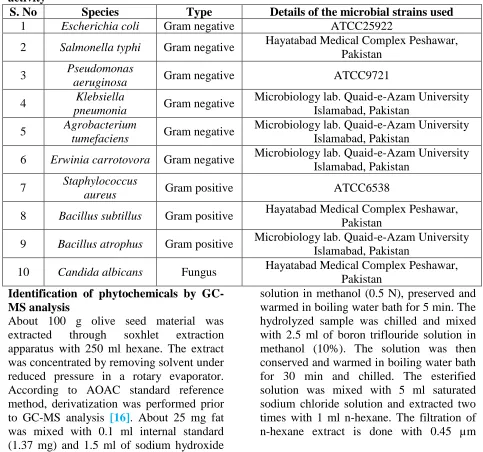
Table 1. List of microorganisms used in this study for Olive (Olea europaea) antimicrobial activity
S. No Species Type Details of the microbial strains used
1 Escherichia coli Gram negative ATCC25922
2 Salmonella typhi Gram negative Hayatabad Medical Complex Peshawar,
Pakistan
3 Pseudomonas
aeruginosa Gram negative ATCC9721
4 Klebsiella
pneumonia Gram negative
Microbiology lab. Quaid-e-Azam University Islamabad, Pakistan
5 Agrobacterium
tumefaciens Gram negative
Microbiology lab. Quaid-e-Azam University Islamabad, Pakistan
6 Erwinia carrotovora Gram negative Microbiology lab. Quaid-e-Azam University
Islamabad, Pakistan
7 Staphylococcus
aureus Gram positive ATCC6538
8 Bacillus subtillus Gram positive Hayatabad Medical Complex Peshawar,
Pakistan
9 Bacillus atrophus Gram positive Microbiology lab. Quaid-e-Azam University
Islamabad, Pakistan
10 Candida albicans Fungus Hayatabad Medical Complex Peshawar,
Pakistan
Identification of phytochemicals by GC-MS analysis
About 100 g olive seed material was extracted through soxhlet extraction apparatus with 250 ml hexane. The extract was concentrated by removing solvent under reduced pressure in a rotary evaporator. According to AOAC standard reference method, derivatization was performed prior to GC-MS analysis [16]. About 25 mg fat was mixed with 0.1 ml internal standard (1.37 mg) and 1.5 ml of sodium hydroxide
340
membrane filter and injected 1 µl to GC-MS through auto injector system.
GC-MS analysis was performed on GC-MS Model QP 2010 plus (Tokyo, Japan) equipped with an auto-sampler (AOC-20S) and auto-injector (AOC-20i). TRB-FFAP Technokroma capillary column (30 m × 0.35 mm, 0.250 µm film thicknesses) was used under the following conditions:
oven temperature programmed from 50°C to 150°C at 15°C/min, and the final temperature was raised to 220°C and kept constant for 3 min; injector temperature 240°C; carrier gas He, flow rate 1ml/min; the volume of injected sample was 1µl ; ion source temperature 200°C; scan mass range of m/z 85-380 and interface line temperature 300°C [13].
Components were identified by comparing the mass spectra obtained during GC-MS
analysis with those of standard mass spectra from the National Institute of Standard and Technology (NIST) library.
Results and discussion
In this study, phytochemical analysis of local wild variety of olive leaves extracts were investigated using standard protocols
[16, 17]. The results of qualitative
phytochemical tests showed the presence of important phytochemical components. It is clear from qualitative screening that alkaloids, phenols, tannins, flavonoids, saponins, steroids, terpenoids, carbohydrates and proteins were present in almost all
solvent extracts (Table 2). The
phytochemicals reported in this study has been reported in the previous literature [11,
12].
Table 2. Qualitative analysis of local olive plant for phytochemical contents
S. No Plant extracts A G S F P/T S T C P R
1 Aqueous + ++ +++ ++ - ++ + ++ ++ +
2 Hexane + + + + - ++ +++ - - +
3 Methanol ++ + ++ +++ + ++ + ++ ++ ++
4 Ethanol ++ ++ ++ +++ + +++ ++ ++ ++ +
5 Ethyl Acetate ++ - ++ + + ++ ++ + + +
+ = present; ++ or +++= abundant - = absent; A = Alkaloids; G = Glycosides; S = Saponins; F = Flavonoids; P/T = Phenols/Tannins; S = S teroids; T = Terpenoids; C = Carbohydrates; P = Proteins; R = Riboflavin
Further confirmation about phytochemicals was done by spectra obtained using Fourier
Transmission Infra-Red Spectroscopy
(FTIR) model (IRPrestige-21, Shimadzu Corporation Kyoto Japan) (Figure 1) which revealed that polar phytochemicals were separated in high amount with polar solvents and non-polar constituents with non-polar solvents. In this spectra, the phenols, tannins, carbohydrates and proteins were absent in hexane extract due to non-polar nature of hexan. While methanol and ethanol extracts had highest amount of flavonoides and terpenoids due to the polar nature of the solvent which is according the like dissolve like concept of solvents. These
results are supported by the previous literature where different extracts were used for phytochemical study of various plants
[11, 12, 16, 17]. The quantitative
phytochemical estimation (Figure 2) revealed that ethanollic and methanollic extracts have high content of flavonoids 16.36±0.03% and 14.0±0.25%, respectively. Aqueous and ethanollic extracts showed higher saponins percentage 13.50±0.25% and 13.80±0.25%, respectively. Similarly, ethanollic and methanollic extracts contained high Alkaloids percentage
13.20±0.17% and 12.50±0.15%,
341 450 600 750 900 1050 1200 1350 1500 1650 1800 1950 2100 2400 2700 3000 3300 3600 3900 1/cm 75 80 85 90 95 100 105 %T 2 9 1 8 .3 0 2 8 4 8 .8 6 1 7 1 4 .7 2 1 6 8 3 .8 6 1 4 5 6 .2 6 1 3 6 1 .7 4 1 2 6 9 .1 6 1 2 5 9 .5 2 1 1 8 2 .3 6 1 1 6 5 .0 0 1 0 7 6 .2 8 1 0 3 3 .8 5 9 9 7 .2 0 7 6 5 .7 4 6 2 8 .7 9 4 3 2 .0 5 4 2 8 .2 0 ethanol_wild 450 600 750 900 1050 1200 1350 1500 1650 1800 1950 2100 2400 2700 3000 3300 3600 3900 1/cm 65 70 75 80 85 90 95 100 105 %T 3 3 2 9 .1 4 2 9 4 7 .2 3 2 3 5 8 .9 4 1 7 1 4 .7 2 1 6 3 5 .6 4 1 2 6 9 .1 6 1 0 7 2 .4 2 1 0 3 3 .8 5 aquesous_wild
important to be used as antibiotic agents against known pathogens [20, 21]. Chemically, flavonoids are hydroxylated phenol rich compounds which are reported for antimicrobial activities [22]. These phytochemicals are also proved useful antioxidant agents and possessing effective
anticancer characteristics [23]. Saponins are capable of precipitation and coagulation of erythrocytes. Some of the activities of saponins are leather formation in aqueous
solutions, hemolysis, binding with
cholesterols [24].
342
0 2 4 6 8 10 12 14 16 18
Ethyl Acetate Hexane Methanol Aqueous Ethanol
P
e
rc
e
n
tag
e
v
al
u
e
s
Plant Extract
Alkaloids
Flavonoids
Phenols/Tannin s
Figure 2. Quantitative phytochemical estimation (percentage) of olive leaves extracts(The obtained results are the mean of triplicate experiments)
The antimicrobial activities revealed that ethyl acetate and methanol extracts of olive exhibited maximum activity while minimum antimicrobial activities were observed for hexane extracts (Figure 3). It is because of the fact that alcohol and ethyl acetate extracts give rise to flavonoids and phenolic phytochemicals as compared to hexane which were confirmed from the Fourier transmission Infra-red Spectroscopy (FTIR) spectra (Figure 1).
The results from the disc diffusion method, followed by measurement of ZOI, indicated that the methanol extract showed strong inhibitory activity against Bacillus Atrophus,
with the highest inhibition zones
(10±0.40mm) which is in the range with its antibiotic counterpart. (16±0.21mm) (Figure 3). The inhibitory activity of these extracts confirmed the antimicrobial activity and its potential use in the treatment of microbial diseases. The antimicrobial activities of Olive leaves have been reported by Sudjana et al. against different bacteria [19]. Antimicrobial activities of different plant extracts obtained were appeared to be very different in the sense of effectiveness as some bacterial species are found more resistant and some other are found more
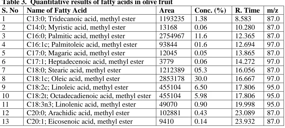
The rest of the fatty acids were present in less than 1% concentration (Table 3). Similar chromatographic analysis has been done for olive plant of different countries and various biomolecules were evaluated for their biopharmaceutical application [9, 11,
12, 19, 25, 26]. GC-MS analysis revealed
that olive oils contain different biologically active compounds like fatty acids thus olive plant, besides its antibacterial and antifungal activities can also be used to produce different pharmaceutical products to cure diseases including cancer.
Figure 3. Antimicrobial activity of olive leaves extracts along with standard antibiotics
344
Table 3. Quantitative results of fatty acids in olive fruit
S. No Name of Fatty Acid Area Conc. (%) R. Time m/z
1 C13:0; Tridecanoic acid, methyl ester 1193235 1.38 8.583 87.0
2 C14:0; Myristic acid, methyl ester 13168 0.06 10.280 87.0
3 C16:0; Palmitic acid, methyl ester 2754967 11.6 12.365 87.0
4 C16:1c; Palmitoleic acid, methyl ester 93844 01.6 12.694 97.0
5 C17:0; Magaric acid, methyl ester 12045 0.05 13.865 87.0
6 C17:1; Heptadecenoic acid, methyl ester 3779 0.06 14.272 97.0
7 C18:0; Stearic acid, methyl ester 1212389 05.3 16.056 87.0
8 C18:1c; Oleic acid, methyl ester 2853178 30.0 16.667 97.0
9 C18:2c; Linoleic acid, methyl ester 455104 6.50 17.806 95.0
10 C18:2t; Octadecadienoic acid, methyl ester 455104 5.98 17.806 95.0
11 C18:3n3; Linolenic acid, methyl ester 49070 0.90 19.998 95.0
12 C20:0; Arachidic acid, methyl ester 102881 0.43 23.089 87.0
13 C20:1; Eicosenoic acid, methyl ester 9410 0.14 23.932 87.0
Conclusions
It is concluded from the study that local olive plant extract contains different phytochemicals such as alkaloids, phenols, tannins, flavonoids, saponins, steroids, terpenoids, riboflavin, carbohydrates and proteins. Olive plant revealed broad spectrum antimicrobial activities against bacterial and fungal strains. It is obvious from GC-MS analysis that Olive fruit oils contain highest concentration of Oleic acid and other related fatty acids which may further be evaluated for bio-pharmaceutical applications.
Author’s contributions
Conceived and designed the experiment: A Waqar, SA Muhammad & A Nawab, Performed the experiments: A Waqar, Analyzed the data: A Waqar, A Nawab, SA Muhammad, R Hazir & A Muhammad, Contributed reagents/ materials/ analysis tools: U Nimat, M Uzair & I Muhammad, Wrote the paper: A Waqar & A Nawab.
Acknowledgements
We are thankful to the technical staff of PCSIR laboratories for expert technical assistance.
References
1. Koehn FE & Carter GT (2005). The evolving role of natural products in drug discovery. Nat Rev Drug Disc 4 (3): 206– 220.
2. Ellof JN (1998). Which extractant should be used for the screening and isolation of antimicrobial components from plants. J. Ethnopharmacol 60: 1-6.
3. Hanbury D (1854). On the febrifuge properties of the olive (Oleaeuropea, L.).
Pharmaceut. J Provin. Trans 353–354. 4. Green PS (2002). A revision of Olea L.
(Oleaceae). Kew Bull 57: 91–140.
5. Nataro JP and Kaper JB (1998).
Diarrheagenic Escherichia coli. Clin Microbio Rev 11(1): 142-201.
6. Sharma R, Sharma C & Kapoor B (2005). Antibacterial resistance; current problems and possible solutions. Ind J Med Sci
59(3): 120-129.
7. Cowan MM (1999). Plant products as antimicrobial agents. Clin Microbiol Rev
12: 564-582.
8. Friedman M (2007). Overview of
antibacterial, antitoxin, antiviral, and antifungal activities of tea flavonoids and teas. Mol. Nutr Food Res 51(1): 116-134. 9. Yang Y, Ferro MD, Cavaco I & Liang Y
extra virgin olive oil adulteration by GC-MS combined with chemometrics. J Agric Food Chem 15: 693-702.
10. Sodipo OA, Akiniyi JA & Ogunbamosu
JU (2000). Studies on certain
characteristics of extracts of bark of
Pansinystalia macruceras (K schemp) picrre Exbeille. Glob. J Pure Appl Sci 6: 83-87.
11. Goldsmith CD, Vuong QV, Sadeqzadeh E, Stathopoulos CE, Roach PD & Scarlett CJ (2015). Phtochemical properties and
antiproliferative activity of Olea
Europaea L. leaf extracts against pancreatic cancer cells. Molecules 20: 12992-13004.
12. Khlif I, Jellali K, Michel T, Halabalaki M, Skaltsounis AL & Allouchi N (2015). Characteristics, Phytochemical analysis and iological activities, of extracts from Tunisian Chetoui Olea euppaea variety. J Chem DOI. 10.1155/2015/418731.
13. Qureshi MN, Siddique M, Rahman IU &
Kanwal F (2011) Analytical
Characterization of Fatty Acids
Composition of Datura Alba Seed Oil by Gas Chromatography Mass Spectrometry.
J Chin Chem Soc 58: 236-240.
14. Vaghasiya Y & Chanda S (2007). Screening of methanol and acetone extracts of fourteen Indian medicinal plants for antimicrobial activity. Turk J Bio 31: 243-248.
15. Murray PR, Baron EJ, Pfaller MA,
Tenover FC & Yolken RH, editors (1999). Manual of clinical microbiology.
7th ed Washington, D.C.: ASM Press 51-59.
16. Ahmad S, Ahmad S, Bibi A, Saqib M, Afridi MS, Kanwal F, Zakir M & Fatima
F (2014) Phytochemical analysis,
Antioxidant activity, fatty acid
composition, and functional group
analysis of Heliotropium bacciferum. Sci world J 1-8
17. Oboh IE, Obasyuf & Akerele JO (2008). Phytochemical and antibacterial studies on the crude ethanol and aqueous extracts
of the leaves of Lacaniodiscus
Cupanoides Planch (Sapindaceae). Drugs Research 65: 565-569.
18. Aida P, Rosa V, Blamea F, Tomas A & Salvador (2001). Antifungal activity of Paraguyan plants used in traditional medicines. J Ethnopharmacol 16: 93-98. 19. Sudjana AN, Orazio CD, Ryan A, Rasool
N, Islam N, Riley TV & Hammer KA
(2009). Antimicrobial activity of
commercial Olea europaea (Olive) leaf extract. Int J Antimicrob Agents 33: 461– 463.
20. Kubmarawa D, Ajoku GA, Enworem NM
& Okorie DA (2007). Roles of
agricultural biotechnology in ensuring adequate food security in developing societies. Afr J Biotech 6: 1690-1696. 21. Mensah JK, Okoli RI, Ohaju-Obodo JO
& Eifediyi K (2008). Aqueous extract of
Telfairia occidentalis leaves reduces blood sugar and increases haematological and reproductive indices in male rats. Afr J Biotech 7: 2304-2309.
22. Marjorie C (1996). Plant products as antimicrobial agents. Clinc Microbiol Rev
12: 564-582.
23. Salah N, Miller NJ, Pagange, Tijburg L, Bolwell GP, Rice E & Evans C (1995). Polyphenolic flavonoids as scavenger of aqueous phase radicals as chai breaking antioxidant. Arc Biochem Broph 2: 339-346.
24. Okwu DE (2004). Phytochemicals and vitamin content of indigenous species of
southeastern Nigeria. J Sustain. Agric Environ 6(1): 30-37.
25. Zoric N, Kopjar N, Kraljic K, Orsolic N, Tomic S & Kosalec I (2016). Olive leaf extract activity against Candida albicans
and C. dubliniensis-the in vitro viability study. Acta Pharm 66: 411-421.
26. Gumgumjee NM & Hajar AS (2014).
Antimicrobial activities and