Open Access
Review
Basic mechanisms for recognition and transport of synaptic cargos
Max A Schlager and Casper C Hoogenraad*
Address: Department of Neuroscience, Erasmus Medical Center, 3015GE, Rotterdam, The Netherlands
Email: Max A Schlager - [email protected]; Casper C Hoogenraad* - [email protected] * Corresponding author
Abstract
Synaptic cargo trafficking is essential for synapse formation, function and plasticity. In order to transport synaptic cargo, such as synaptic vesicle precursors, mitochondria, neurotransmitter receptors and signaling proteins to their site of action, neurons make use of molecular motor proteins. These motors operate on the microtubule and actin cytoskeleton and are highly regulated so that different cargos can be transported to distinct synaptic specializations at both pre- and post-synaptic sites. How post-synaptic cargos achieve specificity, directionality and timing of transport is a developing area of investigation. Recent studies demonstrate that the docking of motors to their cargos is a key control point. Moreover, precise spatial and temporal regulation of motor-cargo interactions is important for transport specificity and cargo recruitment. Local signaling pathways Ca2+ influx, CaMKII signaling and Rab GTPase activity regulate motor activity and cargo release at synaptic locations. We discuss here how different motors recognize their synaptic cargo and how motor-cargo interactions are regulated by neuronal activity.
Introduction
Neurons are highly polarized cells with distinct mem-brane domains, including a single extended axon and multiple dendritic processes which contain thousands of individual synapses. Synapses are composed of a presyn-aptic terminal, a synpresyn-aptic cleft, and a postsynpresyn-aptic special-ization and are the structures through which neurons communicate [1,2]. Presynaptic boutons transmit signals by releasing neurotransmitters from synaptic vesicles and postsynaptic sites receive information using neurotrans-mitter receptors. Most excitatory glutamate receptors, such as N-methyl D-aspartate (NMDA) receptors and α -amino-3-hydroxy-5-methyl-4-isoxazole propionate (AMPA) receptors are located at dendritic spines, whereas inhibi-tory glycine receptors (GlyRs) and γ-aminobutyric acid type A receptors (GABAARs) are mainly located at the shaft of dendrites. Recent studies have identified the molecular components of synapses, particularly by using
proteomic strategies and have revealed that the specifica-tion of synaptic funcspecifica-tion, e.g. excitatory or inhibitory, at both pre- and post-synapses is achieved via the recruit-ment and assembly of distinct receptors and their associ-ated proteins [1,3-6]. The assembly requires selective localization or stabilization of receptors at synaptic mem-branes [7], as well as the highly coordinated intracellular transport of synapse-associated proteins [8]. In Drosophila, selective synaptic cargo transport has been shown to be essential for synapse formation at the neuromuscular junction while axon outgrowth and guidance were unaf-fected [9].
The most widely used mechanism for intracellular traf-ficking involves molecular motor proteins that carry cargo directionally along a cytoskeletal track myosins along actin and kinesins and dyneins along microtubules. Vari-ous organelles and membranVari-ous structures have been Published: 4 August 2009
Molecular Brain 2009, 2:25 doi:10.1186/1756-6606-2-25
Received: 30 June 2009 Accepted: 4 August 2009
This article is available from: http://www.molecularbrain.com/content/2/1/25
© 2009 Schlager and Hoogenraad; licensee BioMed Central Ltd.
shown to move bidirectionally along the cytoskeletal net-work some cargos can even switch between actin and microtubules [10]. Despite extensive knowledge on the complete genomic inventories of molecular motors in several diverse organisms [11,12] and significant progress in our understanding of neuronal transport in general [13], the cellular mechanisms responsible for synaptic cargo trafficking remain unclear. It is not fully understood how a limited number of motor proteins carries the wide variety of synaptic cargos, such as neurotransmitter
recep-tors, ion channels, integral membrane proteins, signaling complexes, mRNAs, synaptic vesicle precursors (SVPs), Piccolo-Bassoon transport vesicles (PTVs), mitochondria or other organelles (Table 1). Emerging evidence suggests that the docking of motors to their cargos via adaptor molecules is an important mechanism to achieve trans-port specificity. Specific transtrans-port mechanisms are crucial for neuronal development and plasticity, such as synap-togenesis [5,9] and synaptic plasticity [14,15]. Moreover, alterations in the trafficking of synaptic receptors and
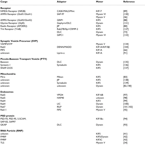
Table 1: Motors and adaptors for transport of synaptic cargos
Cargo Adaptor Motor Reference
Receptor
NMDA Receptor (NR2B) CASK/MALS/Mint KIF17 [89]
AMPA Receptor (GluR1/GluA1) SAP-97 Myosin VI [130]
Myosin V [46]
AMPA Receptor (GluR2/GluA2) GRIP1 KIF5 [88]
Glycine Receptor (GlyR) Gephyrin/DLC Dynein [131]
Reelin Receptor (APOER2) JIP KIF5 [80]
Trk Receptor (TrkB) Rab27B/Slp1/CRMP-2 KIF5 [127]
DLC Dynein [73]
GIPC1 Myosin VI [133]
Synaptic Vesicle Precursor (SVP)
VAMP2/SYP Myosin V [134]
Rab3 DENN/MADD KIF1A/KIF1Bβ [104]
PIP2 KIF1A [66]
unknown Liprin-α KIF1A [91]
Piccolo-Bassoon Transport Vesicle (PTV)
Bassoon DLC Dynein [135]
Syntaxin-1 Syntabulin KIF5 [136]
SNAP-23/25 KIF5 [137]
Mitochondria
Miro Milton KIF5 [83]
unknown JIP KIF5 [138]
unknown Syntabulin KIF5 [139]
unknown unknown Dynein [82,140]
Endosomes
Rab5 VPS34 KIF16B [97]
Rab5 HAP40 unkown [98]
Rab4 KIF3 [99]
Rab4 LIC Dynein [100]
Rab7 RILP Dynein [101,102]
Rab11 FIP2 Myosin V [45]
PSD protein
PSD-93, PSD-95, S-SCAM, KIF1Bα [94]
SAP102, SAP97
GKAP DLC Dynein [93]
RNA Particle (RNP)
unkown KIF5 [41]
FMRP KIF5/Dynein [42]
FMRP KIF3C [43]
associated proteins may contribute to pathologic changes in neurological and psychiatric disease [15-17].
In this review we focus on the basic principles of synaptic cargo transport. We discuss how different motors of the kinesin, dynein and myosin families recognize their cargo and how motor-cargo interactions are regulated. Not cov-ered in this review are detailed descriptions of the differ-ent motor protein families [12,18] and specific pathways in neuronal trafficking [14], but we will discuss briefly the current knowledge of general cargo transport mechanisms [10,19,20]. Other excellent reviews cover the role of cytoskeletal organization in cargo trafficking [21,22], receptor trafficking through lateral diffusion in the plasma membrane [23,24], transport mechanisms during neuro-nal polarization [25] and discuss the relationship between trafficking and neurodegenerative disease [26,27]. We aim to give an overview of the molecular trafficking mecha-nisms important for the delivery and removal of synaptic proteins. Studying the basic machinery for recognition and transport of synaptic cargos might help us to under-stand fundamental operational principles of synapse for-mation, function and plasticity.
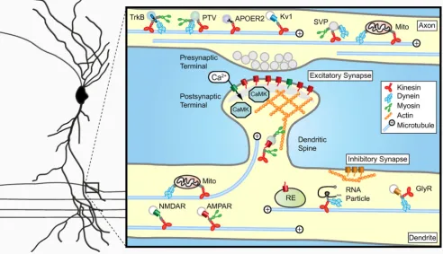
Basic mechanisms regulating motor protein transport All eukaryotic cells rely on the cytoskeleton to facilitate active transport. For neurons it is especially important to have efficient and well-regulated transport to establish and maintain polarized neuronal morphology and synap-tic specializations. Two types of cytoskeletal networks are involved in neuronal transport; the microtubule and actin cytoskeleton. Microtubules are dynamic structures with rapidly growing and shrinking plus-ends and a more sta-ble minus-ends [28]. Microtubules are mainly present in the shafts of the axon and dendrites whereas actin is more abundant in highly dynamic structures such as growth cones and dendritic spines [22,29,30]. In general, cargos are transported over long distances along microtubules before transferring to the actin cytoskeleton for the final part of their journey. A common feature of both actin and microtubule based transport is the fact that force is gener-ated by motor proteins through ATP hydrolysis [12,18,31,32]). The motor proteins that use microtubules as tracks are the minus-end directed dynein and plus-end directed kinesins, whereas myosins move along actin fila-ments. Neuronal cargo trafficking is achieved by the con-certed efforts of both microtubule-based and actin-based motors [8,33-35] (Figure 1). Several classes of myosin motors participate in neural cargo transport in axon and dendrites most commonly used are myosin V and myosin VI (Table 1). Although the basic mechanisms for microtu-bule- and actin-dependent transport in neurons are well established, how neuronal cargos achieve specificity and directionality to their site of action is an emerging field of investigation.
Microtubule organization in neurons
In neurons, the microtubule cytoskeleton is organized dif-ferently in dendrites and axons. Dendritic microtubules have mixed polarity i.e. plus-ends can face either away from or towards the cell body, whereas the microtubules in the axon have a uniform polarity with all plus-ends fac-ing outwards [36,37] (Figure 1). Recently, by imagfac-ing microtubules using fluorescently tagged α-tubulin and microtubule plus-end binding proteins it was demon-strated that neuronal microtubules are very dynamic and regularly enter dendritic spines [38,39]. Microtubules grow with their plus-ends directed towards the spine and are able to repeatedly enter the same spine [39] (Figure 1). In contrast, microtubule nucleation within spines and subsequent polymerization into the dendrite shaft has not been observed. Therefore, polarized growth of microtu-bules into spines could facilitate delivery of synaptic cargo directly to the postsynaptic site (Figure 1). Although microtubule-dependent cargo transport in dendritic spines has not been directly visualized yet, it will be inter-esting to investigate whether the translocation of excita-tory postsynaptic proteins or other cellular components, such as organelles and RNA particles (RNPs), rely on microtubule based transport [40-43] (Table 1). On the other hand, several studies directly demonstrate the importance of the actin-based transport mechanisms at excitatory synapses. Myosin V, for example, mediates RNP translocation into dendritic spines [34], is required for the spine localization of smooth endoplasmic reticulum at Purkinje cell synapses [44] and transports AMPA receptor containing recycling endosomes during synaptic potenti-ation [45,46].
Post-translational modification of microtubules
Regulation of motor protein activity
The motor domains responsible for motility and force generation are connected by a stalk to the tail domain, which directly or indirectly binds to their cargo [12]. Another possible mode of transport regulation is direct modulation of motor protein activity. Tremendous progress has been made in the mechanistic understanding of molecular motors both in force generation and direc-tional movement [12,32,51,52]. For instance, a recent study shows that the specific structure of dynein, with six AAA+ ATPase domains, a linker and a microtubule bind-ing stalk, explain the characteristic dynein steppbind-ing pat-tern [53]. The stepping behavior of kinesin was shown to be regulated by intramolecular strain, allowing the kinesin motor to maintain its plus-end directed move-ment and also coordinate the two motor domains [54].
Moreover, several studies demonstrate that myosin and kinesin motors can fold back on themselves in order to regulate their own motor activity [39]. In the absence of bound cargo, the tail domain interacts with the motor domain and inhibits its activity [55,56]. Furthermore, motor accessory proteins such as dynactin have been shown to increase the processivity of dynein and kinesin-2 [57-59].
Regulation of bidirectional transport
Another intriguing aspect of transport regulation lies in the use of multiple motors by a single cargo (Figure 1). Various synaptic cargos were shown to move bidirection-ally along the microtubule network and some can even switch between actin and microtubules (Table 1). Several models have been proposed to explain bidirectional Basic organization of synaptic cargo transport in the axon and dendrites
Figure 1
transport. The "tug-of-war" and "coordination" models both describe a situation where different types of motors are simultaneously bound to one cargo [20]. In the "tug-of-war" model, motors of both minus-end and plus-end directionality will create a force that is decisive for the direction of cargo transport. The back and forth move-ment of synaptic cargo can be explained by motor loss or changes in activities of motors, causing the opposing motors to take the lead [60]. The "coordination" model predicts that when one motor is actively engaged on the microtubule, the opposing motor is turned off. Here, dis-ruption of one set of motors causes transport defects in both directions [61]. Both transport machineries are most likely controlled by large regulatory complexes present on the cargo, consisting of Rab GTPases, scaffolding and sig-naling proteins [10,20]. In this way, sigsig-naling proteins can turn one of the motors "on" and the opposing motor "off" and thus coordinate the activity of both motors. It was known from in vitro studies that cargos that contain mul-tiple motors were able to move further and faster than car-gos which engaged only one motor [62,63]. However, a recent study using Drosophila embryos showed that one cargo that engages multiple motors in fact does not travel faster nor does it travel a greater distance [64]. These data suggest that in vivo regulation of motor proteins requires additional factors, possibly in the form of motor-adaptor complexes on moving cargo.
Motor-cargo interactions
Members of all three types of cytoskeletal motors are involved in synaptic cargo trafficking [8,33,35]. To under-stand cargo transport it is essential to determine how motors and associated proteins link up to distinct cargos and how cargo-specific attachment is coordinated and regulated. The search for cellular components that link motors to cargos yields a wide spectrum of attachment mechanisms (Figure 2A)(Table 1).
Motor-lipid binding
The most direct mechanism of membrane association is attachment to the phospholipid bilayer. Myosin I motors that possess a basic tail domain bind to acidic phospholi-pids [65] and kinesin-3 family members bind to liphospholi-pids via their pleckstrin homology domain in the tail region [66]. Dynein cannot directly bind to protein-free liposomes, it requires dynactin and spectrin to link to acidic phosphol-ipids in the membrane [59,67].
Motor-receptor binding
Motor proteins can interact with receptors or integral membrane proteins on membrane cargo vesicles. Kinectin is a membrane protein in the endoplasmic reticulum and was the first proposed KIF5 receptor to anchor motors on cargo membranes [68,69]. KIF5 has been suggested to bind directly to amyloid precursor protein (APP), a
trans-membrane protein of which proteolytic fragments give rise to amyloid plaques in Alzheimer's disease patients [70], although some other studies were unable to find evi-dence for a direct interaction [71]. In photoreceptor cells, the light chain of dynein, Tctex-1, directly binds to the G-protein coupled receptor rhodopsin [72]. Rhodopsin mutations responsible for retinitis pigmentosa inhibit the interaction with dynein. Tctex-1 also interacts with neuro-trophin Trk receptors, suggesting that the dynein motor complex is responsible for retrograde transport of Trk receptors [73]. Consistently, real-time imaging revealed that dynein is required for rapid transport of internalized, activated Trk receptors from axon terminals to the cell bodies [74].
Motor-motor binding
Synaptic cargos engage multiple types of motors for both actin and microtubule based transport. There is strong evi-dence that the cooperation of multiple motors on a single cargo is important to regulate cargo transport [19]. Com-munication between different types of motor on a cargo is most easily envisaged if motors are in direct physical con-tact. Myosin V and KIF5 have been shown to bind directly and enhance each other's processivity [75,76]. This motor-motor interaction may represent a mechanism by which the transition of vesicles from microtubules to actin filaments is regulated. Moreover, physical interactions between dynein and kinesin motor proteins have also been reported direct binding between cytoplasmic dynein and KIF5 [77] and Xenopus kinesin-2 associated protein (XKAP) and dynactin has been described [78]. Motor-motor associations may also be a mechanism to coordi-nate anterograde and retrograde cargo movements along microtubules.
Motor-signaling protein binding
Motor proteins can also attach to cargo through binding of signaling proteins and associated components. Jun N-terminal kinase (JNK) interacting proteins (JIPs) are of key importance for axonal cargo trafficking [79] in Dro-sophila, JIP mutants cause aberrant accumulation of axonal cargos, closely resembling the phenotype of kinesin-1/KIF5 mutants. JIP functions as an adaptor pro-tein by binding directly to kinesin light chain and carries Reelin receptor ApoER2-containing vesicles [80]. Moreo-ver, activation of the JNK signaling pathway triggers cargo release [81].
Regulation of motor-cargo binding
Figure 2
Regulation of motor-cargo binding. A) Basic modes of motor-cargo interaction. Several distinct mechanisms are found that dock molecular motors onto cargos interaction with phospholipids, receptors or integral membrane proteins, other motor proteins, scaffolding proteins, signaling proteins, such as kinases and phosphatases, and small Rab GTPases and their effector proteins. (B-D) Regardless of the cargo or the nature of the motor-cargo interaction, it is clear that a very close con-nection exists between the regulation of membrane trafficking and the docking of motor proteins to specific cargo. Three recent examples show that tight regulation of the motor-cargo interaction facilitates transport specificity and cargo binding/ release at synaptic locations. (B) Ca2+ dependent cargo recruitment and release. Activity stimulated influx of Ca2+ allows
unfolding of myosin-V and subsequent binding to the Rab11 effector FIP2 on recycling endosomes containing AMPA receptors. Another example of Ca2+ regulated transport is Miro/Milton dependent transport of mitochondria. Upon binding of Ca2+, Miro
Milton function display similar phenotypes abnormal clustering of mitochondria in the cell body and impaired synaptic function at the neuromuscular junction [84,85].
Motor-scaffold protein binding
Synaptic scaffolds are multifunctional proteins with sev-eral protein-protein interaction modules [1,3,7]. Sevsev-eral studies demonstrate that scaffolding proteins bind directly to motors and function as motor-cargo linkers [10,86,87]. The motor-cargo complex on glutamate recep-tor-containing vesicles consists of glutamate receptor sub-units, scaffolding proteins and kinesin motors [88,89]. The NMDA receptor NR2B subunit interacts with KIF17 through a scaffolding protein complex that consists of Lin7 (also known as MALS/Velis), Lin2 (CASK) and Lin10 (Mint1). Consistently, NR2B and KIF17 have been local-ized on the same vesicle and move within the dendritic shaft [90]. A similar motor-cargo model can be made for AMPA receptor transport AMPA receptor subunits GluA2/ 3 (also named GluR2/3) interact with KIF5 through the multiple PDZ-domain containing scaffolding protein GRIP/ABP [88]. Interestingly, GRIP can also bind to KIF1A through GRIP interacting protein liprin-α [91], sug-gesting that different microtubule-dependent motors are involved in AMPA receptor cargo transport. Moreover, GRIP contains up to seven PDZ domains and might trans-port many other interacting proteins, such as EphB recep-tors, their ephrin ligands and transmembrane protein Fraser syndrome 1 (FRAS1). Recently, GRIP has been implicated in dendrite morphogenesis by serving as an adaptor protein for EphB receptors [92]. Synaptic scaf-folding protein GKAP interacts with dynein light chain [93] and PSD-95 and related PDZ domain containing scaffolding proteins associate with KIF1Bα, however their function as a motor-cargo adaptors has not been shown directly [94] (Table 1). However, PDZ domain containing protein SAP102 has been implicated in NMDA receptor trafficking [95].
Motor-Rab GTPase binding
Motor proteins also make use of small Rab GTPases, often with the help of Rab effector molecules, to attach to their cargo. Several secretory and endosomal Rab proteins have been shown to bind to specific motors [96-103] (Table 1). Recently, it was shown that Rab3 effector DENN/MADD is an important linker between KIF1Bβ/KIF1A motors and Rab3-containing synaptic vesicle precursors [104]. Sur-prisingly, the number of synaptic vesicles is not reduced in quadruple Rab3A-D knockout mice [105], suggesting that redundant synaptic vesicle trafficking mechanisms are present. Recently, Rab6 was shown to regulate transport and targeting of secretory vesicles [106]. Rab6 binds the dynein/dynactin motor complex through its effector pro-tein Bicaudal-D (BICD) and regulates microtubule-minus end directed transport [92,107,108]. Moreover, recycling
endosome GTPase Rab11 was shown to interact with myosin Vb and regulate AMPA receptor trafficking [45,109]. Myosin Vb binds to Rab11 through its effector Rab11-family interacting protein 2 (Rab11-FIP2) allow-ing the transport of AMPAR containallow-ing recyclallow-ing endo-somes into the dendritic spine [45].
Synaptic cargo trafficking regulated at the level of motor-cargo binding
Regulation of membrane trafficking is tightly correlated with the docking of motor proteins to specific cargo. It is crucial to understand how cargo trafficking is regulated at early transport events, during motor-cargo binding and motor activation and late transport events including cargo release and retention at the final destination. Several sign-aling pathways have been found that influence synaptic cargo movement and distribution, such as phosphoryla-tion-dependent signaling [110-113]. Here we focus on the recent studies demonstrating how motor-cargo interac-tions are regulated by neuronal activity.
Ca2+ levels regulate motor-cargo binding
One way to control intracellular trafficking is to regulate motor-cargo interactions by responding to changes in local ionic concentrations. Activation of NMDA receptors causes a rapid influx of Ca2+ in dendritic spines [114]. A
recent study shows that Myosin Vb is a "Ca2+ sensor" for
actin-based postsynaptic AMPA receptor trafficking [45]. Increased Ca2+ levels lead to unfolding of Myosin Vb
motors and allows for binding to Rab11-FIP2 adaptors on recycling endosomes [115,116] (Figure 2B). The associa-tion of myosin Vb with Rab11-FIP2 recruits AMPA recep-tor containing recycling endosomes into spines. Thus, elevated Ca2+ levels in spines promote actin-based
postsy-naptic trafficking. On the other hand, Ca2+ influx reduces
mitochondrial motility [117]. Recent studies suggest that the mitochondrial Miro-Milton adaptor complex is essen-tial for the Ca2+-dependent regulation of mitochondria
trafficking [118]. Elevated Ca2+ levels permit Miro to
inter-act directly with the motor domain of KIF5 (Figure 2B). The interesting aspect of this model is that KIF5 remains associated with mitochondria regardless of whether they are moving or stationary. In the "moving" state, KIF5 is bound to mitochondria by binding to Milton, which in turn interacts with Miro on the mitochondrial surface. In the "stationary" state, in presence of high Ca2+ levels, the
KIF5 motor domain interacts directly with Miro and pre-vents microtubule interactions. Both findings imply the existence of "Ca2+ sensors" that detect neuronal activity
stimuli and convert Ca2+ influx into mechanisms
regulat-ing cargo traffickregulat-ing.
CaMKII signaling regulates motor-cargo binding
Ca2+ influx also triggers synaptic signaling cascades
calcium/calmod-ulin-dependent protein kinase II (CaMKII), a calcium-activated serine/threonine kinase that is present at synap-tic terminals. Recent findings link CaMKII activity with regulated motor-cargo binding. For instance, CaMKII has been shown to phosphorylate KIF17, which induces the dissociation of the Mint1 scaffolding protein complex and release of NMDA receptor-containing cargo near the post-synaptic membrane [90] (Figure 2C). In this way, regu-lated CaMKII activity provides an attractive mechanism for targeting NMDA receptor complexes to active synapses where the kinase is switched "on". CaMKII has also been shown to modulate myosin V cargo binding in Xenopus melanophores [121]. It will be interesting to test whether myosin V is important for NMDA receptor-containing ves-icle transport. Another recent study shows that CaMKII controls the trafficking of leukocyte common antigen-related (LAR) family of receptor protein tyrosine phos-phatases (Figure 2C) [122]. LAR associates with KIF1A via the adaptor protein liprin-α1 [91,123,124]. Interestingly, liprin-α1 is degraded in response to CaMKII phosphoryla-tion and addiphosphoryla-tionally regulated by the ubiquitin-proteas-ome system [122]. At synapses where CAMKII is active, LAR-liprin-α1 containing vesicles would be "unloaded" due to CaMKII-mediated degradation of liprin-α1. An attractive idea is that the regulated degradation of cargo-adaptors or motor proteins is a general molecular mecha-nism for directed motor trafficking.
Rab GTPases regulate motor-cargo binding
Rab proteins are molecular switches which cycle between a GDP-bound and a GTP-bound state and represent the inactive and active states, respectively [125]. Activation of Rab GTPases is mediated by predominantly membrane-associated Rab guanine nucleotide exchange factor GEFs, inactivation of Rabs is a consequence of their intrinsic capacity to hydrolyse GTP. However, this activity is low and requires stimulation by Rab GTPase-activating pro-teins (GAPs). An additional level of regulation exists through guanine nucleotide dissociation inhibitors (GDIs), which prevent activation of GTPases by blocking their interaction with GEFs and effector proteins. Recent studies show that motor-cargo interactions are controlled by regulated Rab GTPase activity.
Collapsin response mediator protein-2 (CRMP-2) is cru-cial for axon formation in hippocampal neurons and functions as a motor-adaptor protein to link KIF5 to neu-rotrophin Trk receptors [126]. CRMP-2 forms a complex with Rab27 and its effector Slp1, associates with the cyto-plasmic region of TrkB and regulates anterograde trans-port of TrkB [127]. Interestingly, the direct binding of TrkB to Slp1 is dependent on the GTPase activity of Rab27. The binding between Slp1 and GTP-bound Rab27 might be regulated by a Rab27 GAP mediated block of Rab inactivation (Figure 2D). On the other hand it is pos-sible that Rab27 specific GDIs and GEFs regulate the
recruitment to TrkB-containing vesicles. Another study shows that the nucleotide-bound Rab3 regulates traffick-ing of KIF1Bβ/KIF1A-positive synaptic vesicle precursors through the interaction with its effector protein DENN/ MADD [104]. GTP-Rab3 is more effectively transported than GDP-Rab3 in axons and Rab3 activity can regulate the interaction between KIF1Bβ/KIF1A-DENN/MADD and Rab3-containing synaptic vesicle precursors (Figure 2D). It will be interesting to find signaling pathways lead-ing to the regulation of Rab3 GTPase activity. Interest-ingly, DENN/MADD has been shown to function as a GEF for Rab3 [128]. Identifying GEFs and GAPs for specific Rabs will be a major future challenge to understand how Rab GTPases regulate motor-cargo binding and transport specificity.
Concluding remarks and future perspectives
During the last decade a number of factors have been identified that interact with specific motor proteins. Bio-chemical and proteomics approaches and high-through-put yeast two-hybrid screens have identified more than 100 proteins that bind to kinesin-1/KIF5 in mammals, flies and worms [129]. Most of these proteins act as cargo molecules themselves, function as motor-adaptor teins (scaffolding proteins, Rab GTPases, signaling pro-teins) or are regulators of motor activity. It is becoming increasingly clear that motor-adaptor-cargo interactions play a key role in identifying synaptic cargos and regulat-ing synaptic cargo traffickregulat-ing. As such, these complexes function as important regulators of synaptic structure and function.signaling mechanisms. It even makes it possible to pre-cisely tune transport in such a way that synaptic cargos can seamlessly switch from microtubules to actin. This might be crucial for positioning synaptic cargos at their specific place of action microtubule dependent motors deliver synaptic cargo to the entrance of synaptic sites and myosin motors facilitate the transport into synapses. Interestingly, by imaging super-ecliptic pHluorin-tagged AMPA recep-tors, GluA1 insertions were seen at the extrasynaptic sur-faces [141,142]. If so, synaptic delivery of AMPA receptors may require an additional trafficking step involving lateral diffusion from extrasynaptic pools to synaptic sites [24]. Similar three-step mechanisms may be responsible for the delivery of other synaptic components at both pre- and postsynaptic sites. The cooperation between different motor proteins in synaptic cargo transport raises entirely new questions about motor protein regulation and synap-tic cargo docking. Identification of new motor-adaptor-cargo regulators will increase our understanding of trans-port regulation. Since adaptor proteins play a crucial role in the transport of synaptic cargo, they can serve as excel-lent starting points to identify other components of the machinery.
One major challenge is to visualize synaptic cargo trans-port along single microtubule tracks. This is imtrans-portant since dendrites do not have a uniform microtubule orien-tation and many microtubules are aligned anti-parallel to each other (Figure 1). In this way, minus- and plus-end directed motors move towards both ends of a dendrite. As a consequence, visualizing synaptic cargo trafficking alone does not identify the motor driving cargo motility. The unique microtubule organization in dendrites raises additional questions about the coordination of directed postsynaptic cargo trafficking. To better understand cargo trafficking in dendrites, more knowledge is needed on dendritic microtubule organization as well as novel approaches to visualize synaptic cargo transport. Recent studies started to address the role of dynamic microtubule organization in dendrites [39], investigate the influence of neuronal activity on microtubule PTM [132] and study the effect of long-term microtubule stabilization on neu-ronal polarity [143]. The spatial and temporal arrange-ment of transport complexes and the underlying cytoskeleton is likely to be important to understand how multiple motors can regulate specific synaptic cargo trans-port and delivery.
Competing interests
The authors declare that they have no competing interests.
Authors' contributions
Both authors participated in developing the ideas, the writing, discussion and integration of the information. Both authors read and approved the final manuscript.
Authors' information
M.A.S is a PhD student in the lab of C.C.H. He graduated with a B.Sc and M.Sc in Biology from Leiden University, The Netherlands, and a M.Sc in Neuroscience from Eras-mus University Rotterdam, The Netherlands. C.C.H is an Associate Professor at the Erasmus Medical Center in Department of Neuroscience. He has been a postdoctoral fellow in the lab of Morgan Sheng at the Picower Center for Learning and Memory at the Massachusetts Institute of Technology, USA. He graduated with a B.Sc in Biochemis-try from the Hogeschool Rotterdam in The Netherlands, followed by a M.Sc in Biology from Utrecht University, The Netherlands, and a Ph.D in Molecular and Cellular Biology from Erasmus University, Rotterdam, The Nether-lands.
Acknowledgements
We thank members of the Hoogenraad lab for their comments on the man-uscript. C.C.H is supported by The Prinses Beatrix Fonds, Netherlands Organization for Scientific Research (NWO-ALW and NWO-ECHO), Netherlands Organization for Health Research and Development (ZonMw-VIDI, ZonMw-TOP), Human Frontier Science Program Career Develop-ment Award (HFSP-CDA) and European Science Foundation (European Young Investigators (EURYI) Award).
References
1. Sheng M, Hoogenraad CC: The postsynaptic architecture of excitatory synapses: a more quantitative view. Annu Rev Bio-chem 2007, 76:823-847.
2. Bourne JN, Harris KM: Balancing structure and function at hip-pocampal dendritic spines. Annu Rev Neurosci 2008, 31:47-67. 3. Jacob TC, Moss SJ, Jurd R: GABA(A) receptor trafficking and its
role in the dynamic modulation of neuronal inhibition. Nat Rev Neurosci 2008, 9:331-343.
4. Kneussel M, Loebrich S: Trafficking and synaptic anchoring of ionotropic inhibitory neurotransmitter receptors. Biol Cell 2007, 99:297-309.
5. Margeta MA, Shen K, Grill B: Building a synapse: lessons on syn-aptic specificity and presynsyn-aptic assembly from the nema-tode C. elegans. Curr Opin Neurobiol 2008, 18:69-76.
6. Ziv NE, Garner CC: Cellular and molecular mechanisms of presynaptic assembly. Nat Rev Neurosci 2004, 5:385-399. 7. Kim E, Sheng M: PDZ domain proteins of synapses. Nat Rev
Neu-rosci 2004, 5:771-781.
8. Hirokawa N, Takemura R: Molecular motors and mechanisms of directional transport in neurons. Nat Rev Neurosci 2005, 6:201-214.
9. Goldstein AY, Wang X, Schwarz TL: Axonal transport and the delivery of pre-synaptic components. Curr Opin Neurobiol 2008, 18:495-503.
10. Karcher RL, Deacon SW, Gelfand VI: Motor-cargo interactions: the key to transport specificity. Trends Cell Biol 2002, 12:21-27. 11. Miki H, Okada Y, Hirokawa N: Analysis of the kinesin
super-family: insights into structure and function. Trends Cell Biol 2005, 15:467-476.
12. Vale RD: The molecular motor toolbox for intracellular trans-port. Cell 2003, 112:467-480.
13. Hirokawa N, Noda Y: Intracellular transport and kinesin super-family proteins, KIFs: structure, function, and dynamics. Physiol Rev 2008, 88:1089-1118.
14. Kennedy MJ, Ehlers MD: Organelles and trafficking machinery for postsynaptic plasticity. Annu Rev Neurosci 2006, 29:325-362. 15. Shepherd JD, Huganir RL: The cell biology of synaptic plasticity:
AMPA receptor trafficking. Annu Rev Cell Dev Biol 2007, 23:613-643.
17. Lau CG, Zukin RS: NMDA receptor trafficking in synaptic plas-ticity and neuropsychiatric disorders. Nat Rev Neurosci 2007, 8:413-426.
18. Schliwa M, Woehlke G: Molecular motors. Nature 2003, 422:759-765.
19. Gross SP, Vershinin M, Shubeita GT: Cargo transport: two motors are sometimes better than one. Curr Biol 2007, 17:R478-486.
20. Welte MA: Bidirectional transport along microtubules. Curr Biol 2004, 14:R525-537.
21. Conde C, Caceres A: Microtubule assembly, organization and dynamics in axons and dendrites. Nat Rev Neurosci 2009, 10:319-332.
22. Dent EW, Gertler FB: Cytoskeletal dynamics and transport in growth cone motility and axon guidance. Neuron 2003, 40:209-227.
23. Renner M, Specht CG, Triller A: Molecular dynamics of postsyn-aptic receptors and scaffold proteins. Curr Opin Neurobiol 2008, 18:532-540.
24. Choquet D, Triller A: The role of receptor diffusion in the organization of the postsynaptic membrane. Nat Rev Neurosci 2003, 4:251-265.
25. Witte H, Bradke F: The role of the cytoskeleton during neuro-nal polarization. Curr Opin Neurobiol 2008, 18:479-487.
26. Chevalier-Larsen E, Holzbaur EL: Axonal transport and neurode-generative disease. Biochim Biophys Acta 2006, 1762:1094-1108. 27. Duncan JE, Goldstein LS: The genetics of axonal transport and
axonal transport disorders. PLoS Genet 2006, 2:e124.
28. Akhmanova A, Steinmetz MO: Tracking the ends: a dynamic pro-tein network controls the fate of microtubule tips. Nat Rev Mol Cell Biol 2008, 9:309-322.
29. Tada T, Sheng M: Molecular mechanisms of dendritic spine morphogenesis. Curr Opin Neurobiol 2006, 16:95-101.
30. Ethell IM, Pasquale EB: Molecular mechanisms of dendritic spine development and remodeling. Prog Neurobiol 2005, 75:161-205. 31. Hook P, Vallee RB: The dynein family at a glance. J Cell Sci 2006,
119:4369-4371.
32. Koonce MP, Samso M: Of rings and levers: the dynein motor comes of age. Trends Cell Biol 2004, 14:612-619.
33. Desnos C, Huet S, Darchen F: 'Should I stay or should I go?': myosin V function in organelle trafficking. Biol Cell 2007, 99:411-423.
34. Yoshimura A, Fujii R, Watanabe Y, Okabe S, Fukui K, Takumi T: Myosin-Va facilitates the accumulation of mRNA/protein complex in dendritic spines. Curr Biol 2006, 16:2345-2351. 35. Bridgman PC: Myosin-dependent transport in neurons. J
Neuro-biol 2004, 58:164-174.
36. Baas PW, Deitch JS, Black MM, Banker GA: Polarity orientation of microtubules in hippocampal neurons: uniformity in the axon and nonuniformity in the dendrite. Proc Natl Acad Sci USA 1988, 85:8335-8339.
37. Jaworski J, Hoogenraad CC, Akhmanova A: Microtubule plus-end tracking proteins in differentiated mammalian cells. Int J Bio-chem Cell Biol 2008, 40:619-637.
38. Hu X, Viesselmann C, Nam S, Merriam E, Dent EW: Activity-dependent dynamic microtubule invasion of dendritic spines. J Neurosci 2008, 28:13094-13105.
39. Jaworski J, Kapitein LC, Gouveia SM, Dortland BR, Wulf PS, Grigoriev I, Camera P, Spangler SA, Di Stefano P, Demmers J, Krugers H, Defilippi P, Akhmanova A, Hoogenraad CC: Dynamic microtu-bules regulate dendritic spine morphology and synaptic plas-ticity. Neuron 2009, 61:85-100.
40. Bassell GJ, Warren ST: Fragile X syndrome: loss of local mRNA regulation alters synaptic development and function. Neuron 2008, 60:201-214.
41. Kanai Y, Dohmae N, Hirokawa N: Kinesin transports RNA: isola-tion and characterizaisola-tion of an RNA-transporting granule. Neuron 2004, 43:513-525.
42. Ling SC, Fahrner PS, Greenough WT, Gelfand VI: Transport of Drosophila fragile X mental retardation protein-containing ribonucleoprotein granules by kinesin-1 and cytoplasmic dynein. Proc Natl Acad Sci USA 2004, 101:17428-17433.
43. Davidovic L, Jaglin XH, Lepagnol-Bestel AM, Tremblay S, Simonneau M, Bardoni B, Khandjian EW: The fragile X mental retardation protein is a molecular adaptor between the neurospecific
KIF3C kinesin and dendritic RNA granules. Hum Mol Genet 2007, 16:3047-3058.
44. Miyata M, Finch EA, Khiroug L, Hashimoto K, Hayasaka S, Oda SI, Inouye M, Takagishi Y, Augustine GJ, Kano M: Local calcium release in dendritic spines required for long-term synaptic depression. Neuron 2000, 28:233-244.
45. Wang Z, Edwards JG, Riley N, Provance DW Jr, Karcher R, Li XD, Davison IG, Ikebe M, Mercer JA, Kauer JA, Ehlers MD: Myosin Vb mobilizes recycling endosomes and AMPA receptors for postsynaptic plasticity. Cell 2008, 135:535-548.
46. Correia SS, Bassani S, Brown TC, Lise MF, Backos DS, El-Husseini A, Passafaro M, Esteban JA: Motor protein-dependent transport of AMPA receptors into spines during long-term potentiation. Nat Neurosci 2008, 11:457-466.
47. Westermann S, Weber K: Post-translational modifications reg-ulate microtubule function. Nat Rev Mol Cell Biol 2003, 4:938-947. 48. Verhey KJ, Gaertig J: The tubulin code. Cell Cycle 2007,
6:2152-2160.
49. Ikegami K, Heier RL, Taruishi M, Takagi H, Mukai M, Shimma S, Taira S, Hatanaka K, Morone N, Yao I, Campbell PK, Yuasa S, Janke C, Mac-gregor GR, Setou M: Loss of alpha-tubulin polyglutamylation in ROSA22 mice is associated with abnormal targeting of KIF1A and modulated synaptic function. Proc Natl Acad Sci USA 2007, 104:3213-3218.
50. Konishi Y, Setou M: Tubulin tyrosination navigates the kinesin-1 motor domain to axons. Nat Neurosci 2009, kinesin-12:559-567. 51. Hackney DD: Biochemistry. Processive motor movement.
Sci-ence 2007, 316:58-59.
52. Block SM: Kinesin motor mechanics: binding, stepping, track-ing, gattrack-ing, and limping. Biophys J 2007, 92:2986-2995. 53. Roberts AJ, Numata N, Walker ML, Kato YS, Malkova B, Kon T,
Ohkura R, Arisaka F, Knight PJ, Sutoh K, Burgess SA: AAA+ Ring and linker swing mechanism in the dynein motor. Cell 2009, 136:485-495.
54. Yildiz A, Tomishige M, Gennerich A, Vale RD: Intramolecular strain coordinates kinesin stepping behavior along microtu-bules. Cell 2008, 134:1030-1041.
55. Coy DL, Hancock WO, Wagenbach M, Howard J: Kinesin's tail domain is an inhibitory regulator of the motor domain. Nat Cell Biol 1999, 1:288-292.
56. Thirumurugan K, Sakamoto T, Hammer JA 3rd, Sellers JR, Knight PJ: The cargo-binding domain regulates structure and activity of myosin 5. Nature 2006, 442:212-215.
57. King SJ, Schroer TA: Dynactin increases the processivity of the cytoplasmic dynein motor. Nat Cell Biol 2000, 2:20-24. 58. Berezuk MA, Schroer TA: Dynactin enhances the processivity of
kinesin-2. Traffic 2007, 8:124-129.
59. Schroer TA: Dynactin. Annu Rev Cell Dev Biol 2004, 20:759-779. 60. Muller MJ, Klumpp S, Lipowsky R: Tug-of-war as a cooperative
mechanism for bidirectional cargo transport by molecular motors. Proc Natl Acad Sci USA 2008, 105:4609-4614.
61. Gross SP, Welte MA, Block SM, Wieschaus EF: Coordination of opposite-polarity microtubule motors. J Cell Biol 2002, 156:715-724.
62. Mallik R, Petrov D, Lex SA, King SJ, Gross SP: Building complexity: an in vitro study of cytoplasmic dynein with in vivo implica-tions. Curr Biol 2005, 15:2075-2085.
63. Vershinin M, Carter BC, Razafsky DS, King SJ, Gross SP: Multiple-motor based transport and its regulation by Tau. Proc Natl Acad Sci USA 2007, 104:87-92.
64. Shubeita GT, Tran SL, Xu J, Vershinin M, Cermelli S, Cotton SL, Welte MA, Gross SP: Consequences of motor copy number on the intracellular transport of kinesin-1-driven lipid droplets. Cell 2008, 135:1098-1107.
65. Tang N, Lin T, Ostap EM: Dynamics of myo1c (myosin-ibeta) lipid binding and dissociation. J Biol Chem 2002, 277:42763-42768.
66. Klopfenstein DR, Tomishige M, Stuurman N, Vale RD: Role of phos-phatidylinositol(4,5)bisphosphate organization in mem-brane transport by the Unc104 kinesin motor. Cell 2002, 109:347-358.
68. Plitz T, Pfeffer K: Intact lysosome transport and phagosome function despite kinectin deficiency. Mol Cell Biol 2001, 21:6044-6055.
69. Toyoshima I, Yu H, Steuer ER, Sheetz MP: Kinectin, a major kinesin-binding protein on ER. J Cell Biol 1992, 118:1121-1131. 70. Kamal A, Stokin GB, Yang Z, Xia CH, Goldstein LS: Axonal
trans-port of amyloid precursor protein is mediated by direct bind-ing to the kinesin light chain subunit of kinesin-I. Neuron 2000, 28:449-459.
71. Lazarov O, Morfini GA, Lee EB, Farah MH, Szodorai A, DeBoer SR, Koliatsos VE, Kins S, Lee VM, Wong PC, Price DL, Brady ST, Sisodia SS: Axonal transport, amyloid precursor protein, kinesin-1, and the processing apparatus: revisited. J Neurosci 2005, 25:2386-2395.
72. Tai AW, Chuang JZ, Bode C, Wolfrum U, Sung CH: Rhodopsin's carboxy-terminal cytoplasmic tail acts as a membrane receptor for cytoplasmic dynein by binding to the dynein light chain Tctex-1. Cell 1999, 97:877-887.
73. Yano H, Lee FS, Kong H, Chuang J, Arevalo J, Perez P, Sung C, Chao MV: Association of Trk neurotrophin receptors with compo-nents of the cytoplasmic dynein motor. J Neurosci 2001, 21:RC125.
74. Heerssen HM, Pazyra MF, Segal RA: Dynein motors transport activated Trks to promote survival of target-dependent neu-rons. Nat Neurosci 2004, 7:596-604.
75. Ali MY, Lu H, Bookwalter CS, Warshaw DM, Trybus KM: Myosin V and Kinesin act as tethers to enhance each others' processiv-ity. Proc Natl Acad Sci USA 2008, 105:4691-4696.
76. Huang JD, Brady ST, Richards BW, Stenolen D, Resau JH, Copeland NG, Jenkins NA: Direct interaction of microtubule- and actin-based transport motors. Nature 1999, 397:267-270.
77. Ligon LA, Tokito M, Finklestein JM, Grossman FE, Holzbaur EL: A direct interaction between cytoplasmic dynein and kinesin I may coordinate motor activity. J Biol Chem 2004, 279:19201-19208.
78. Deacon SW, Serpinskaya AS, Vaughan PS, Lopez Fanarraga M, Vernos I, Vaughan KT, Gelfand VI: Dynactin is required for bidirectional organelle transport. J Cell Biol 2003, 160:297-301.
79. Koushika SP: "JIP"ing along the axon: the complex roles of JIPs in axonal transport. Bioessays 2008, 30:10-14.
80. Verhey KJ, Meyer D, Deehan R, Blenis J, Schnapp BJ, Rapoport TA, Margolis B: Cargo of kinesin identified as JIP scaffolding pro-teins and associated signaling molecules. J Cell Biol 2001, 152:959-970.
81. Horiuchi D, Collins CA, Bhat P, Barkus RV, Diantonio A, Saxton WM: Control of a kinesin-cargo linkage mechanism by JNK path-way kinases. Curr Biol 2007, 17:1313-1317.
82. Pilling AD, Horiuchi D, Lively CM, Saxton WM: Kinesin-1 and Dynein are the primary motors for fast transport of mito-chondria in Drosophila motor axons. Mol Biol Cell 2006, 17:2057-2068.
83. Glater EE, Megeath LJ, Stowers RS, Schwarz TL: Axonal transport of mitochondria requires milton to recruit kinesin heavy chain and is light chain independent. J Cell Biol 2006, 173:545-557.
84. Guo X, Macleod GT, Wellington A, Hu F, Panchumarthi S, Schoenfield M, Marin L, Charlton MP, Atwood HL, Zinsmaier KE: The GTPase dMiro is required for axonal transport of mitochondria to Drosophila synapses. Neuron 2005, 47:379-393.
85. Stowers RS, Megeath LJ, Gorska-Andrzejak J, Meinertzhagen IA, Schwarz TL: Axonal transport of mitochondria to synapses depends on milton, a novel Drosophila protein. Neuron 2002, 36:1063-1077.
86. Kneussel M: Postsynaptic scaffold proteins at non-synaptic sites. The role of postsynaptic scaffold proteins in motor-protein-receptor complexes. EMBO Rep 2005, 6:22-27. 87. Klopfenstein DR, Vale RD, Rogers SL: Motor protein receptors:
moonlighting on other jobs. Cell 2000, 103:537-540.
88. Setou M, Seog DH, Tanaka Y, Kanai Y, Takei Y, Kawagishi M, Hirokawa N: Glutamate-receptor-interacting protein GRIP1 directly steers kinesin to dendrites. Nature 2002, 417:83-87. 89. Setou M, Nakagawa T, Seog DH, Hirokawa N: Kinesin superfamily
motor protein KIF17 and mLin-10 in NMDA receptor-con-taining vesicle transport. Science 2000, 288:1796-1802.
90. Guillaud L, Setou M, Hirokawa N: KIF17 dynamics and regulation of NR2B trafficking in hippocampal neurons. J Neurosci 2003, 23:131-140.
91. Shin H, Wyszynski M, Huh KH, Valtschanoff JG, Lee JR, Ko J, Streuli M, Weinberg RJ, Sheng M, Kim E: Association of the kinesin motor KIF1A with the multimodular protein liprin-alpha. J Biol Chem 2003, 278:11393-11401.
92. Hoogenraad CC, Wulf P, Schiefermeier N, Stepanova T, Galjart N, Small JV, Grosveld F, de Zeeuw CI, Akhmanova A: Bicaudal D induces selective dynein-mediated microtubule minus end-directed transport. EMBO J 2003, 22:6004-6015.
93. Naisbitt S, Valtschanoff J, Allison DW, Sala C, Kim E, Craig AM, Wein-berg RJ, Sheng M: Interaction of the postsynaptic density-95/ guanylate kinase domain-associated protein complex with a light chain of myosin-V and dynein. J Neurosci 2000, 20:4524-4534.
94. Mok H, Shin H, Kim S, Lee JR, Yoon J, Kim E: Association of the kinesin superfamily motor protein KIF1Balpha with postsyn-aptic density-95 (PSD-95), synapse-associated protein-97, and synaptic scaffolding molecule PSD-95/discs large/zona occludens-1 proteins. J Neurosci 2002, 22:5253-5258.
95. Sans N, Wang PY, Du Q, Petralia RS, Wang YX, Nakka S, Blumer JB, Macara IG, Wenthold RJ: mPins modulates PSD-95 and SAP102 trafficking and influences NMDA receptor surface expres-sion. Nat Cell Biol 2005, 7:1179-1190.
96. Jordens I, Marsman M, Kuijl C, Neefjes J: Rab proteins, connecting transport and vesicle fusion. Traffic 2005, 6:1070-1077. 97. Hoepfner S, Severin F, Cabezas A, Habermann B, Runge A, Gillooly
D, Stenmark H, Zerial M: Modulation of receptor recycling and degradation by the endosomal kinesin KIF16B. Cell 2005, 121:437-450.
98. Pal A, Severin F, Lommer B, Shevchenko A, Zerial M: Huntingtin-HAP40 complex is a novel Rab5 effector that regulates early endosome motility and is up-regulated in Huntington's dis-ease. J Cell Biol 2006, 172:605-618.
99. Imamura T, Huang J, Usui I, Satoh H, Bever J, Olefsky JM: Insulin-induced GLUT4 translocation involves protein kinase C-lambda-mediated functional coupling between Rab4 and the motor protein kinesin. Mol Cell Biol 2003, 23:4892-4900. 100. Bielli A, Thornqvist PO, Hendrick AG, Finn R, Fitzgerald K, McCaffrey
MW: The small GTPase Rab4A interacts with the central region of cytoplasmic dynein light intermediate chain-1. Bio-chem Biophys Res Commun 2001, 281:1141-1153.
101. Jordens I, Fernandez-Borja M, Marsman M, Dusseljee S, Janssen L, Calafat J, Janssen H, Wubbolts R, Neefjes J: The Rab7 effector pro-tein RILP controls lysosomal transport by inducing the recruitment of dynein-dynactin motors. Curr Biol 2001, 11:1680-1685.
102. Cantalupo G, Alifano P, Roberti V, Bruni CB, Bucci C: Rab-interact-ing lysosomal protein (RILP): the Rab7 effector required for transport to lysosomes. EMBO J 2001, 20:683-693.
103. Seabra MC, Coudrier E: Rab GTPases and myosin motors in organelle motility. Traffic 2004, 5:393-399.
104. Niwa S, Tanaka Y, Hirokawa N: KIF1Bbeta- and KIF1A-medi-ated axonal transport of presynaptic regulator Rab3 occurs in a GTP-dependent manner through DENN/MADD. Nat Cell Biol 2008, 10:1269-1279.
105. Schluter OM, Schmitz F, Jahn R, Rosenmund C, Sudhof TC: A com-plete genetic analysis of neuronal Rab3 function. J Neurosci 2004, 24:6629-6637.
106. Grigoriev I, Splinter D, Keijzer N, Wulf PS, Demmers J, Ohtsuka T, Modesti M, Maly IV, Grosveld F, Hoogenraad CC, Akhmanova A: Rab6 regulates transport and targeting of exocytotic carri-ers. Dev Cell 2007, 13:305-314.
107. Matanis T, Akhmanova A, Wulf P, Del Nery E, Weide T, Stepanova T, Galjart N, Grosveld F, Goud B, De Zeeuw CI, Barnekow A, Hoogen-raad CC: Bicaudal-D regulates COPI-independent Golgi-ER transport by recruiting the dynein-dynactin motor complex. Nat Cell Biol 2002, 4:986-992.
Publish with BioMed Central and every scientist can read your work free of charge "BioMed Central will be the most significant development for disseminating the results of biomedical researc h in our lifetime."
Sir Paul Nurse, Cancer Research UK
Your research papers will be:
available free of charge to the entire biomedical community
peer reviewed and published immediately upon acceptance
cited in PubMed and archived on PubMed Central
yours — you keep the copyright
Submit your manuscript here:
http://www.biomedcentral.com/info/publishing_adv.asp
BioMedcentral 109. Park M, Penick EC, Edwards JG, Kauer JA, Ehlers MD: Recycling
endosomes supply AMPA receptors for LTP. Science 2004, 305:1972-1975.
110. Toda H, Mochizuki H, Flores R 3rd, Josowitz R, Krasieva TB, Lamorte VJ, Suzuki E, Gindhart JG, Furukubo-Tokunaga K, Tomoda T: UNC-51/ATG1 kinase regulates axonal transport by mediating motor-cargo assembly. Genes Dev 2008, 22:3292-3307. 111. Yeh TY, Peretti D, Chuang JZ, Rodriguez-Boulan E, Sung CH:
Regu-latory dissociation of Tctex-1 light chain from dynein com-plex is essential for the apical delivery of rhodopsin. Traffic 2006, 7:1495-1502.
112. Rong J, Li S, Sheng G, Wu M, Coblitz B, Li M, Fu H, Li XJ: 14-3-3 pro-tein interacts with Huntingtin-associated propro-tein 1 and reg-ulates its trafficking. J Biol Chem 2007, 282:4748-4756.
113. Dorner C, Ullrich A, Haring HU, Lammers R: The kinesin-like motor protein KIF1C occurs in intact cells as a dimer and associates with proteins of the 14-3-3 family. J Biol Chem 1999, 274:33654-33660.
114. Wong RO, Ghosh A: Activity-dependent regulation of den-dritic growth and patterning. Nat Rev Neurosci 2002, 3:803-812. 115. Hales CM, Vaerman JP, Goldenring JR: Rab11 family interacting protein 2 associates with Myosin Vb and regulates plasma membrane recycling. J Biol Chem 2002, 277:50415-50421. 116. Krementsov DN, Krementsova EB, Trybus KM: Myosin V:
regula-tion by calcium, calmodulin, and the tail domain. J Cell Biol 2004, 164:877-886.
117. Chang DT, Honick AS, Reynolds IJ: Mitochondrial trafficking to synapses in cultured primary cortical neurons. J Neurosci 2006, 26:7035-7045.
118. Wang X, Schwarz TL: The mechanism of Ca2+ -dependent reg-ulation of kinesin-mediated mitochondrial motility. Cell 2009, 136:163-174.
119. Sheng M, Kim MJ: Postsynaptic signaling and plasticity mecha-nisms. Science 2002, 298:776-780.
120. Kennedy MB, Beale HC, Carlisle HJ, Washburn LR: Integration of biochemical signalling in spines. Nat Rev Neurosci 2005, 6:423-434.
121. Karcher RL, Roland JT, Zappacosta F, Huddleston MJ, Annan RS, Carr SA, Gelfand VI: Cell cycle regulation of myosin-V by calcium/ calmodulin-dependent protein kinase II. Science 2001, 293:1317-1320.
122. Hoogenraad CC, Feliu-Mojer MI, Spangler SA, Milstein AD, Dunah AW, Hung AY, Sheng M: Liprinalpha1 degradation by calcium/ calmodulin-dependent protein kinase II regulates LAR receptor tyrosine phosphatase distribution and dendrite development. Dev Cell 2007, 12:587-602.
123. Spangler SA, Hoogenraad CC: Liprin-alpha proteins: scaffold molecules for synapse maturation. Biochem Soc Trans 2007, 35:1278-1282.
124. Miller KE, DeProto J, Kaufmann N, Patel BN, Duckworth A, Van Vac-tor D: Direct observation demonstrates that Liprin-alpha is required for trafficking of synaptic vesicles. Curr Biol 2005, 15:684-689.
125. Pfeffer S, Aivazian D: Targeting Rab GTPases to distinct mem-brane compartments. Nat Rev Mol Cell Biol 2004, 5:886-896. 126. Arimura N, Kaibuchi K: Neuronal polarity: from extracellular
signals to intracellular mechanisms. Nat Rev Neurosci 2007, 8:194-205.
127. Arimura N, Kimura T, Nakamuta S, Taya S, Funahashi Y, Hattori A, Shimada A, Menager C, Kawabata S, Fujii K, Iwamatsu A, Segal RA, Fukuda M, Kaibuchi K: Anterograde transport of TrkB in axons is mediated by direct interaction with Slp1 and Rab27. Dev Cell 2009, 16:675-686.
128. Yamaguchi K, Tanaka M, Mizoguchi A, Hirata Y, Ishizaki H, Kaneko K, Miyoshi J, Takai Y: A GDP/GTP exchange protein for the Rab3 small G protein family up-regulates a postdocking step of synaptic exocytosis in central synapses. Proc Natl Acad Sci USA 2002, 99:14536-14541.
129. Gindhart JG: Towards an understanding of kinesin-1 depend-ent transport pathways through the study of protein-protein interactions. Brief Funct Genomic Proteomic 2006, 5:74-86. 130. Wu H, Nash JE, Zamorano P, Garner CC: Interaction of SAP97
with minus-end-directed actin motor myosin VI. Implica-tions for AMPA receptor trafficking. J Biol Chem 2002, 277:30928-30934.
131. Fuhrmann JC, Kins S, Rostaing P, El Far O, Kirsch J, Sheng M, Triller A, Betz H, Kneussel M: Gephyrin interacts with Dynein light chains 1 and 2, components of motor protein complexes. J Neurosci 2002, 22:5393-5402.
132. Maas C, Belgardt D, Lee HK, Heisler FF, Lappe-Siefke C, Magiera MM, van Dijk J, Hausrat TJ, Janke C, Kneussel M: Synaptic activation modifies microtubules underlying transport of postsynaptic cargo. Proc Natl Acad Sci USA 2009, 106:8731-8736.
133. Yano H, Ninan I, Zhang H, Milner TA, Arancio O, Chao MV: BDNF-mediated neurotransmission relies upon a myosin VI motor complex. Nat Neurosci 2006, 9:1009-1018.
134. Prekeris R, Terrian DM: Brain myosin V is a synaptic vesicle-associated motor protein: evidence for a Ca2+-dependent interaction with the synaptobrevin-synaptophysin complex. J Cell Biol 1997, 137:1589-1601.
135. Fejtova A, Davydova D, Bischof F, Lazarevic V, Altrock WD, Romorini S, Schone C, Zuschratter W, Kreutz MR, Garner CC, Ziv NE, Gundelfinger ED: Dynein light chain regulates axonal traf-ficking and synaptic levels of Bassoon. J Cell Biol 2009, 185:341-355.
136. Cai Q, Pan PY, Sheng ZH: Syntabulin-kinesin-1 family member 5B-mediated axonal transport contributes to activity-dependent presynaptic assembly. J Neurosci 2007, 27:7284-7296.
137. Diefenbach RJ, Diefenbach E, Douglas MW, Cunningham AL: The heavy chain of conventional kinesin interacts with the SNARE proteins SNAP25 and SNAP23. Biochemistry 2002, 41:14906-14915.
138. Horiuchi D, Barkus RV, Pilling AD, Gassman A, Saxton WM: APLIP1, a kinesin binding JIP-1/JNK scaffold protein, influ-ences the axonal transport of both vesicles and mitochon-dria in Drosophila. Curr Biol 2005, 15:2137-2141.
139. Cai Q, Gerwin C, Sheng ZH: Syntabulin-mediated anterograde transport of mitochondria along neuronal processes. J Cell Biol 2005, 170:959-969.
140. Russo GJ, Louie K, Wellington A, Macleod GT, Hu F, Panchumarthi S, Zinsmaier KE: Drosophila Miro is required for both antero-grade and retroantero-grade axonal mitochondrial transport. J Neu-rosci 2009, 29:5443-5455.
141. Lin DT, Makino Y, Sharma K, Hayashi T, Neve R, Takamiya K, Huganir RL: Regulation of AMPA receptor extrasynaptic insertion by 4.1N, phosphorylation and palmitoylation. Nat Neurosci 2009, 12:879-887.
142. Petrini EM, Lu J, Cognet L, Lounis B, Ehlers MD, Choquet D: Endo-cytic Trafficking and Recycling Maintain a Pool of Mobile Surface AMPA Receptors Required for Synaptic Potentia-tion. Neuron 2009, 63:92-105.