R E V I E W
Open Access
Spinal cord trauma and the molecular point of
no return
Ping K Yip
*and Andrea Malaspina
*Abstract
A mechanical trauma to the spinal cord can be followed by the development of irreversible and progressive neurodegeneration, as opposed to a temporary or partially reversible neurological damage. An increasing body of experimental and clinical evidence from humans and animal models indicates that spinal cord injury may set in motion the development of disabling and at times fatal neuromuscular disorders, whose occurrence is not normally associated with any major environmental event. This outcome appears to be dependent on the co-occurrence of a particular form of mechanical stress and of a genetically-determined vulnerability. This increased vulnerability to spinal cord injury may depend on a change of the nature and of the timing of activation of a number of neuroprotective and neurodestructive molecular signals in the injured cord. Among the main determinants, we could mention an altered homeostasis of lipids and neurofilaments, an earlier inflammatory response and the failure of the damaged tissue to rein in oxidative damage and apoptotic cell death. These changes could force injured tissue beyond a point of no return and precipitate an irreversible neurodegenerative process. A better knowledge of the molecular signals activated in a state of increased vulnerability to trauma can inform future treatment strategies and the prediction of the neurological outcome after spinal cord injury.
Introduction
Acute or chronic compressive radiculopathies and/or mye-lopathies are associated with a wide range of transitory or permanent neurological disturbances [1,2]. Less commonly, as a result of these traumatic events, the development and progression of pain, loss of power and muscle wasting can be observed over time. These neurological features are more typical of amyotrophic neuralgias, neuromuscular disorders better known as idiopathic or genetically-induced conditions [3]. Different modalities of neurotraumas have also been linked to the development of either localised muscle wasting (focal amyotrophy), or to the development of a more widespread form of muscle weakness and wast-ing which become clinically indistwast-inguishable from motor neuron disease (MND), an irreversible and generally fatal neurodegenerative disorder associated with a survival of approximately 3 to 5 years from disease onset and to the loss of motor cells in the cortex, brain stem and spinal cord [4]. Case studies have indicated how amyotrophic lat-eral sclerosis (ALS), a clinical form of MND, may have a higher occurrence in individuals exposed to hard physical
contact, including mechanical traumas to the head, neck or back [5-18]. The potential role of trauma in engendering ALS also emerges in association with other stressors, like bone fractures and surgical intervention [19,20].
From a molecular perspective, the clinical observations reported above suggest that a neurotrauma may mobilise molecular processes leading to a progressive neurodegen-erative disorder normally occurring as an idiopathic or genetically-induced condition. The development of a pro-gressive neurodegenerative disorder following spinal cord injury (SCI) may be facilitated by a genetic trait which renders certain individuals more vulnerable to the pre-existence of a sub-clinical degenerative process in the affected tissue, which is more likely to be present with aging and to be made worse and/or precipitated by neuro-trauma. The recognition of molecular factors determining the hitherto unforeseen consequences of neurotrauma con-stitute an important step towards the understanding of neurodegeneration and towards the development of novel treatment strategies and biomarkers. This paper will review the molecular response to spinal trauma and its temporal unravelling, as well as those states known to modulate spinal cord tissue vulnerability to trauma.
* Correspondence: [email protected]; [email protected]
Centre for Neuroscience and Trauma, Blizard Institute, Barts and The London School of Medicine and Dentistry, Queen Mary University of London, UK
Spinal cord injury: early and lateinjury genesand tissue regeneration
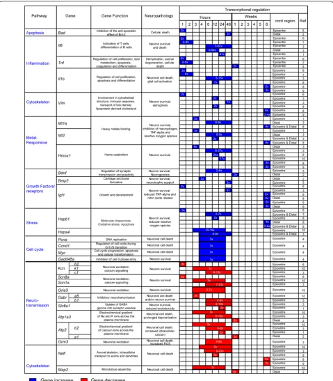
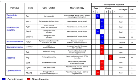
Different molecules and their spatio-temporal activation in the injured tissue may have diverging effects on total cell loss and on tissue regeneration. As such, they deter-mine the outcome of SCI. Mechanical injuries cause necrosis of those neurons directly affected by the force of impact (primary injury phase). A secondary injury phase is characterised by a protracted neuronal loss driven by changes in oxygen, glucose, neuroactive lipids and eicosa-noids homeostasis, by the release of free radicals and bio-genic amines, endogenous opioids and excitatory amino acids [21-27]. The use of large-scale spinal cord tran-scriptional analysis in well-established animal models of SCI has shown the rapid differential regulation of a num-ber of genes, here referred to as earlyinjury genes(within a few hours from injury), and the slower response of others termed as lateinjury genes(more than 48 hours from injury). Expression profiling of injured spinal cord tissue is a powerful method for unearthing the molecular consequences of trauma, particularly if gene expression changes are considered in light of the associated func-tional and histopathological alterations. In Figures 1 and 2 we have reported the main molecular responses which have been described in the rat spinal cord following injury, according to recent pathway analyses of gene expression studies and to other relevant transcriptomic studies of SCI [28,29]. Within each molecular pathway, we have selected some of the most representative differ-entially regulated genes with a very early and a late acti-vation (genes activated in the first few hours from injury and after 48 hours respectively). The figures display the levels of transcriptional regulation and the position of the reported gene expression change with respect to the epi-centre of injury, along with the functional role and the neuropathological changes which have been associated with the differential regulation of each gene.
A number of molecular pathways become activated in an early post-SCI phase (less than 48 hours from SCI; Figure 1) [29-39]. This early response, mostly reported at the epicentre of injury, encompasses biological signals
and early injury geneswhich have opposite effects on
cell survival. Whilst apoptotic and pro-inflammatory responses are likely to be detrimental to cell survival other more protracted growth signals facilitate tissue repair. Among the latter, metallothioneins promote angiogenesis and neuronal re-growth [30,37,38]. Cytos-keletal proteins impact variably on tissue survival. Loss of neurofilaments like the microtubule-associated pro-teins (Map2) for example, prevents neurotoxic protein aggregates disrupting axonal transport, whilst vimentin up-regulation reduces the protracted release from macrophages of toxic reactive oxygen species (ROS) [37,38,40,41]. A reduction in Ca2+ ATPase activity in
the injured tissue causes a neurotoxic increase in intra-cellular calcium and up-regulation of genes modulating cell cycle mostly resulting in neuronal death. In contrast, down-regulation of genes involved in neurotransmission via regulation of sodium and potassium channels as well as AMPA receptors exert an anti-apoptotic effect [30-32,39,42-48]. The change of neurons and axons membrane excitability has been associated to neuronal degeneration in animal models and in neurophysiologi-cal studies conducted on patients with ALS [49,50]. The predominant down-regulation of these signals may thus be seen to play a part in the cell-survival drive.
Delayed molecular responses, mostly identified distally from the injury epicentre, involve the differential regula-tion of lateinjury geneswhich modulate apoptosis, growth, neurotransmission, the homeostasis of the extracellular matrix and of cell metabolism (Figure 2) [28,31,34]. Some of these late responses can have an effect on lipid metabo-lism. For example, the differential regulation of glycerol-3-phosphate dehydrogenase(Gpd1), a mitochondrial enzyme bridging carbohydrate and lipid metabolism, reduces ROS generation whilst the dioxygenase 12-lipoxygenase
(Alox12)may work along the same lines, incorporating oxygen into specific positions of polyunsaturated fatty acids [31,51,52]. The down-regulation of anti-apoptotic genes such ascat,Bag1andBcl2can exert an increase in neuronal cell death [53]. As already mentioned, the late activation of key modulators of membrane excitability also reported to become over-expressed in ALS, such as the hyperpolarization-activated cyclic nucleotide-gated cation channel(Hcn), is likely to impair the functional recovery by enhancement of axonal excitability [54] Similarly to growth factors, heat shock proteins exert a neurorestora-tive effect for neurons, glial and muscle cells, both as a rapid and as a delayed response [32,35,37-39,55].
Increased vulnerability to spinal trauma: what does it hide?
The degree of tissue destruction and the residual neuro-logical disability following SCI depend primarily on the nature of the mechanical stress (e.g. penetrating injuries versus compressive and/or traction type of impact) [29-39], on the different spatial distributions and tem-poral activations that different neurorestorative and neu-rodestructive molecular signals may have, in line with those reported in animal models of SCI (Figures 1 and 2). Certain states modify the response to SCI, including a) the pre-existence of a subclinical neurodegenerative pro-cess, a situation that becomes more likely with aging, and b) the presence of a specific genetic trait which increases the vulnerability to trauma.
Neurodegeneration and the effects of trauma
Figure 1Differentially regulated genes that become activated or inhibited within the first few hours from injury, reported as early
DNA-binding protein 43 (Tardbp)in the brain, a hall-mark of ALS pathology, in individuals who will later be affected by a neuromuscular disorder indistinguishable from ALS [13]. Hence, neurotrauma could initiate an ALS-like neuropathology or worsen a pre-existing sub-clinical ALS state. This concept has been investigated using pre-symptomatic rodent models of ALS, engi-neered using the mutant human superoxide dismutase 1
(SOD1) gene which is found in up to 10% of familial
cases of ALS [28,56,57]. Both pre-symptomatic SOD1
mutated rats and mice showed a poor post-injury loco-motor recovery, compared to wild type littermates, fol-lowing mild compression SCI and sciatic nerve injury respectively [28,56]. In the post-injury phase, the trans-genic rat cord displayed a more robust activation of sev-eral pro-apoptotic genes, cytochrome-C release, a high level of expression of neurofilaments and an early acti-vation of a wide range of inflammatory signals [28]. It has also been possible to identify a significant activation of molecules involved in lipid metabolism, in isoprenoid biosynthesis and in the proteasome ubiquination system, along with a late up-regulation of lysosomal cysteine
proteases and of genes involved in neurotransmission. A more subdued surge of growth-promoting signals at the epicenter of injury is another characteristic of the injured transgenic SOD1spinal cord [28,58-61]. Whilst the post-injury transgenic spinal cord displays an altered transcriptional profile compared to wild type tissue, there are no overt histopathological differences between these tissues with regard to the extension of myelin destruction, motor cell loss and the inflammatory infil-trates caudal to the epicenter of injury [28]. This obser-vation illustrates how SCI in pre-symptomatic animals
carrying a SOD1 gene mutation may not necessarily
cause more structural changes compared to wild type animals, although the trauma may be disruptive enough at a molecular level to instigate functional disruption.
Aging and SCI
Elderly patients have a 5 to 8-fold higher mortality rate fol-lowing SCI compared to younger patients [62-66]. The vul-nerability to SCI in the elderly may be linked to a process of senescence of the brain, involving beta amyloid deposi-tion in neurons and microglia [67-69]. Aging is also one of the most important risk factors for the development of
most neurodegenerative disorders, which manifest clinically after the progressive accumulation of microscopic tissue alterations in the CNS has overcome a certain threshold. Acute or chronic traumatisms may accelerate this process of abnormal protein deposition, leading to the premature surfacing of neurodegenerative conditions. Trauma to the neuroaxis can also enhance the level of protein aggregation, a process that causes the appearance of the histological hallmarks of idiopathic and genetically induced neurode-generative disorders [40,70,71]. The spectrum of protein aggregates observed in neurodegenerative disorders whose expression could be conditioned by trauma includes beta amyloid and phosphorylated tau proteins normally observed within neurofibrillary tangles in Alzheimer’s dis-ease [72], alpha-synuclein within Lewy bodies found in
Par-kinson’s disease [73], neurofilaments in bunina and
spheroids bodies typical of ALS neuropathology and prion protein in Prion disease [71]. Trauma may further impair axonal transport and the functioning of the proteasome system, two molecular functions at the origin of the forma-tion of most toxic protein aggregates.
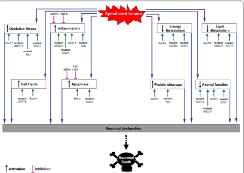
Genes modifying the molecular response to trauma
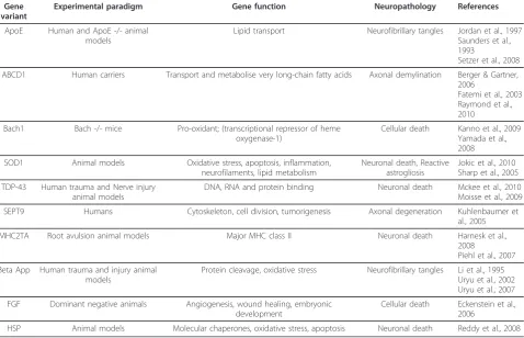
Recent experimental data show how a number of genes may act as modifiers of animals and humans response to
SCI, thus collectively or independently increasing one’s susceptibility to injury (Table 1 and Figure 3). Allelic var-iants of these genes or mutations causing loss or gain of function condition the unraveling of various molecular cascades which are key components of the response to injury (Figure 3). An altered protein cleavage, one of the main driving forces behind protein aggregation in neuro-degenerative disorders, can be further enhanced by trauma in the presence of specific Apolipoprotein E (Apoe) and beta amyloid precursor protein (App) var-iants. TheApoE4allele has been unanimously linked to an increased risk of late onset Alzheimer’s disease and to the development of other neurodegenerative disorders with professional boxing [74]. Loss of Apoe reduces recovery following neurotrauma or ischemic insults, as
shown in Apoe-deficient mice whereas carriers of the
ApoE4 allele have also a 4 to 6-fold increased risk of developing cervical spondylotic myelopathy (CSM) in a situation of chronic spinal cord compression [2,75].Apoe
fragments produced by the trauma-induced proteolytic cleavage of this protein may disrupt the cell’s cytoskele-ton by phosphorylation of tau and promote neurofibril-lary tangles which ultimately cause neuronal death [76,77]. Hence the detrimental effect of theApoE4allele Table 1 Gene modifiers of the response to spinal cord injury and/or to neurotrauma.
Gene variant
Experimental paradigm Gene function Neuropathology References
ApoE Human and ApoE -/- animal models
Lipid transport Neurofibrillary tangles Jordan et al., 1997 Saunders et al., 1993
Setzer et al., 2008
ABCD1 Human carriers Transport and metabolise very long-chain fatty acids Axonal demylination Berger & Gartner, 2006
Fatemi et al., 2003 Raymond et al., 2010
Bach1 Bach -/- mice Pro-oxidant; (transcriptional repressor of heme oxygenase-1)
Cellular death Kanno et al., 2009 Yamada et al., 2008
SOD1 Animal models Oxidative stress, apoptosis, inflammation,
neurofilaments, lipid metabolism
Neuronal death, Reactive astrogliosis
Jokic et al., 2010 Sharp et al., 2005
TDP-43 Human trauma and Nerve injury animal models
DNA, RNA and protein binding Neuronal death Mckee et al., 2010 Moisse et al., 2009
SEPT9 Humans Cytoskeleton, cell division, tumorigenesis Axonal degeneration Kuhlenbaumer et
al., 2005
MHC2TA Root avulsion animal models Major MHC class II Neuronal death Harnesk et al.,
2008
Piehl et al., 2007
Beta App Human trauma and injury animal models
Protein cleavage, oxidative stress Neurofibrillary tangles Li et al., 1995 Uryu et al., 2002 Uryu et al., 2007
FGF Dominant negative animals Angiogenesis, wound healing, embryonic development
Cellular death Eckenstein et al., 2006
HSP Animal models Molecular chaperones, oxidative stress, apoptosis Neuronal death Reddy et al., 2008
in neurodegeneration may be partly due to its higher sus-ceptibility to proteolytic cleavage compared to E2 or E3 isoforms [78]. Similarly, a derangement of proteolysis may explain the increased level of deposition of beta amyloid following trauma, as demonstrated neuropatho-logically in humans and transgenic mice (Tg2576) expressing mutant human beta amyloid precursor pro-tein [52,72,79].
Change in lipid metabolism and in the homeostasis of lipid mediators is another route through which genes are thought to modulate the nervous tissue susceptibility to trauma, similarly to what was previously discussed for
theSOD1gene. For example, beta amyloid is known to
modulate lipid peroxidation whilst Apoe is a lipid-bind-ing protein that is important in the redistribution of lipids among cells in the CNS and in cholesterol trans-port [72,74,78-80]. Mutations of the ATP-binding
cas-sette transporter subfamily D1 (ABCD1) gene, which
encode for defective protein transporters in the peroxi-somal membrane, affect the homeostasis of saturated
and unbranched very long-chain fatty acids. Traumas can precipitate X-linked adrenoleukodystrophy (X-ALD) in young males or a milder variant of this condition
named adrenomyeloneuropathy (AMN) in ABCD1
mutation carriers, a slowly progressive axonopathy in the spinal cord tracts and in the peripheral nerves [81-84]. In some cases, neuroimaging has shown how the pathological expression of the disease following trauma remains confined to the CNS and to the spinal cord areas more directly affected by the mechanical injury [83,85-89].
Several of the genes mentioned above are likely to mod-ulate tissue vulnerability to mechanical trauma through oxidative stress, an important determinant of SCI-induced secondary injury neuronal loss. X-ALD post mortem brains [90] and mouse model of X-ALD [91] show signifi-cant levels of oxidative damage. Mice deficient in Bach1, a transcriptional repressor of the heme oxygenase-1
(Hmox1)gene which has a cytoprotective and anti-oxidant effect, showed a better profile of functional recovery
following moderate SCI and a significant smaller area of injury [92,93].ABCD1can also give rise to inflammatory-related demyelination [81]. Trauma-induced lipid
peroxi-dation in mutantAppanimals is also pointing towards
oxidative stress as well as a deranged lipid metabolism as important factors in the determination of susceptibility to trauma [79].
Variants of genes exerting control over the inflammatory response, like polymorphisms of theCiita (alternative symbol: Mhc2ta)have been reported to be associated with both lower expression of MHC class II-associated genes and with an increased neurodestruction in animal models of root avulsion injury [94,95].ApoE4increases the inflam-matory tone following neurotrauma with a significant surge of Il6, Tnf and nitric oxide in the injured tissue [96]. Heat shock proteins (HSPs) are intracellular stress-respon-sive molecular chaperons, which participate in the second-ary injury phase by scavenging damaged proteins. Whilst universally known to provide an effective clearance of abnormal proteins, their recognized motor-cell sparing effect in SCI effect is linked to their ability to prevent chronic inflammation, once these proteins are released by acutely stressed microglial, endothelial, and ependymal cells, [97].
The silencing or ablation in dominant-negative animals of the fibroblast growth factor receptor (Fgfr), which is known to inhibit fibroblast growth factor (Fgf) signaling, does not appear to cause any overt neurological disorder. However, this genetic manipulation seems to induce a higher level of neuronal vulnerability to a stab injury to the spinal cord in mice [98]. Endogenous Fgf released by astrocytes and neurons after mechanical injury is thought to counteract the excitotoxic or ischemic damage by acti-vating anti-apoptotic signals in stressed neurons [98].
Mannose binding lectin (Mbl1/2)-deficient mice have been described to show exacerbated CA3 cell death and remarkable behavioral changes after traumatic brain injury, compared to wild type mice [99]. Mannose bind-ing lectin is a glycoprotein of the collectin family that plays an important role in the host’s initial response to infection by initiating complement activation and pro-moting phagocytosis by leukocytes [100]. The septin-9
(SEPT9)gene has been associated to an increased sus-ceptibility to develop a form of brachial plexus pathology as a result of different stressors including immunizations and traumas [3].SEPT9belongs to the septin family of proteins, GTPases active on cell cycle and on cytoskeletal components, including microtubules and actin [3].
Conclusions: SCI and the molecular point of no return
The neurological impairment induced by SCI may gra-dually subside or, despite comprehensive rehabilitative efforts over a period of time, turn into an irreversible
functional deficits. More atypical post-injury clinical pic-tures include localized, non-progressive as well as dif-fused and evolving forms of amyotrophy, neurological pictures very close to what observed in MND [101]. In some other cases, protracted and repetitive mechanical stress like the strenuous use of a limb due to particular occupational exposures or professional sports have been linked to the development of recurrent painful brachial plexus neuropathies, with features of muscle weakness and atrophy as well as sensory loss, similarly to what seen in hereditary neuralgic amyotrophies [3]. Whether permanent or progressive, the neurological conse-quences of trauma reflect a complex interplay of genetic and environmental factors, which condition an indivi-dual’s susceptibility to withstand injury. This paper has embraced the body of experimental data describing genes which may potentially modulate susceptibility to trauma, in order to dissect those molecular events that may be responsible of the establishment of irreversible neurodegeneration in the post-injury phase, here defined as the“point of no return”.
lipid and inflammatory mediators, as well as of neurofila-ments are examples of complex molecular signals involved in the modulation of irreversible neurodegeneration in dif-ferent pathological contexts, particularly in ALS [102,103]. The up-regulation of lipids in the post-injury phase is in line with what has been reported in ALS patients and in animal models of ALS, where an early derangement of mediators of lipid homeostasis is a distinctive feature of the pathology and may be part of a rescue mechanism of degenerating neurons [102].
The modality of mechanical force applied to the spinal cord and the level of tissue penetration account also for
the different post-injury behaviour in the same SOD1
gene mutated rat model. Compression and stabbing spinal cord injuries on the pre-symptomatic G93A-SOD1 rat model of ALS, for example, evoke completely differ-ent tissue responses at both molecular and cellular level [28,57]. Surviving motor neurons in the G93A-SOD1 rodents subjected to compression SCI undergo signifi-cant atrophy when compared to wild type littermates, a feature not seen using penetrating injuries in the same animal model [28,57].
Both in animal models of most neurodegenerative disor-ders and in real life, neurotrauma may precipitate the pathological process which is already altering the fine structure and the function of a macroscopically intact tis-sue. The injury may simply accelerate the course of neuro-degeneration, which would have otherwise followed a different time line. Aging is clearly an important factor in this interaction, as it is an important risk factor for the development of neurodegenerative disorders and of the subtle molecular changes that pre-date the main clinical manifestations of most neurological conditions.
Understanding the molecular framework of the response to SCI in relationship to aging and to the pre-sence of a potential underlying genetic vulnerability is an essential precondition for the development of disease-modifying treatments, of prognostic biomarkers and to monitor the response to a targeted and timely treatment strategy. A better knowledge of the molecular framework which conditions the outcome from neurotrauma is also an ideal ground for a better understanding of the wider concept of both idiopathic and genetically-induced neurodegeneration.
List of abbreviations
ALS: Amyotrophic lateral sclerosis; ALD: Adrenoleukodystrophy;Alox12: 12-Lipoxygenase; AMN: Adrenomyeloneuropathy; Apoe: Apolipoprotein;ApoE4: Apolipoprotein E4 allele;App: Amyloid beta (A4) precursor protein;Atp1a3: ATPase, Na+/K+ transporting, alpha 3 polypeptide;Atp2a1: ATPase, Ca++ transporting, cardiac muscle, fast twitch 1;Atp2b2: ATPase, Ca++
transporting, plasma membrane 2;Bad: Bcl2 associated agonist of cell death; Bach1: BTB and CNC homology 1, basic leucine zipper transcription factor 1; Bag1: Bcl2-associated athanogene;Bcl2: B-cell CLL/lymphoma 2;Bdnf: Brain-derived neurotrophic factor;Bmp2: Bone morphogenetic protein 2;Bmpr1a:
Bone morphogenetic protein receptor, type IA;Cat: Catalase;Ccnd1: cyclin D1;Ciita: Class II, major histocompatibility complex, transactivator; CSM: Cervical spondylotic myelopathy;Dcn: Decorin; fgf: Fibroblast growth factor; Fgfr1: Fibroblast growth factor receptor 1; GABA: gamma-aminobutyric acid; Gabbr1: GABA B receptor;Gabra5: GABA A receptor, alpha 5;Gabrb1: GABA A receptor, beta 1Gabbr2: GABA B receptor 2;Gadd45aGrowth arrest and DNA-damage-inducible gene 45a;Gpd1: Glycerol-3-phosphate dehydrogenase 1;Gria3: ionotropic glutamate receptor 3;Grm3:
Metabotropic glutamate receptor 3;Hcn2: Hyperpolarization activated cyclic nucleotide-gated potassium channel 2;Hmox1: Heme oxygenase (decycling) 1; HSPs: Heat shock proteins;Hspb1: Heat shock 27kDa protein 1;Hspa4: Heat shock protein 70KDa protein 4;Igf1: Insulin-like growth factor 1;Il1b: Interleukin 1 beta;Il6: Interleukin 6;Kcnc1: Potassium voltage gated channel, Shaw-related subfamily, member 1;Kcnh2: Potassium voltage-gated channel, subfamily H (eag-related), member 2;Kcnk1: Potassium channel, subfamily K, member 1;Map2: Microtubule-associated protein 2;Mbl1/2: Mannose-binding lectin (protein A and C) 1 and 2; MND: Motor neurons disease; MMP: Matrix metalloproteinase;Mt1a: Metallothionein 1a; Mt2: Metallothionein II;Myc: Myelocytomatosis oncogene;Nefl: Neurofilament light polypeptide;Ngfr: Low-affinity nerve growth factor;Ntrk2; Neurotrophic tyrosine kinase receptor type 2;Pcna: Proliferating cell nuclear antigen; ROS: Reactive oxygen species;SEPT9; Septin 9;Scn1a: sodium channel, voltage-gated, type I, alpha;Scn8a: odium channel, voltage gated, type VIII, alpha subunit;Slc6a1: solute carrier family 6 (neurotransmitter transporter, GABA), member 1;SOD1: Superoxide dismutase 1;Tardbp: TAR DNA binding protein; Tnf: Tumor necrosis factor;Vim: Vimentin.
Acknowledgements
We are grateful to The Royal London Hospital Charitable Foundation and to the Motor Neuron Disease Association UK for its financial support to our research projects on ALS.
Authors’contributions
PKY & AM: Equal contribution to the writing of this manuscript and approval of the final submitted text.
Competing interests
The authors declare that they have no competing interests.
Received: 17 August 2011 Accepted: 8 February 2012 Published: 8 February 2012
References
1. Ghatak NR, Campbell WW, Lippman RH, Hadfield MG:Anterior horn changes of motor neuron disease associated with demyelinating radiculopathy.J Neuropathol Exp Neurol1986,45:385-395. 2. Setzer M, Hermann E, Seifert V, Marquardt G:Apolipoprotein E gene
polymorphism and the risk of cervical myelopathy in patients with chronic spinal cord compression.Spine (Phila Pa 1976)2008,33:497-502. 3. Kuhlenbaumer G, Hannibal MC, Nelis E, Schirmacher A, Verpoorten N,
Meuleman J, Watts GD, De VE, Young P, Stogbauer F,et al:Mutations in SEPT9 cause hereditary neuralgic amyotrophy.Nat Genet2005, 37:1044-1046.
4. Leigh PN, Abrahams S, Al-Chalabi A, Ampong MA, Goldstein LH, Johnson J, Lyall R, Moxham J, Mustfa N, Rio A,et al:The management of motor neurone disease.J Neurol Neurosurg Psychiatry2003,74(Suppl 4):iv32-iv47. 5. Abel EL:Football increases the risk for Lou Gehrig’s disease, amyotrophic
lateral sclerosis.Percept Mot Skills2007,104:1251-1254.
6. Binazzi A, Belli S, Uccelli R, Desiato MT, Talamanca IF, Antonini G, Corsi FM, Scoppetta C, Inghilleri M, Pontieri FE,et al:An exploratory case-control study on spinal and bulbar forms of amyotrophic lateral sclerosis in the province of Rome.Amyotroph Lateral Scler2009,10:361-369.
7. Bracco L, Antuono P, Amaducci L:Study of epidemiological and etiological factors of amyotrophic lateral sclerosis in the province of Florence, Italy.Acta Neurol Scand1979,60:112-124.
8. Chen H, Richard M, Sandler DP, Umbach DM, Kamel F:Head injury and amyotrophic lateral sclerosis.Am J Epidemiol2007,166:810-816. 9. Chio A, Benzi G, Dossena M, Mutani R, Mora G:Severely increased risk of
10. Chio A, Calvo A, Dossena M, Ghiglione P, Mutani R, Mora G:ALS in Italian professional soccer players: the risk is still present and could be soccer-specific.Amyotroph Lateral Scler2009,10:205-209.
11. Kondo K, Tsubaki T:Case-control studies of motor neuron disease: association with mechanical injuries.Arch Neurol1981,38:220-226. 12. Matser JT, Kessels AG, Lezak MD, Troost J:A dose-response relation of
headers and concussions with cognitive impairment in professional soccer players.J Clin Exp Neuropsychol2001,23:770-774.
13. McKee AC, Gavett BE, Stern RA, Nowinski CJ, Cantu RC, Kowall NW, Perl DP, Hedley-Whyte ET, Price B, Sullivan C,et al:TDP-43 proteinopathy and motor neuron disease in chronic traumatic encephalopathy.J Neuropathol Exp Neurol2010,69:918-929.
14. Riggs JE:Antecedent trauma and amyotrophic lateral sclerosis in young adult men.Mil Med1993,158:55-57.
15. Riggs JE:The latency between traumatic axonal injury and the onset of amyotrophic lateral sclerosis in young adult men.Mil Med2001, 166:731-732.
16. Schmidt S, Kwee LC, Allen KD, Oddone EZ:Association of ALS with head injury, cigarette smoking and APOE genotypes.J Neurol Sci2010, 291:22-29.
17. Strickland D, Smith SA, Dolliff G, Goldman L, Roelofs RI:Physical activity, trauma, and ALS: a case-control study.Acta Neurol Scand1996,94:45-50. 18. Wicks P, Ganesalingham J, Collin C, Prevett M, Leigh NP, Al-Chalabi A:Three
soccer playing friends with simultaneous amyotrophic lateral sclerosis. Amyotroph Lateral Scler2007,8:177-179.
19. Kihira T, Kanno S, Miwa H, Okamoto K, Kondo T:The role of exogenous risk factors in amyotrophic lateral sclerosis in Wakayama, Japan. Amyotroph Lateral Scler2007,8:150-156.
20. Yamada M, Furukawa Y, Hirohata M:Amyotrophic lateral sclerosis: frequent complications by cervical spondylosis.J Orthop Sci2003, 8:878-881.
21. Bareyre FM, Schwab ME:Inflammation, degeneration and regeneration in the injured spinal cord: insights from DNA microarrays.Trends Neurosci 2003,26:555-563.
22. DeWitt DS, Prough DS, Taylor CL, Whitley JM:Reduced cerebral blood flow, oxygen delivery, and electroencephalographic activity after traumatic brain injury and mild hemorrhage in cats.J Neurosurg1992, 76:812-821.
23. Kruman II, Mattson MP:Pivotal role of mitochondrial calcium uptake in neural cell apoptosis and necrosis.J Neurochem1999,72:529-540. 24. Pedersen MO, Jensen R, Pedersen DS, Skjolding AD, Hempel C, Maretty L,
Penkowa M:Metallothionein-I+II in neuroprotection.Biofactors2009, 35:315-325.
25. Takahashi H, Manaka S, Sano K:Changes in extracellular potassium concentration in cortex and brain stem during the acute phase of experimental closed head injury.J Neurosurg1981,55:708-717. 26. Yamakami I, McIntosh TK:Effects of traumatic brain injury on regional
cerebral blood flow in rats as measured with radiolabeled microspheres. J Cereb Blood Flow Metab1989,9:117-124.
27. Zemper ED:Analysis of cerebral concussion frequency with the most commonly used models of football helmets.J Athl Train1994,29:44-50. 28. Jokic N, Yip PK, Michael-Titus A, Priestley JV, Malaspina A:The human
G93A-SOD1 mutation in a pre-symptomatic rat model of amyotrophic lateral sclerosis increases the vulnerability to a mild spinal cord compression.BMC Genomics2010,11:633.
29. Malaspina A, Jokic N, Huang WL, Priestley JV:Comparative analysis of the time-dependent functional and molecular changes in spinal cord degeneration induced by the G93A SOD1 gene mutation and by mechanical compression.BMC Genomics2008,9:500.
30. Aimone JB, Leasure JL, Perreau VM, Thallmair M:Spatial and temporal gene expression profiling of the contused rat spinal cord.Exp Neurol 2004,189:204-221.
31. Bareyre FM, Haudenschild B, Schwab ME:Long-lasting sprouting and gene expression changes induced by the monoclonal antibody IN-1 in the adult spinal cord.J Neurosci2002,22:7097-7110.
32. Carmel JB, Galante A, Soteropoulos P, Tolias P, Recce M, Young W, Hart RP: Gene expression profiling of acute spinal cord injury reveals spreading inflammatory signals and neuron loss.Physiol Genomics2001,7:201-213. 33. Di Giovanni S, Knoblach SM, Brandoli C, Aden SA, Hoffman EP, Faden AI:
Gene profiling in spinal cord injury shows role of cell cycle in neuronal death.Ann Neurol2003,53:454-468.
34. Fan M, Mi R, Yew DT, Chan WY:Analysis of gene expression following sciatic nerve crush and spinal cord hemisection in the mouse by microarray expression profiling.Cell Mol Neurobiol2001,21:497-508. 35. Nesic O, Svrakic NM, Xu GY, McAdoo D, Westlund KN, Hulsebosch CE, Ye Z,
Galante A, Soteropoulos P, Tolias P,et al:DNA microarray analysis of the contused spinal cord: effect of NMDA receptor inhibition.J Neurosci Res 2002,68:406-423.
36. Pan JZ, Ni L, Sodhi A, Aguanno A, Young W, Hart RP:Cytokine activity contributes to induction of inflammatory cytokine mRNAs in spinal cord following contusion.J Neurosci Res2002,68:315-322.
37. Resnick DK, Schmitt C, Miranpuri GS, Dhodda VK, Isaacson J, Vemuganti R: Molecular evidence of repair and plasticity following spinal cord injury. Neuroreport2004,15:837-839.
38. Schmitt C, Miranpuri GS, Dhodda VK, Isaacson J, Vemuganti R, Resnick DK: Changes in spinal cord injury-induced gene expression in rat are strain-dependent.Spine J2006,6:113-119.
39. Song G, Cechvala C, Resnick DK, Dempsey RJ, Rao VL:GeneChip analysis after acute spinal cord injury in rat.J Neurochem2001,79:804-815. 40. Lin H, Schlaepfer WW:Role of neurofilament aggregation in motor
neuron disease.Ann Neurol2006,60:399-406.
41. Mor-Vaknin N, Punturieri A, Sitwala K, Markovitz DM:Vimentin is secreted by activated macrophages.Nat Cell Biol2003,5:59-63.
42. Lees GJ:Inhibition of sodium-potassium-ATPase: a potentially ubiquitous mechanism contributing to central nervous system neuropathology. Brain Res Brain Res Rev1991,16:283-300.
43. Noh KM, Yokota H, Mashiko T, Castillo PE, Zukin RS, Bennett MV:Blockade of calcium-permeable AMPA receptors protects hippocampal neurons against global ischemia-induced death.Proc Natl Acad Sci USA2005, 102:12230-12235.
44. Palmer AM, Carter N:The role of sodium channels in disease.Drug News Perspect2001,14:568-576.
45. Spillson AB, Russell JW:Metabotropic glutamate receptor regulation of neuronal cell death.Exp Neurol2003,184(Suppl 1):S97-105.
46. Yu SP, Yeh CH, Sensi SL, Gwag BJ, Canzoniero LM, Farhangrazi ZS, Ying HS, Tian M, Dugan LL, Choi DW:Mediation of neuronal apoptosis by enhancement of outward potassium current.Science1997,278:114-117. 47. Zeevalk GD, Nicklas WJ:Attenuation of excitotoxic cell swelling and GABA
release by the GABA transport inhibitor SKF 89976A.Mol Chem Neuropathol1996,29:27-36.
48. Zeevalk GD, Nicklas WJ:Activity at the GABA transporter contributes to acute cellular swelling produced by metabolic impairment in retina. Vision Res1997,37:3463-3470.
49. Meehan CF, Moldovan M, Marklund SL, Graffmo KS, Nielsen JB, Hultborn H: Intrinsic properties of lumbar motor neurones in the adult G127insTGGG superoxide dismutase-1 mutant mouse in vivo: evidence for increased persistent inward currents.Acta Physiol (Oxf)2010,200:361-376. 50. Shibuya K, Misawa S, Arai K, Nakata M, Kanai K, Yoshiyama Y, Ito K, Isose S,
Noto Y, Nasu S,et al:Markedly reduced axonal potassium channel expression in human sporadic amyotrophic lateral sclerosis: An immunohistochemical study.Exp Neurol2011,232:149-153. 51. Brand MD:The sites and topology of mitochondrial superoxide
production.Exp Gerontol2010,45:466-472.
52. Li Y, Maher P, Schubert D:A role for 12-lipoxygenase in nerve cell death caused by glutathione depletion.Neuron1997,19:453-463.
53. Pedersen MO, Larsen A, Stoltenberg M, Penkowa M:Cell death in the injured brain: roles of metallothioneins.Prog Histochem Cytochem2009, 44:1-27.
54. Chu HY, Zhen X:Hyperpolarization-activated, cyclic nucleotide-gated (HCN) channels in the regulation of midbrain dopamine systems.Acta Pharmacol Sin2010,31:1036-1043.
55. Brown IR:Heat shock proteins and protection of the nervous system. Ann N Y Acad Sci2007,1113:147-158.
56. Sharp PS, Dick JR, Greensmith L:The effect of peripheral nerve injury on disease progression in the SOD1(G93A) mouse model of amyotrophic lateral sclerosis.Neuroscience2005,130:897-910.
57. Suzuki M, Klein S, Wetzel EA, Meyer M, McHugh J, Tork C, Hayes A, Svendsen CN:Acute glial activation by stab injuries does not lead to overt damage or motor neuron degeneration in the G93A mutant SOD1 rat model of amyotrophic lateral sclerosis.Exp Neurol2010,221:346-352. 58. Ferraiuolo L, Heath PR, Holden H, Kasher P, Kirby J, Shaw PJ:Microarray
progression of motor neuron injury in the SOD1 G93A mouse model of familial ALS.J Neurosci2007,27:9201-9219.
59. Kabashi E, Durham HD:Failure of protein quality control in amyotrophic lateral sclerosis.Biochim Biophys Acta2006,1762:1038-1050.
60. Kudo LC, Parfenova L, Vi N, Lau K, Pomakian J, Valdmanis P, Rouleau GA, Vinters HV, Wiedau-Pazos M, Karsten SL:Integrative gene-tissue microarray-based approach for identification of human disease biomarkers: application to amyotrophic lateral sclerosis.Hum Mol Genet 2010,19:3233-3253.
61. Lobsiger CS, Boillee S, Cleveland DW:Toxicity from different SOD1 mutants dysregulates the complement system and the neuronal regenerative response in ALS motor neurons.Proc Natl Acad Sci USA 2007,104:7319-7326.
62. Fassett DR, Harrop JS, Maltenfort M, Jeyamohan SB, Ratliff JD, Anderson DG, Hilibrand AS, Albert TJ, Vaccaro AR, Sharan AD:Mortality rates in geriatric patients with spinal cord injuries.J Neurosurg Spine2007,7:277-281. 63. Furlan JC, Fehlings MG:The impact of age on mortality, impairment, and
disability among adults with acute traumatic spinal cord injury.J Neurotrauma2009,26:1707-1717.
64. Jackson AP, Haak MH, Khan N, Meyer PR:Cervical spine injuries in the elderly: acute postoperative mortality.Spine (Phila Pa 1976)2005, 30:1524-1527.
65. Kuhne CA, Ruchholtz S, Kaiser GM, Nast-Kolb D:Mortality in severely injured elderly trauma patients–when does age become a risk factor? World J Surg2005,29:1476-1482.
66. Scivoletto G, Morganti B, Ditunno P, Ditunno JF, Molinari M:Effects on age on spinal cord lesion patients’rehabilitation.Spinal Cord2003,41:457-464. 67. Rodrigue KM, Kennedy KM, Park DC:Beta-amyloid deposition and the
aging brain.Neuropsychol Rev2009,19:436-450.
68. Scheibel ME, Lindsay RD, Tomiyasu U, Scheibel AB:Progressive dendritic changes in aging human cortex.Exp Neurol1975,47:392-403.
69. Streit WJ, Sammons NW, Kuhns AJ, Sparks DL:Dystrophic microglia in the aging human brain.Glia2004,45:208-212.
70. Anderton BH:Changes in the ageing brain in health and disease.Philos Trans R Soc Lond B Biol Sci1997,352:1781-1792.
71. Lindner AB, Demarez A:Protein aggregation as a paradigm of aging. Biochim Biophys Acta2009,1790:980-996.
72. Uryu K, Chen XH, Martinez D, Browne KD, Johnson VE, Graham DI, Lee VM, Trojanowski JQ, Smith DH:Multiple proteins implicated in
neurodegenerative diseases accumulate in axons after brain trauma in humans.Exp Neurol2007,208:185-192.
73. Eller M, Williams DR:alpha-Synuclein in Parkinson disease and other neurodegenerative disorders.Clin Chem Lab Med2011,49:403-408. 74. Jordan BD, Relkin NR, Ravdin LD, Jacobs AR, Bennett A, Gandy S:
Apolipoprotein E epsilon4 associated with chronic traumatic brain injury in boxing.JAMA1997,278:136-140.
75. Jha A, Lammertse DP, Coll JR, Charlifue S, Coughlin CT, Whiteneck GG, Worley G:Apolipoprotein E epsilon4 allele and outcomes of traumatic spinal cord injury.J Spinal Cord Med2008,31:171-176.
76. Strittmatter WJ, Weisgraber KH, Huang DY, Dong LM, Salvesen GS, Pericak-Vance M, Schmechel D, Saunders AM, Goldgaber D, Roses AD:Binding of human apolipoprotein E to synthetic amyloid beta peptide: isoform-specific effects and implications for late-onset Alzheimer disease.Proc Natl Acad Sci USA1993,90:8098-8102.
77. Xu Q, Walker D, Bernardo A, Brodbeck J, Balestra ME, Huang Y:Intron-3 retention/splicing controls neuronal expression of apolipoprotein E in the CNS.J Neurosci2008,28:1452-1459.
78. Mahley RW:Apolipoprotein E: cholesterol transport protein with expanding role in cell biology.Science1988,240:622-630. 79. Uryu K, Laurer H, McIntosh T, Pratico D, Martinez D, Leight S, Lee VM,
Trojanowski JQ:Repetitive mild brain trauma accelerates Abeta deposition, lipid peroxidation, and cognitive impairment in a transgenic mouse model of Alzheimer amyloidosis.J Neurosci2002,22:446-454. 80. Saunders AM, Strittmatter WJ, Schmechel D, George-Hyslop PH,
Pericak-Vance MA, Joo SH, Rosi BL, Gusella JF, Crapper-MacLachlan DR, Alberts MJ, et al:Association of apolipoprotein E allele epsilon 4 with late-onset familial and sporadic Alzheimer’s disease.Neurology1993,43:1467-1472. 81. Berger J, Gartner J:X-linked adrenoleukodystrophy: clinical, biochemical and pathogenetic aspects.Biochim Biophys Acta2006,1763:1721-1732. 82. Moser HW, Raymond GV, Dubey P:Adrenoleukodystrophy: new
approaches to a neurodegenerative disease.JAMA2005,294:3131-3134.
83. Raymond GV, Seidman R, Monteith TS, Kolodny E, Sathe S, Mahmood A, Powers JM:Head trauma can initiate the onset of
adreno-leukodystrophy.J Neurol Sci2010,290:70-74.
84. Schaumburg HH, Powers JM, Raine CS, Suzuki K, Richardson EP Jr: Adrenoleukodystrophy. A clinical and pathological study of 17 cases. Arch Neurol1975,32:577-591.
85. Carmant L, Decarie JC, Fon E, Shevell MI:Transient visual symptoms as the initial manifestation of childhood adrenoleukodystrophy.Pediatr Neurol1998,19:62-64.
86. Fatemi A, Barker PB, Ulug AM, Nagae-Poetscher LM, Beauchamp NJ, Moser AB, Raymond GV, Moser HW, Naidu S:MRI and proton MRSI in women heterozygous for X-linked adrenoleukodystrophy.Neurology 2003,60:1301-1307.
87. Turpin JC, Paturneau-Jouas M, Sereni C, Pluot M, Baumann N:Adult disclosure of a case of familial adrenoleukodystrophy.Rev Neurol (Paris) 1985,141:289-295.
88. Weller M, Liedtke W, Petersen D, Opitz H, Poremba M:Very-late-onset adrenoleukodystrophy: possible precipitation of demyelination by cerebral contusion.Neurology1992,42:367-370.
89. Wilkinson IA, Hopkins IJ, Pollard AC:Can head injury influence the site of demyelination in adrenoleukodystrophy?Dev Med Child Neurol1987, 29:797-800.
90. Gilg AG, Singh AK, Singh I:Inducible nitric oxide synthase in the central nervous system of patients with X-adrenoleukodystrophy.J Neuropathol Exp Neurol2000,59:1063-1069.
91. Powers JM, Pei Z, Heinzer AK, Deering R, Moser AB, Moser HW, Watkins PA, Smith KD:Adreno-leukodystrophy: oxidative stress of mice and men.J Neuropathol Exp Neurol2005,64:1067-1079.
92. Kanno H, Ozawa H, Dohi Y, Sekiguchi A, Igarashi K, Itoi E:Genetic ablation of transcription repressor Bach1 reduces neural tissue damage and improves locomotor function after spinal cord injury in mice.J Neurotrauma2009,26:31-39.
93. Yamada K, Tanaka N, Nakanishi K, Kamei N, Ishikawa M, Mizuno T, Igarashi K, Ochi M:Modulation of the secondary injury process after spinal cord injury in Bach1-deficient mice by heme oxygenase-1.J Neurosurg Spine2008,9:611-620.
94. Harnesk K, Swanberg M, Ockinger J, Diez M, Lidman O, Wallstrom E, Lobell A, Olsson T, Piehl F:Vra4 congenic rats with allelic differences in the class II transactivator gene display altered susceptibility to experimental autoimmune encephalomyelitis.J Immunol2008, 180:3289-3296.
95. Piehl F, Swanberg M, Lidman O:The axon reaction: identifying the genes that make a difference.Physiol Behav2007,92:67-74.
96. Colton CA, Brown CM, Vitek MP:Sex steroids, APOE genotype and the innate immune system.Neurobiol Aging2005,26:363-372.
97. Reddy SJ, La MF, Park P:The role of heat shock proteins in spinal cord injury.Neurosurg Focus2008,25:E4.
98. Eckenstein FP, McGovern T, Kern D, Deignan J:Neuronal vulnerability in transgenic mice expressing an inducible dominant-negative FGF receptor.Exp Neurol2006,198:338-349.
99. Yager PH, You Z, Qin T, Kim HH, Takahashi K, Ezekowitz AB, Stahl GL, Carroll MC, Whalen MJ:Mannose binding lectin gene deficiency increases susceptibility to traumatic brain injury in mice.J Cereb Blood Flow Metab 2008,28:1030-1039.
100. Takahashi K, Ip WE, Michelow IC, Ezekowitz RA:The mannose-binding lectin: a prototypic pattern recognition molecule.Curr Opin Immunol 2006,18:16-23.
101. Cerami C, Valentino F, Piccoli F, La BV:A cervical myelopathy with a Hirayama disease-like phenotype.Neurol Sci2008,29:451-454. 102. Dupuis L, Corcia P, Fergani A, Gonzalez De Aguilar JL,
Bonnefont-Rousselot D, Bittar R, Seilhean D, Hauw JJ, Lacomblez L, Loeffler JP,et al: Dyslipidemia is a protective factor in amyotrophic lateral sclerosis. Neurology2008,70:1004-1009.
103. Kim SM, Kim H, Kim JE, Park KS, Sung JJ, Kim SH, Lee KW:Amyotrophic lateral sclerosis is associated with hypolipidemia at the presymptomatic stage in mice.PLoS One2011,6:e17985.
doi:10.1186/1750-1326-7-6
Cite this article as:Yip and Malaspina:Spinal cord trauma and the