Potentiation of Antimicrobial Activity of Roxithromycin -An In Vitro Study
Sumi Sebastian, V. M. Subrahmanyam*, J. Venkata Rao, C. S. Shreedhara and B. V. Kamath.
Manipal College of Pharmaceutical Sciences, Manipal University, Manipal-576104
Summary
Since the time of antibiotic discovery and use of antibiotics as chemotherapeutic agents, there has been a belief that this would lead to the eradication of infectious diseases. However, diseases that were once thought to have been controlled by antibiotics are returning in new forms and are becoming resistant to antibiotic therapies. Finding agents which potentiate antimicrobial activity could circumvent this problem. Present study was envisaged to study the in vitro activity of potentiating agents viz., caffeine, EDTA, citric acid, tartaric acid, theophylline and trisodium citrate in combination with Roxithromycin, a macrolide antibiotic. It acts by blocking the 50S ribosomal subunit required for protein synthesis. The combination was tested against both Gram-positive (Bacillus subtilis, Staphylococcus aureus) and Gram-negative (Escherichia coli, Pseudomonas aeruginosa) organisms. Effect of potentiating agents on Roxithromycin was examined by determination of MIC, agar diffusion technique and turbidimetric analysis. Among the potentiating agents tested, it was found that EDTA and caffeine showed maximum activity in reducing MIC, increasing the zone diameter and reducing growth.
Key words: Antibiotic Resistance, Macrolide, Potentiating agents, Minimum Inhibitory
Concentration
Address of communication, *corresponding author
Dr. V. M. Subrahmanyam, Professor,
Department of Pharmaceutical Biotechnology,
Manipal College of Pharmaceutical Sciences,
Manipal University, Manipal-576104, Karnataka, India.
Email: [email protected], [email protected]
Telephone: 0091 820 2922482. Fax: 0091 820 2571998
Introduction
Since the time of antibiotic discovery and use of antibiotics as chemotherapeutic agents, there has been a belief that this would lead to the eradication of infectious diseases. However, diseases that were once thought to have been controlled by antibiotics are returning in new forms and are becoming resistant to antibiotic therapies2.
Infectious microbes have a remarkable ability to adapt and develop drug resistance in an unpredictable and dynamic manner. Bacterial resistance to an antimicrobial agent could mainly be due to drug not reaching its site due to impermeability factor3 or alteration in target site4. To overcome this problem, pharmaceutical companies and several research institutions are nowadays hunting for new methodologies to meet the demand to fight troublesome infections, namely by the use of potentiating agents.
As a step in this direction a study was carried out to examine the action of potentiating agents on Roxithromycin, a macrolide antibiotic produced by various strains of Streptomyces. This antibiotic acts by blocking the 50S ribosomal subunit needed for protein synthesis.
Material and methods
Antimicrobial and potentiating agents: Standard laboratory powder of Roxithromycin
(Wockhardt, Mumbai) was used in the study. The potentiating agents used were caffeine (Loba chemie, Mumbai), citric acid (Sigma Aldrich, Germany), theophylline (Genuine chemical co ., Mumbai Ltd.), Disodium EDTA (S.D fine chem. Ltd., Mumbai), trisodium citrate (Nice chemicals Pvt Ltd., Cochin) and tartaric acid (Merck Ltd., Mumbai).
Preparation of antibiotic solution: Stock solution of Roxithromycin was prepared using Ethanol (95%) as a solvent. Aliquots were distributed into 2 mL eppendorff tubes and stored. Working standards were prepared using water as a diluent.
Microorganisms and media: The organisms employed for the purpose were Escherichia coli (NCIM 2345), Bacillus subtilis (NCIM 2063), Staphylococcus aureus (NCIM 2079) and Pseudomonas aeruginosa (NCIM 2036). The identities of the microorganisms were confirmed by standard biochemical tests and maintained as per standard guidelines.
Muller Hinton broth (MH broth), nutrient broth, nutrient agar, Muller Hinton (MH) agar, EMB agar, antibiotic assay medium, mannitol salt agar (Hi-media lab Pvt. Ltd., Mumbai) were used in the study.
Determination of Minimum Inhibitory Concentration for antimicrobial and potentiating agents: Minimum Inhibitory Concentration (MIC) of potentiating agents and Roxithromycin alone were determined by tube dilution method. The inocula contained 5x10⁵ CFU/mL. The concentration range used for the study were Roxithromycin 0.39-50 µg/mL, theophylline 156.25-10,000 µg/mL, EDTA 3.125-200 µg/mL, caffeine 78.125-5,000 µg/mL, Trisodium citrate 156.25-10,000 µg/mL, tartaric acid 156.25-10,000 µg/mL and citric acid 156.25-10,000 µg/mL.
less than the obtained MIC values to avoid the chance of these agents themselves showing antibacterial activity.
Agar diffusion method: The bacterial inoculum was swabbed uniformly onto nutrient agar plate and the compounds to be checked were placed in wells (6 mm dia.). Initially, potentiating agents in three different concentrations (1,000, 500 and 250 µg/mL for citric acid), (200, 100 and 50 µg/mL for EDTA), (1,000, 500 and 250 µg/mL for tartaric acid), (2,000, 1,000 and 500 µg/mL for tri sodium citrate), (2,000, 1,000 and 500 µg/mL for caffeine) and (2,000, 1,000 and 500 µg/mL for theophylline) were checked by agar diffusion method. Further study was carried out with the concentration at which no zone of inhibition was observed.
The study was carried out by two designs:
1) In the first design, the medium mixed with potentiating agent was seeded and plated. The antibiotic alone was transferred into the wells.
2) In the second design, antibiotic mixed with potentiating agent was added to wells bored into seeded agar medium.
The concentration of Roxithromycin used was 20µg/mL. The plates were kept at 4°C for 1 h to allow diffusion of the antibiotic. This was followed by incubation at 37°C for 12 h. Zone diameters were measured by using an antibiotic zone reader.
Turbidimetric analysis: Turbidity of samples was measured employing both the potentiating agent and antibiotic alone and in combination. Potentiating agents in half the concentration of their MIC values were added to 50mL nutrient broth medium contained in 250mL Erlenmeyer flask. This concentration of potentiating agent was chosen as it would give turbidity almost close to the turbidity on medium without potentiating agent. Roxithromycin in concentration just below their MIC values were then added. Flasks having Roxithromycin without potentiating agent was also prepared. All the flasks were inoculated and kept for incubation at 37°C for 12 h. Turbidity was measured using nephlo turbidimeter.
Results and discussion
The present study was carried out in order to examine the effect of some potentiating agents on the activity of Roxithromycin. The first step towards achieving the aim was to determine the MIC values for Roxithromycin and potentiating agents both alone and in combination.
Zone diameter of Roxithromycin did not alter to a large extent in combination with potentiating agents, except with caffeine, tartaric acid, citric acid and theophylline all of which were active against Staphylococcus aureus (Tables 2 & 3). Among the four organisms tested, Escherichia coli and Bacillus subtilis showed more susceptibility towards the action of Roxithromycin and potentiating agent combination.
In the final stage, potentiation of Roxithromycin was studied by turbidimetric analysis. Potentiating agents and Roxithromycin were used in concentrations below their MIC values (Table 4). Escherichia coli showed maximum reduction in turbidity when Roxithromycin and EDTA were combined. Caffeine was effective against Pseudomonas aeruginosa and Bacillus subtilis (Tables 5 & 6). Remaining combinations did not show any significant change.
Among the potentiating agents used in combination with Roxithromycin, EDTA and caffeine showed maximum activity in reducing the MIC, increasing the zone diameter and reducing the growth. It has been reported that caffeine inhibits Staphylococcus penicillanase enzyme5. The study can be extended to clinical strains. This approach of using a combination of antibiotic and potentiating agent can reduce the cost of therapy and can be effective for containing microbial resistance.
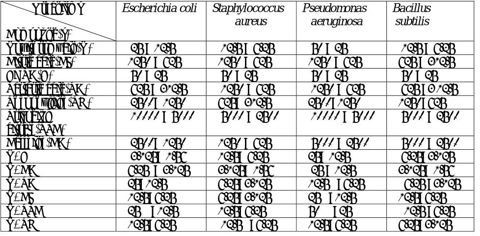
Table 1: MIC values for antimicrobial and potentiating agents (both alone and combined)
Range of MIC values (µg/mL) against test organisms
Organisms
Compound(s)
Escherichia coli Staphylococcus aureus
Pseudomonas
aeruginosa Bacillus subtilis
Roxithromycin(R) ≤ 25 > 12.5 ≤ 12.5 > 6.25 ≤ 50 > 25 ≤ 12.5 > 6.25 Citric acid(CI) ≤1250 > 625 ≤1250 > 625 ≤1250 > 625 ≤625 > 312.5 EDTA(E) ≤ 50 > 25 ≤ 50 > 25 ≤ 50 > 25 ≤ 50 > 25 Tartaric acid(TA) ≤ 625 > 312.5 ≤ 1250 > 625 ≤ 1250 > 625 ≤ 625 >3 12.5 Theophylline(TH) ≤ 2500> 1250 ≤625> 312.5 ≤2500>1250 ≤1250>625 Trisodium
citrate(TSC)
≤ 10000 > 5000 ≤ 5000 > 2500 ≤ 10000 > 5000 ≤ 5000 > 2500
Caffeine(CA) ≤ 2500> 1250 ≤1250 > 625 ≤ 5000 > 2500 ≤ 5000 > 2500
R+E ≤ 3.125> 1.56 ≤12.5> 6.25 ≤25> 12.5 ≤ 6.25>3.125
Table 2: Zone diameters after addition of potentiating agents to the medium
Organisms Zone diameter(mm)
R R+E R+CA R+TA R+CI R+TSC R+TH
B. subtilis 23 28 25 25 24 24 26
E. coli 22 22 29 26 22 25 26
P. aeruginosa 17 20 17 19 19 21 19
S. aureus 12 13 28 27 22 14 26
Table 3: Zone diameters when Roxithromycin was mixed with potentiating agents Organism Zone diameter(mm) R R+E R+CA R+TA R+CI R+TSC R+TH B. subtilis 23 23 24 24 23 22 22
E. coli 22 24 23 23 22 23 23
P. aeruginosa 12 14 14 13 13 13 13
S. aureus 12 12 13 12 12 11 11
Table 4: Concentration of Roxithromycin and Potentiating agents used for turbidimetric analysis Organism Concentration of potentiating agents and Roxithromycin (µg/mL) R E CA TA CI TSC TH B. subtilis 6.25 12.5 1250 78.125 156.25 1250 312.5 E. coli 12.5 12.5 625 156.25 312.5 2500 625
P. aeruginosa 25 12.5 625 156.25 312.5 2500 625
Table 5: Turbidity measurements of Roxithromycin and potentiating agents alone
Organism Nepheloturbidimetric units (NTU)
R E CA TA CI TSC TH control
+ -
B. subtilis 12.5 19.6 25.2 26.4 27.6 23.6 21.8 37.7 0.3
E. coli 9.8 18.4 22.1 24.6 31.5 25.1 22.8 39.6 1.2
P. aeruginosa 11.1 24.1 24.1 23.5 28.1 21.2 21.2 34.5 0.5
S. aureus 10.2 22 22.7 28.3 26.3 24.5 23.6 36.4 0.6
Table 6: Turbidity measurements of Roxithromycin in combination with potentiating agents
Organism Nepheloturbidimetric units (NTU)
R+E R+CA R+TA R+CI R+TSC R+TH
B. subtilis 6.6 2.7 8.7 4.5 10.5 4.9
E. coli 0.6 4.0 8.4 6.4 9.6 6.3
P. aeruginosa 4.3 2.9 4.4 10.6 8.5 11.2
S. aureus 7.8 3.1 8.1 9.8 7.9 7.2
Acknowledgement
References
1) Ayres, H. M., Furr, J. R. and Russel, A. D. Effect of permeabilizers on antibiotic sensitivity of Pseudomonas aeruginosa. Letters in applied microbiology, 1999; 28: 13-16.
2) Levy, S. B and Marshall, B. Antibacterial resistance worldwide: causes, challenges and responses, Nature Medicine Supplement, 2004, Vol. 10, No. 12, PP: S122-S129.
3) Davies, J. Inactivation of antibiotics and the dissemination of resistance genes. Science, 1994; 264: 375-384.
4) Spartt, B. G. Resistance to antibiotics mediated by target alterations. Science, 1994; 264:388-393.